The layout of the system can be seen in figure 1. The workstation has two dual output graphics adapters (nVidia Quadro. FX 1500) that provide the overlays to ...
Augmented Reality Image Guidance for Minimally Invasive Coronary Artery Bypass Michael Figl1 , Daniel Rueckert1 , David Hawkes3 , Roberto Casula4 , Mingxing Hu3 , Ose Pedro1 , Dong Ping Zhang1 , Graeme Penney5 , Fernando Bello2 , Philip Edwards2 1
2
Department of Computing, Imperial College London, UK Department of Biosurgery and Surgical Technology, Imperial College London, UK 3 Centre of Medical Image Computing, University College London, UK 4 Cardiothoracic Surgery, St. Mary’s Hospital, London, UK 5 Division of Imaging Sciences, King’s College London, UK ABSTRACT
We propose a novel system for image guidance in totally endoscopic coronary artery bypass (TECAB). A key requirement is the availability of 2D-3D registration techniques that can deal with non-rigid motion and deformation. Image guidance for TECAB is mainly required before the mechanical stabilization of the heart, thus the most dominant source of non-rigid deformation is the motion of the beating heart. To augment the images in the endoscope of the da Vinci robot, we have to find the transformation from the coordinate system of the preoperative imaging modality to the system of the endoscopic cameras. In a first step we build a 4D motion model of the beating heart. Intraoperatively we can use the ECG or video processing to determine the phase of the cardiac cycle. We can then take the heart surface from the motion model and register it to the stereo-endoscopic images of the da Vinci robot using 2D-3D registration methods. We are investigating robust feature tracking and intensity-based methods for this purpose. Images of the vessels available in the preoperative coordinate system can then be transformed to the camera system and projected into the calibrated endoscope view using two video mixers with chroma keying. It is hoped that the augmented view can improve the efficiency of TECAB surgery and reduce the conversion rate to more conventional procedures. Keywords: Image-Guided Therapy, Registration, Medical Robotics, Localization & Tracking Technologies, Endoscopic Procedures
1. INTRODUCTION 1.1 Augmented Reality Applications in Surgery Augmented reality (AR) systems applied to surgery aim to overlay additional information, most often in form of images or renderings, onto the real view of the surgeon. Using a stereoscopic device has the potential advantage of enabling 3-D perception of both the surgical field and overlays, potentially allowing virtual structures appear beneath the real surface as though the tissue were transparent. Some stereo AR devices have been developed for applications in medicine. Head mounted displays (HMD), for instance, have been proposed by Fuchs et al,1 Birkfellner et al2 or Wendt et al.3 HMDs generally suffer from fast head movements resulting in misregistration. Only few have therefore ever left their laboratories, and most appear in latency or stereo perception measurements, phantom, cadaver or animal studies.4–8 The use of AR in devices that remain in fixed positions for an amount of time, such as operating microscopes9, 10 or those that undergo tremor reduced movements such as the stereo-endoscopes used with the da Vinci robotic operating system are more promising to proceed to the operating theatre.
1.2 Purpose In this paper we describe a system for image-guided robotic surgical treatment of coronary artery disease. We aim to enhance the endoscopic view provided by the da Vinci robot with information from preoperative imaging. This requires construction of a fully 4D model of the patient from coronary CT, both temporal and spatial registration of this model to physical space and visualisation as overlays on the endoscopic view.
1.3 Clinical Need Totally endoscopic coronary artery bypass (TECAB) has the potential to treat coronary artery disease without the need for invasive sternotomy or heart-lung bypass. However there is still a conversion rate to more invasive methods of 20-30%. 11–13 This can occur if there is misidentification of the target vessel or difficulty in locating the artery if it is hidden by fat. We have identified two critical points in the procedure that might gain from intraoperative guidance. During harvesting of the left internal mammary artery the position of the bifurcation would be useful to know to allow surgery to progress rapidly to this point. After opening of the pericardium overlay of the target vessel will allow accurate location and identification. It is hoped that such guidance will make surgery more efficient and reduce the conversion rate for TECAB.
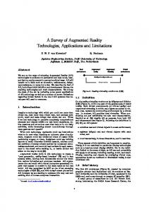
2. METHODS The layout of the system can be seen in figure 1. The workstation has two dual output graphics adapters (nVidia Quadro FX 1500) that provide the overlays to each eye and a multiple input framegrabbing device (Active Silicon, Uxbridge, UK) for the purposes of registration. Workstation Quad input frame grabber
GUI
Monitor
Dual output graphics
Monitor
da Vinci stack in
Left
out Right Camera controller
in
Video mixer out
in
Left
da Vinci out
master
Right Video converter
console da Vinci slave robot Endoscope
Control
Figure 1. The layout of the system in theatre.
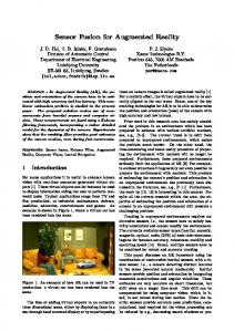
Overlay on the view through the da Vinci is provided using two video mixers (Panasonic WJ-MX 50) with chromakeying functionality. This ensures that there is no increased lag introduced by the system. An idea of the quality of chroma keyed overlay can be found in figure 2 To achieve guidance we require a 4D model of the beating heart. This must be both temporally and spatially registered to the patient. Finally the model must be visualised using the calibrated endoscope view.
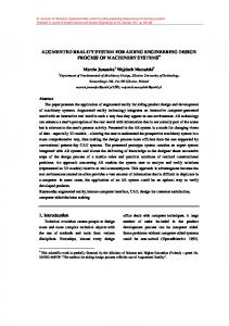
2.1 4D Model Construction The preoperative model of the patient comes from coronary CT, which provides a fully 4D representation of the patient. The CT can be reconstructed at up to 20 multiple even phases throughout the cardiac cycle, see figure 3 for an example. The relevant vessels must be segmented along with the surface of the myocardium. The motion of the heart can potentially be obtained from image registration.14, 15 We are investigating the use of a 4D statistical model to produce a segmentation16 and also the use of a subdivision surface representation of the heart with
a
b
Figure 2. The surface of a heart phantom and the overlay using chroma keying. a shows the rendering of the surface. The surface is truncated to visualise the difference to the heart phantom underneath as can be seen in b.
volume preservation to track the motion of the myocardium. 17 We will also apply the registration method in18 to our volumes. An example of a patient model can be seen in figure 5(a).
Figure 3. The phases of a heart displayed using the software rview. Only about half of the 20 phases are shown.
2.2 Model-based 2D-3D registration techniques Having obtained a preoperative model of the patient we now need to align this model to the video view through the da Vinci endoscope. In this step we will develop suitable non-rigid 2D-3D registration algorithms. Our strategy in performing registration will be to separate the temporal and spatial alignment of the preoperative model. Temporal alignment may be obtained using the ECG signal. However, there may be residual lag between the ECG and the video and we are investigating whether feature tracking in the video images could be used for this purpose. Feature tracking has been proposed as a means of reconstructing the surface viewed through the endoscope and tracking its motion.19 If corresponding features can be tracked in the left and right views through the da Vinci stereo-endoscope, these can provide 3D tracked points on the viewed surface. We propose this technique to measure the motion of the heart and to separate cardiac and respiratory motion. We are also examining whether geometry constraints can be used as an accurate means of finding the period of the heart cycle, in the present of rigid motion of the camera or near rigid motion due to breathing. Having established temporal registration, the remaining motion will be rigid apart from possible deformation of the heart due to breathing. Preliminary work has shown that 3D-3D non-rigid registration can be used to build separate models of respiratory motion of the liver 20 and heart.21 Since both respiratory and cardiac motion are smooth and continuous, we will develop 4D parametric motion models based on 4D B-splines. These motion models will provide compact representations of cardiac motions. To establish correspondence we are investigating two approaches. We are adopting a similar approach to that of 19 to reconstruct the motion of the viewed surface, which can then be registered to the preoperative 4D model. Secondly we are investigating whether intensity-based similarity metrics can be developed. We are using the concept of photoconsistency22, 23 as a similarity measure using the calibrated stereo views that are available on the da Vinci system. It is hoped that a combination of these techniques will be able to provide alignment of the 4D model from the cardiac CT images with the series of 2D video images grabbed through the endoscope.
2.3 Visualisation The da Vinci system provides the surgeon with endoscopic stereo video images of the surgical scene during coronary artery bypass. The goal of this step is to provide an augmented reality facility for the da Vinci system during this procedure. In order to achieve this we need to establish the relationship between camera coordinates and image coordinates (determined by the intrinsic transformation parameters) as well as the relationship between world coordinates and camera coordinates (determined by the extrinsic transformation parameters). Since the intrinsic parameters describe the internal properties of the stereo endoscopic cameras of the da Vinci system, we use an offline camera calibration technique to determine these parameters. 24, 25 As the image fusion is done by use of chroma keying with two video mixers, an additional 2D-2D transformation from the graphical output to the input channels of the mixer is needed. This is achieved by point to point registration. We will then use the model-based 2D-3D registration algorithm described previously to estimate the extrinsic transformation parameters. The resulting visualisation will be able to guide the surgeon during critical parts of the procedure.
3. RESULTS 3.1 4D Model Construction For preoperative model building we use coronary CT images reconstructed at 20 even phases throughout the cardiac cycle. To produce a 4D model we segment the first phase by hand and propagate this to the other 19 phases by non-rigid registration.18 This is similar to the method used by Szpala et al,15 but here it is demonstrated on clinical coronary CT scans rather than phantom data. Figure 4 gives an idea of the quality of the registration.
3.2 Visualisation An example of retrospectively aligned images is shown in figure 5. We have a number of clinical coronary CT images that are being used to investigate preoperative model construction. Registration algorithms using both feature tracking and intensity-based techniques are being developed.
a
b
Figure 4. Non-rigid registration was used to register the first image to the other images. In a the deformation of the ventricle is displayed. b shows the registered image slices from different phases (0% and 60 %) of the cardiac cycle.
4. DISCUSSION We propose a system for augmented reality image guidance of totally endoscopic coronary artery bypass. Previous work has suggested the use of image guidance in TECAB surgery and demonstrated its feasibility on a heart phantom.15 Falk et al have demonstrated a system for AR guidance based on multi-planar x-ray angiography. 26 We describe the first such results using clinical coronary CT scans to provide the 4D patient model using non-rigid registration. We also propose two novel strategies for alignment of this model to the endoscopic view. The first uses robust feature tracking to reconstruct the viewed surface, which can then be matched to the preoperative model. The second strategy uses intensity-based methods for registration. For augmentation of the endoscopic view we use video mixers, which does not introduce any lag to the surgeons view of the real surface. We use chroma-keying for the image fusion, which limits the range of available colours. This is not a significant limitation as we want colour separation between the overlaid view and the largely red surgical view. It is hoped that such information can improve the efficiency of TECAB surgery and reduce the conversion rate to more invasive procedures.
5. ACKNOWLEDGEMENTS We would like to thank the EPSRC for funding this project. We are also grateful to the theatre staff at St Mary’s hospital, London and to the radiology staff in St Mary’s and the Royal Brompton hospitals for their cooperation.
a
b
c
Figure 5. A rendering of the preoperative model showing the myocardial surface, left internal mammary artery, left anterior descending artery and a diagonal branch (a), an aligned rendering (b) and its corresponding endoscope view (c).
REFERENCES [1] H. Fuchs, M. A. Livingston, R. Raskar, D. Colucci, K. Keller, A. State, J. R. Crawford, P. Rademacher, S. H. Drake, and A. A. Meyer, “Augmented reality visualization for laparoscopic surgery,” in Proc. Medical Image Computation and Computer-Assisted Intervention, pp. 934–943, 1998. [2] W. Birkfellner, M. Figl, K. Huber, F. Watzinger, F. Wanschitz, J. Hummel, R. Hanel, W. Greimel, P. Homolka, R. Ewers, and H. Bergmann, “A head-mounted operating binocular for augmented reality visualization in medicine design and initial evaluation,” IEEE Trans. Med. Imaging 21, pp. 991–997, 2002. [3] M. Wendt, F. Sauer, A. Khamene, B. Bascle, S. Vogt, and F. K. Wacker, “A head-mounted display system for augmented reality: Initial evaluation for interventional mri,” R¨ofo-Fortschr. Gebiet R¨ontgenstrahlen Bildgeb. Verfahr. 175, pp. 418–421, 2003. [4] W. Birkfellner, M. Figl, C. Matula, J. Hummel, R. Hanel, H. Imhof, F. Wanschitz, A. Wagner, F. Watzinger, and H. Bergmann, “Computer-enhanced stereoscopic vision in a head-mounted operating binocular,” Phys. Med. Biol. 48, pp. N49–N57, 2003. [5] F. Wanschitz, W. Birkfellner, M. Figl, S. Patruta, A. Wagner, F. Watzinger, K. Yerit, K. Schicho, R. Hanel, F. Kainberger, H. Imhof, H. Bergmann, and R. Ewers, “Computer-enhanced stereoscopic vision in a head-mounted display for oral implant surgery,” Clin. Oral Implants Res. 13(6), pp. 610–6, 2002. [6] M. Figl, C. Ede, J. Hummel, F. Wanschitz, R. Ewers, H. Bergmann, and W. Birkfellner, “A fully automated calibration method for an optical see-through head-mounted operating microscope with variable zoom and focus,” IEEE Trans. Med. Imaging 24(11), pp. 1492–9, 2005. [7] F. Wacker, S. Vogt, A. Khamene, J. Jesberger, S. Nour, D. Elgort, F. Sauer, J. Duerk, and J. Lewin, “An augmented reality system for mr image-guided needle biopsy: initial results in a swine model,” Radiology 238(2), pp. 497–504, 2006. [8] M. Das, F. Sauer, U. Schoepf, A. Khamene, S. Vogt, S. Schaller, R. Kikinis, E. vanSonnenberg, and S. Silverman, “Augmented reality visualization for ct-guided interventions: system description, feasibility, and initial evaluation in an abdominal phantom.,” Radiology 240(1), pp. 230–5, 2006. [9] P. J. Edwards, A. P. King, C. R. Maurer, Jr., D. A. de Cunha, D. J. Hawkes, D. L. G. Hill, R. P. Gaston, M. R. Fenlon, A. Jusczyzck, A. J. Strong, C. L. Chandler, and M. J. Gleeson, “Design and evaluation of a system for microscope-assisted guided interventions (MAGI),” IEEE Trans. Med. Imaging 19(11), pp. 1082–1093, 2000. [10] P. J. Edwards, L. G. Johnson, D. J. Hawkes, M. R. Fenlon, A. J. Strong, and M. J. Gleeson, “Clinical experience and perception in stereo augmented reality surgical navigation,” in Proceedings of Medical Imaging and Augmented Reality, Lecture Notes in Computer Science (3150), pp. 369–376, Springer-Verlag, 2004. [11] U. Kappert, R. Cichon, J. Schneider, V. Gulielmos, T. Ahmadzade, J. Nicolai, S. M. Tugtekin, and S. Schueler, “Technique of closed chest coronary artery surgery on the beating heart,” Eur. J. Cardio-Thorac. Surg. 20, pp. 765– 769, 2001. [12] S. Dogan, T. Aybek, E. Andressen, C. Byhahn, S. Mierdl, K. Westphal, G. Matheis, A. Moritz, and G. WimmerGreinecker, “Totally endoscopic coronary artery bypass grafting on cardiopulmonary bypass with robotically enhanced telemanipulation: Report of forty-five cases,” J. Thorac. Cardiovasc. Surg. 123, pp. 1125–1131, 2002. [13] V. Falk, A. Diegeler, T. Walther, J. Banusch, J. Brucerius, J. Raumans, R. Autschbach, and F. W. Mohr, “Total endoscopic computer enhanced coronary artery bypass grafting,” Eur. J. Cardio-Thorac. Surg. 17, pp. 38–45, 2000. [14] M. Wierzbicky and T. Peters, “Determining epicardial surface motion using elastic registration: Towards virtual reality guidance of minimally invasive cardiac interventions,” in MICCAI 2003—Medical Image Computation and Computer-Assisted Intervention, Lecture Notes in Computer Science 2878, pp. 722–9, Springer-Verlag, 2003. [15] S. Szpala, M. Wierzbicki, G. Guiraudon, and T. M. Peters, “Real-time fusion of endoscopic views with dynamic 3-d cardiac images: A phantom study,” IEEE Trans. Med. Imaging 24, pp. 1207–1215, 2005. [16] D. Perperidis, R. Mohiaddin, P. Edwards, D. Rueckert, and Hill, “Segmentation of cardiac mr and ct image sequences using model-based registration of a 4d statistical model,” in Proc. SPIE Medical Imaging 2007, 6512, 2007. [17] R. Chandrashekara, R. Mohiaddin, R. Razavi, and R. Rueckert, “Nonrigid image registration with subdivision lattices: Application to cardiac mr image analysis,” in MICCAI 2007—Medical Image Computation and Computer-Assisted Intervention, Lecture Notes in Computer Science (1679), pp. 335–42, Springer-Verlag, 2007. [18] D. Rueckert, L. I. Sonoda, C. Hayes, D. L. G. Hill, M. O. Leach, and D. J. Hawkes, “Nonrigid registration using free-form deformations: Application to breast MR images,” IEEE Trans. Med. Imaging 18(8), pp. 712–721, 1999.
[19] D. Stoyanov, G. P. Mylonas, F. Deligianni, A. Darzi, and G. Z. Yang, “Soft-tissue motion tracking and structure estimation for robotic assisted mis procedures,” Medical Image Computing And Computer-Assisted Intervention MICCAI 2005 3750, pp. 139–146, 2005. [20] J. M. Blackall, G. P. Penney, A. P. King, and D. J. Hawkes, “Alignment of sparse freehand 3-d ultrasound with preoperative images of the liver using models of respiratory motion and deformation,” Ieee Transactions On Medical Imaging 24, pp. 1405–1416, 2005. [21] K. McLeish, D. L. G. Hill, D. Atkinson, J. M. Blackall, and R. Razavi, “A study of the motion and deformation of the heart due to respiration,” IEEE Trans. Med. Imaging 21, pp. 1142–1150, 2002. [22] M. J. Clarkson, D. Rueckert, D. L. G. Hill, and D. J. Hawkes, “A multiple 2D video-3D medical image registration algorithm,” in Proc. SPIE Medical Imaging 2000, 3979, pp. 342–352, 2000. [23] M. J. Clarkson, D. Rueckert, D. L. G. Hill, and D. J. Hawkes, “Using photo-consistency to register 2d optical images of the human face to a 3D surface model,” IEEE Trans. Pattern Anal. Mach. Intell. 23, pp. 1266–1280, 2001. [24] J. Bouguet, “Camera calibration toolbox for matlab.” Website, 2007. http://www.vision.caltech.edu/ bouguetj. [25] R. Y. Tsai, “A versatile camera calibration technique for high-accuracy 3D machine vision metrology using off-theshelf TV cameras and lenses,” IEEE J. Robotics and Automation 3(4), pp. 323–344, 1987. [26] V. Falk, F. Mourgues, L. Adhami, S. Jacobs, H. Thiele, S. Nitzsche, F. W. Mohr, and T. Coste-Maniere, “Cardio navigation: Planning, simulation, and augmented reality in robotic assisted endoscopic bypass grafting,” Ann. Thorac. Surg. 79, pp. 2040–2048, 2005.