However, excellent sequence can be obtained without band isolation, since multiple bands will have the same origins at the nested primer end of the reaction, and the final nested primer is also used in the cycle sequencing reaction. Therefore, when sequencing reactions were run with products containing several bands, the distant sequences were unaffected and were very readable. Forty-one samples sequenced without band isolation yielded an average of 212 bases/walk. Since the sequence farthest away from the primer is unaffected and can be used to select primers for an additional walk, band isolation is optional. This procedure compares favorably with others. Advantages include its simplicity (since it involves no DNA digestion or ligation), consistent results and speed. Cost is low since the expense of three primers per walk is balanced by a significant savings of technical time and in the cost of other reagents such as restriction enzymes and ligases. Using this procedure and a modification of an “enhancer trap” (8), we have rapidly identified several glucocorticoid-regulated genes in AtT-20 mouse pituitary tumor cells. REFERENCES 1.Arnold, C. and I.J. Hodgson. 1991. Vectorette PCR: a novel approach to genomic walking. PCR Methods Appl. 1:39-42. 2.Devon, R.S., D.J. Porteous and A.J. Brookes. 1995. Splinkerettes—improved vectorettes for greater efficiency in PCR walking. Nucleic Acids Res. 23:1644-1645. 3.Dominguez, O. and C. Lopez-Larrea. 1994. Gene walking by unpredictably primed PCR. Nucleic Acids Res. 22:3247-3248. 4.Fors, L., R.A. Saavedra and L. Hood. 1990. Cloning of the shark Po promoter using a genomic walking technique based on the polymerase chain reaction. Nucleic Acids Res. 18:2793-2799. 5.Ghiso, N.S., H. Parekh and G.G. Lennon. 1993. A subset of 1200 hexamers is sufficient to sequence over 95% of cDNAs by hexamer string primer walking. Genomics 17:798-799. 6.Hamada, H. 1986. Random isolation of gene activator elements from the human genome. Mol. Cell. Biol. 6:4179-4184. 7.Hamada, H. 1986. Activation of an enhancerless gene by chromosomal integration. Mol. Cell. Biol. 6:4185-4194. 8.Harrison, R.W. and J. Dusel. 1996. Functional identification of genes up- and downregulated by glucocorticoids in AtT-20 pituitary cells using an enhancer trap. Endocrinology 137:2758-2765. Vol. 22, No. 4 (1997)
9.Jones, D.H. and S.C. Winistorfer. 1993. Genome walking with 2-4-kb steps using panhandle PCR. PCR Methods Appl. 2:197-203. 10.Rosenthal, A. and D.S. Jones. 1990. Genomic walking and sequencing by oligo-cassette mediated polymerase chain reaction. Nucleic Acids Res. 18:3095-3096. 11.Sarkar, G.S., R.T. Turner and M.E. Bolander. 1993. Restriction-site PCR: a direct method of unknown sequence retrieval adjacent to a known locus by using universal primers. PCR Methods Appl. 2:318-322. 12.Screaton, G.R., C.R. Bangham and J.I. Bell. 1993. Direct sequencing of single primer PCR products: a rapid method to achieve short chromosomal walks. Nucleic Acids Res. 21:2263-2264. 13.Siebert, P.D., A. Chenchik, D.E. Kellogg, K.A. Lukyanov and S.A. Lukyanov. 1995. An improved PCR method for walking in uncloned genomic DNA. Nucleic Acids Res. 23:1087-1088.
Supported by NCI Grant No. CA 42091. The authors thank Ms. Andrea Weinstein for excellent secretarial assistance. Address correspondence to Robert W. Harrison, University of Rochester, 601 Elmwood Avenue, Box 693, Rochester, NY 14642, USA. Internet:
[email protected] Received 20 May 1996; accepted 4 October 1996.
Robert W. Harrison, Janice C. Miller, Mary J. D’Souza and George Kampo University of Rochester School of Medicine and Dentistry Rochester, NY, USA
Automated Fluorescent DNA Sequencing by a Simplified Solid-Phase Chemical Sequencing Method BioTechniques 22:653-656 (April 1997)
DNA sequencing is now done principally by two methods, the chemical degradation method of Maxam and Gilbert (2) and the enzymatic chain termination method of Sanger et al. (9). Although the enzymatic method is overwhelmingly preferred in largescale DNA sequencing projects, the
Benchmarks chemical method remains indispensable under some circumstances, such as the determination of sequences refractory to DNA polymerase reaction. Furthermore, the chemical sequencing method is, at least in principle, robust and cost-effective. However, an obvious drawback has been its tediousness: many lyophilization or DNA precipitation steps for removal of chemical reagents are usually required to complete the sequencing reactions; therefore, this method is avoided if possible. We recently reported a solid-phase chemical DNA sequencing method using streptavidin-coated magnetic beads that eliminated much of the tedium (5). This method enabled us to: (i) purify DNA templates by simple magnetic capture for removal of chemical impurities that affect chemical sequencing reactions; (ii) carry out all the base modification reactions at ambient temperature; and (iii) purify the sequencing reaction products by a simple magnetic capture-washing cycle, without the use of conventional DNA precipitation steps. We now report an improved solid-phase chemical DNA sequencing method suitable for automated fluorescent DNA sequencers. Modifications introduced in the solid-phase chemical sequencing method are as follows: (i) the use of a polymerase chain reaction (PCR) primercarrying biotin and fluorescein moieties (in this order) at the 5′ end, so that sequencing reaction products can be detected by fluorescence and retrieved with streptavidin-coated magnetic beads and (ii) replacement of the timeconsuming piperidine (PIP) evaporation step with a magnetic capturewashing cycle. The protocol is given in Table 1, and the details are described below. A biotin/fluorescein-labeled M13reverse primer (BF-RV primer; 5′-biotin-fluorescein-GAGCGGATAACAATTTCACACAGG-3′) was synthesized with Biotin-ON and FluoresceinON Phosphoramidites (CLONTECH Laboratories, Palo Alto, CA, USA) and purified using reversed-phase, high-performance liquid chromatography (TOYOBO, Osaka, Japan). A template DNA, murine cot gene fragment [ApaIPstI fragment of CZ-21 (4)], was amplified from a subcloned plasmid derived 654 BioTechniques
Table 1. Protocol of the Chemical Degradation Method
1. Generation of labeled DNA templates with biotin and fluorescein a PCR amplification (100 µL) with 10 pmol each of BF-RV primer and unlabeled M13 forward primer. 2. Purification of the labeled DNA templates by magnetic capturea Add 50 µL of the PCR mixture and 12.5 µL of 5 M NaCl to SA-Mg pellet (250 µg). After formation of SA-Mg/DNA complexes, suspend them in 50 µL of deionized H2O (dH2O) after extensive washing with dH2Ob. 3. Base modification reactionsa G reaction: Treat SA-Mg/DNA complexes (10 µL of the suspension) in 30 µL of 0.5% (vol/vol) dimethyl sulfate in 1 mM EDTA for 1 min at room temperature (RT). A reaction: Treat SA-Mg/DNA complexes (10 µL of the suspension) in 30 µL of 3 mM K2PdCl4 in 3.3 mM acetic acid for 5 min at RT, and then add 2 µL of 100 mM 2-mercaptoethanol. T reaction: After denaturation of DNA on SA-Mg (15 µL of the suspension) with NaOH, followed by extensive washing with dH2Oa, treat SA-Mg/DNA complexes in 30 µL of 0.1 mM KMnO4 in 1 mM Tris-HCl (pH 8.8) for 8 min at RT. C reaction: After denaturation of DNA on SA-Mg (15 µL of the suspension) with NaOH, followed by extensive washing with dH2Oa, treat SA-Mg/DNA complexes in 30 µL of 4 M hydroxylammonium chloride for 3 min at RT. 4. PIP cleavage reaction After each base modification reaction, wash SA-Mg/DNA complexes with dH2O extensivelyb, and then resuspend them in 30 µL of dH2O. Add 7.5 µL of 5 M PIP to the suspension and incubate for 30 min at 90°C. 5. Recovery of the sequencing reaction products Transfer the DNA (35 µL), eluted from SA-Mg by PIP reaction, to a new tube. Add 20 µL of 3 M sodium acetate, 15 µL of 5 M NaCl and 5 µL of SA-Mg suspension (10 mg/mL in 1 mM Tris-HCl [pH 7.5] and 0.5 mM EDTA) to the tube in this order. After 15 min incubation at RT, wash the SA-Mg/DNA complexes four times with dH2Ob, and suspend them in 2 µL of 80% formamide/10 mM NaOH for DNA sequencing electrophoresis. aDetails
are described in Reference 3. was washed by repeated cycles of magnetic capture/resuspension as described by the supplier’s instructions.
bSA-Mg
from pBluescript® (Stratagene, La Jolla, CA, USA) with BF-RV primer and unlabeled M13 forward primer (5′GTTGTAAAACGACGGCCAGT-3′) by PCR as described previously (5). After the PCR, the amplified DNA (about 600 bp) was recovered by streptavidincoated magnetic beads (SA-Mg; Dynal, Oslo, Norway) as described in the manufacturer’s instructions and washed extensively with water. Conditions used for the respective chemical modification reactions are shown in Table 1. Although the reaction with KMnO4 at high temperature is known to increase the uniformity of the T-specific reaction (3), we conducted the T-specific reaction at room temperature because ele-
vated temperature results in the dissociation of biotinylated DNAs from streptavidin-coated magnetic beads. We used SA-Mg to remove PIP, since SA-Mg could bind the sequencing reaction products in the presence of PIP after the pH of the PIP reaction mixtures was lowered. Finally, SA-Mg/DNA complexes suspended in 2 µL of formamide solution (80% formamide and 10 mM NaOH) were kept at -20°C until use. Just before running on a sequencing gel, the SA-Mg/DNA complexes were heatdenatured at 95°C for 2 min and briefly centrifuged to collect SA-Mg at the bottom. Two microliters of the resultant formamide solution of each reaction were analyzed on a DSQ-1000 DNA Vol. 22, No. 4 (1997)
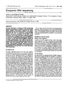
Figure 1. A sequence chart obtained by the DSQ-1000 automated fluorescent DNA Sequencer. Up to 600 bp could be read from the primer, but only the sequence chart including a GC-rich region is shown here. The results of the automated base calling are shown above the fluorescent signal chart, and the numbers indicated are the residue numbers from the primer binding site.
Sequencer (Shimadzu, Kyoto, Japan) using 4.2% Long Ranger Gels (FMC BioProducts, Rockland, ME, USA) containing 7 M urea. Figure 1 shows a typical sequencing chart resulting from the use of this method. Although this DNA contained a relatively GC-rich region (the GC content is 63% around residues 200–250, bottom panel of Figure 1), we could read the whole region of the DNA insert, i.e., up to 600 bases from the primer. The quality of the sequence pattern appeared to be as good as that obtained by the conventional enzymatic method, except that T-specific signals became unusually low at some positions because of the local secondary structure as described in the original paper (8). The overall quality of the sequencing results was due mainly to the successful removal of PIP by the magnetic capture/washing step. Contamination by a trace amount of PIP results in considerably reduced band resolution. The PIP removal step is still a timeconsuming obstacle for large-scale sample processing by chemical sequencing methods using an ion-exchange carrier matrix (7) or microplates (1). However, this new solid-phase method allows us to greatly speed up this step. Although we demonstrated the performance of the new solid-phase chemical sequencing method by using a four-lane, one-dye fluorescent DNA sequencer, this method could be, at least in principle, also applicable to a onelane, four-dye fluorescent DNA seVol. 22, No. 4 (1997)
quencer. However, due to commercial unavailability of a set of the fluorescent labeling reagents, it would be difficult to prepare a set of biotinylated PCR primers labeled with four different dyes suitable for the four-color DNA sequencer. Thus, as an alternative approach, we are now trying to develop a method to label PCR products with biotin and each of the four fluorophores without the use of biotinylated fluorescent primers, so as to make this method readily applicable to the four-color sequencers. On the other hand, it should be emphasized that this method minimizes the potential for exposure of researchers to hazardous chemicals through complete replacement of the tedious, time-consuming reagent-removal steps by the simple magnetic capture-washing step. In particular, the magnetic capture-washing cycle was found to be more rapid and, at the same time, a safer way to remove PIP from many sequencing samples, than by the repeated evaporation or DNA precipitation step in the conventional chemical method. Since the fluorescent chemical method reported so far (6) included at least one reaction that generated degenerated base-specific degradation products, the on-line base-calling software for the enzymatic method could not be used directly for automatic base calling from the charts obtained by the chemical method. However, since our new method generates G-, A-, T- and C-specific cleavage products, the automated DNA sequencers currently available for the enzymatic method could be used
Benchmarks for this chemical sequencing method, maintaining the capability of base calling automatically without any alteration of the system. Furthermore, due to successful removal of contaminating chemicals before gel electrophoresis, the readable length per single run by this method appears to be longer than that by the fluorescent chemical sequencing methods described previously (6). Besides DNA sequencing, this protocol could be extended easily to other applications that involve the same reactions as the chemical DNA sequencing, such as a chemical footprinting experiment. REFERENCES 1.Church, G.M. and S. Kiefer-Higgins. 1988. Multiplex DNA sequencing. Science 240:185188. 2.Maxam, A.M. and W. Gilbert. 1977. A new method for sequencing DNA. Proc. Natl. Acad. Sci. USA 74:560-564. 3.McCarthy, J.G. 1989. An improvement in thymine specific chemical DNA sequencing. Nucleic Acids Res. 17:7541. 4.Ohara, R., J. Miyoshi, M. Aoki and K. Toyoshima. 1993. The murine cot proto-oncogene: genome structure and tissue-specific expression. Jpn. J. Cancer Res. 84:518-525. 5.Ohara, R. and O. Ohara. 1995. A new solidphase chemical DNA sequencing method which uses streptavidin-coated magnetic beads. DNA Res. 2:123-128. 6.Rosenthal, A. 1993. DNA sequencing by chemical degradation using one, two, and four different fluorophores. Methods Mol. Biol. 23:261-280. 7.Rosenthal, A., R. Jung and H.-D. Hunger. 1987. Solid-phase methods for sequencing oligonucleotides and DNA. Methods Enzymol. 155:301-331. 8.Rubin, C.M. and C.W. Schmid. 1980. Pyrimidine-specific chemical reactions useful for DNA sequencing. Nucleic Acids Res. 8:46134619. 9.Sanger, F., S. Nicklen and A.R. Coulson. 1977. DNA sequencing with chain terminating inhibitors. Proc. Natl. Acad. Sci. USA 74:5463-5467.
Microassay for the Assessment of Low Levels of Hydroxyproline BioTechniques 22:656-658 (April 1997)
In connective tissue research, the number of collagenous proteins or their breakdown products is usually determined by assessing the hydroxyproline content. In tissue samples, measurement of this amino acid can be performed with relative ease, but the determination of hydroxyproline-containing peptides in medium conditioned by tissue explants or cells proves to be more difficult (3). Usually the amount of collagenous peptides in conditioned medium is substantially lower than in tissue samples, and measurements are compromised by the interference of medium constituents such as bovine serum albumin (BSA) and serum added to the culture medium. As our research focuses on breakdown processes of collagen in small tissue explants, a reliable assay was required that enabled us to detect hydroxyproline in conditioned medium in the nanomolar range, bypassing the need to set up a comparably sensitive high-pressure liquid chromatography assay (5,7,8). To this end, we have developed a microassay based on the colorimetric assay introduced by Stegemann and Stalder (6) and modified by Guis et al. (4).
Fragments (ca. 4 mm2) of periosteum from the concave and convex aspects of rabbit (New Zealand White, 8 days old) calvariae were isolated and cultured in Iscove’s Modified Dulbecco’s Medium (IMDM) supplemented with amphotericin, streptomycin and penicillin and 10% normal rabbit serum (NRS) (all from Life Technologies, Gaithersburg, MD, USA) or 4 mg/mL BSA (Sigma Chemical, St. Louis, MO, USA) as described previously (2). Media were collected after 24, 48 and 72 h of culture, immediately frozen and stored at -20°C until further processing. For the hydroxyproline assay, duplicate standards were prepared from IMDM supplemented with antibiotics, 10% NRS or 4 mg/mL BSA and 0–5 µg/mL hydroxyproline. Conditioned medium and standards (200 µL) were precipitated overnight at 4°C with 400 µL absolute ethanol in 2-mL microcentrifuge test tubes. Precipitates were pelleted by centrifugation for 15 min at 4°C, 1710× g and washed with 200 µL 70% ethanol. Supernatants were collected with care because the pellets can easily come off, particularly in samples containing serum. Both supernatants were combined in Kimax tubes with Teflon-lined screwcaps (Kimble, Illinois, OH, USA), and the pellets were discarded. Following vacuum-evaporation, the samples were hydrolyzed for 3.5 h at 135°C in 300 µL of 6 N HCl, again desiccated and dissolved in 200
Address correspondence to Osamu Ohara, Kazusa DNA Research Institute, 1532-3 Yana, Kisarazu, Chiba 292, Japan. Internet:
[email protected] Received 24 June 1996; accepted 15 October 1996.
Reiko Ohara, Ayako Tanaka and Osamu Ohara Kazusa DNA Research Institute Chiba, Japan 656 BioTechniques
Figure 1. Standard curves made up in IMDM containing 4 mg/mL BSA or 10% NRS and hydroxyproline added at various concentrations, showing a linear relationship between the extinction and hydroxyproline content. Data are expressed as mean ± standard deviation of 4 measurements. Vol. 22, No. 4 (1997)