www.nature.com/scientificreports
OPEN
received: 17 December 2015 accepted: 30 March 2016 Published: 21 April 2016
Automated microfluidic platform of bead-based electrochemical immunosensor integrated with bioreactor for continual monitoring of cell secreted biomarkers Reza Riahi1,2, Seyed Ali Mousavi Shaegh1,2, Masoumeh Ghaderi1,2, Yu Shrike Zhang1,2,3, Su Ryon Shin1,2,3, Julio Aleman1,2, Solange Massa1,2, Duckjin Kim1,2, Mehmet Remzi Dokmeci1,2,3 & Ali Khademhosseini1,2,3,4,5 There is an increasing interest in developing microfluidic bioreactors and organs-on-a-chip platforms combined with sensing capabilities for continual monitoring of cell-secreted biomarkers. Conventional approaches such as ELISA and mass spectroscopy cannot satisfy the needs of continual monitoring as they are labor-intensive and not easily integrable with low-volume bioreactors. This paper reports on the development of an automated microfluidic bead-based electrochemical immunosensor for in-line measurement of cell-secreted biomarkers. For the operation of the multi-use immunosensor, disposable magnetic microbeads were used to immobilize biomarker-recognition molecules. Microvalves were further integrated in the microfluidic immunosensor chip to achieve programmable operations of the immunoassay including bead loading and unloading, binding, washing, and electrochemical sensing. The platform allowed convenient integration of the immunosensor with liver-on-chips to carry out continual quantification of biomarkers secreted from hepatocytes. Transferrin and albumin productions were monitored during a 5-day hepatotoxicity assessment in which human primary hepatocytes cultured in the bioreactor were treated with acetaminophen. Taken together, our unique microfluidic immunosensor provides a new platform for in-line detection of biomarkers in low volumes and longterm in vitro assessments of cellular functions in microfluidic bioreactors and organs-on-chips. Microfluidic bioreactors and organs-on-chips are emerging devices to model physiological functions of tissues and organs in a controlled environment1. These devices produce biomimetic units that can recapitulate tissue- and organ-level functions for applications such as disease modeling, and pre-clinical in vitro drug screening1–6. In order to perform a precise analysis, often alteration of cellular behavior and response to the changes in the microenvironment should be continuously monitored, ideally using integrated in-line sensors1. Such sensors should be capable of continuous measurement of single or multiple analytes in small sample volumes for extended periods of time particularly for applications that deal with chronic or retarded cellular reactions to certain drugs or conditions. In addition, a sensing platform should allow for convenient integration with a microfluidic bioreactor with the capability of automated interface and integrated control7, which can improve the accuracy of measurements. Conventional approaches such as ELISA and mass spectroscopy cannot fulfill the requirements of continual monitoring because they are labor-intensive and not easily integrable with low-volume bioreactors. Thus, development of
1 Harvard-MIT Division of Health Sciences and Technology, Massachusetts Institute of Technology, Cambridge, MA 02139, USA. 2Biomaterials Innovation Research Center, Department of Medicine, Brigham and Women’s Hospital, Harvard Medical School, Cambridge, MA 02139, USA. 3Wyss Institute for Biologically Inspired Engineering, Harvard University, Boston, MA 02115 USA. 4Department of Physics, King Abdulaziz University, Jeddah 21569, Saudi Arabia. 5 College of Animal Bioscience and Technology, Department of Bioindustrial Technologies, Konkuk University, Hwayang-dong, Kwangjin-gu, Seoul 143-701, Republic of Korea. Correspondence and requests for materials should be addressed to A.K. (email:
[email protected])
Scientific Reports | 6:24598 | DOI: 10.1038/srep24598
1
www.nature.com/scientificreports/ microfluidic platforms with integrated in-line sensing capabilities for long-term continual analysis plays a vital role in the advancement of organs-on-chip devices for in vitro assessments of cellular functions. Integration of analytical detection methods with microfluidics can potentially improve the detection performance by reducing the analysis time, decreasing the consumption of liquid samples and reagents, and increasing reliability through standardization and automation8,9. Among the available analytical methods integrated with microscale systems to measure biomolecules10–19, electrochemical (EC) techniques20–23 are highly suited for microfluidic systems. This is mainly due to easy miniaturization of detection elements and high degree of integration ability with analytical functions for analysis of small-volume biochemical samples at low cost22. These features of EC methods allow for fabrication of compact sensing platforms that are capable of continuously detecting biomolecules with high selectivity, as well as, convenient access by end users. In particular, EC immunosensors for bimolecular detection or kinetic analysis take advantage of the high sensitivity and specificity of the antibody-antigen interactions, whereby antibodies are usually immobilized on the surface of the EC electrode for antigen detection8,22. However, such a surface immobilization strategy hinders the use of the sensor for continual analysis of biomolecules due to the saturation of the electrodes over repeated detection cycles23. An approach to construct multi-use EC immunosensors relies on the use of disposable microbeads to immobilize antigen-recognition molecules24. Once a cycle of measurement is completed, the microbeads can be replaced with new ones. This approach enables the microelectrode to be used repeatedly over many measurements. Microbeads provide a large surface area to immobilize recognition molecules in large quantities, which is conveniently modulated by the number of microbeads introduced into the system. In particular, magnetic microbeads (MBs) can be efficiently manipulated in a microfluidic chip in the presence of an external magnetic field and easily flushed out by the liquid flow upon deactivation of the magnet25. These features provide necessary capabilities to realize microfluidic MBs-based immunosensors in combination with EC detection methods for biomarker detections8 such as Immunoglobulin G (IgG)26,27, cancer biomarkers28, and bacteriophage MS229. Here, we introduce a MBs-based EC immunosensor contained in a microfluidic perfusion circuitry integrated with a microfluidic liver bioreactor for continual on-chip detection of cell-secreted biomarkers (Fig. 1). Initially, an off-chip arrangement using a well plate was conducted to optimize different experimental parameters affecting the sensitivity of the EC immunoassay. The immunoassay was then implemented in a microfluidic chip with integrated microvalves for automated on-chip detection of a target biomarker (Fig. 1a,b). On-chip manipulation of the required aqueous reagents for biomarker detection was carried out using an automated valve controller system. The microfluidic sensing platform could accurately perform loading and unloading of the MBs, liquid handling for antibody-antigen interactions, covalent binding of streptavidin horseradish peroxidase (SA-HRP) to secondary antibody, as well as sample extraction from the bioreactor. The sensing platform was characterized against HepG2 liver bioreactors for the wide range detection of transferrin (TF) as a known liver biomarker30. This protein is mainly synthesized in the liver and it has a critical role to transport and deliver iron in various metabolic processes. The sensing system was further fully integrated with the microfluidic bioreactor containing bioprinted primary human hepatocyte spheroids (Figs 1c and S1), demonstrating its capability of long-term continual monitoring of changes in the level of secreted liver biomarkers including TF and albumin (ALB). Finally, drug toxicity assessment was conducted using this integrated system by monitoring the effect of different acetaminophen (APAP) concentrations on the production rate of the cell-secreted biomarkers.
Results and Discussion
Optimization of the bead-based sensing method. Prior to conducting on-chip detection of cell-secreted biomarkers, the immunosensing assay was characterized and optimized off-chip using standard biomarker solutions based on a chessboard titration method. The effect of operating parameters including the number of MBs as well as amount of secondary antibody and SA-HRP on the overall assay time was investigated, as indicated in Fig. 2. The effect of each parameter was evaluated one at a time, and the optimized parameter was adopted for the subsequent experiments. Initially, the quantity of the primary antibody-coated MBs was optimized in the range of 500 to 10,000 beads. Other parameters, including the concentration of the secondary antibody, concentration of SA-HRP, and the incubation time were maintained constant at 2.5 μg/mL, 1 μg/mL, and 30 min, respectively. Incubation process was required for each of the following binding steps: (1) capturing of the target biomarker (antigen) by primary antibody-coated MBs, (2) binding of the secondary antibody to the biomarker, and (3) attachment of SA-HRP to the biotinylated secondary antibody residing on the MBs. The required time for HRP-catalyzed TMB oxidation was fixed at 15 min. TF with a concentration of 4,000 ng/mL was used as a standard solution for all amperometric optimization measurements to cover the wide physiological range. Figure 2a illustrates the measured current corresponding to TF detection at different quantities of captured MBs obtained at − 100 mV applied potential. While the EC signal improved by 40% as the number of beads increased from 5,000 to 7,500 beads, non-significant signal enhancement was observed for subsequent increase in the number of beads. Thus, the number of 7,500 beads was selected for the following experiments. As shown in Fig. 2b, the current response as a function of the secondary antibody concentration was studied within the range of 1.0–10.0 μg/mL. When the concentration of the secondary antibody was increased from 1.0 μg/mL to 2.5 μg/mL, the signal improved more than 1.6 fold from 0.43 μA to 0.71 μA. However, a further increase in antibody concentration resulted in negligible signal improvements. In addition, at concentrations above 2.5 μg/mL, the deviation of the signal response from the average value was large due to non-specific protein binding to the channel surface. Consequently, to reduce the overall operation cost of the sensor associated with the amount of the antibody and maintain a high signal-to-noise ratio, a detection antibody concentration of 2.5 μg/mL was selected as the optimal value for further experiments. The EC immunosensor was subsequently characterized to find a proper value for SA-HRP concentration and to determine the lowest limit of detection. As shown in Fig. 2c, current response was measured using SA-HRP within the range of 0.01–10.0 μg/mL. As the concentration of SA-HRP was increased from 1.0 μg/mL to 10.0 μg/mL, the signal response showed only 15% improvement, whereas, there Scientific Reports | 6:24598 | DOI: 10.1038/srep24598
2
www.nature.com/scientificreports/
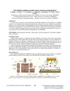
Figure 1. Design, fabrication, and detection principle of the bead-based microfluidic EC sensing system. (a) Illustration of the immunosensing principle with the EC sensor for detection of target biomarkers. (b) Configuration of the fabricated microfluidic sensing chip, (i) photograph of the microfluidic chip with an integrated microelectrode, (ii) photograph of the microelectrode, (iii) photograph of the reaction chamber with oxidized TMB, (iv) transfer of oxidized TMB to the detection chamber. (c) Schematic of the microfluidic sensing system integrated with organs-on-chip for continual monitoring of a target biomarker. was a pronounced increase in signal intensity between 0.01 μg/mL and 1.0 μg/mL. More importantly, the increase in the sensor response at 10.0 μg/mL was accompanied by a large deviation from the average signal value causing low assay sensitivity. Thus, a concentration of 1.0 μg/mL was selected as the optimal value for SA-HRP. In addition to optimization of reaction parameters, the effect of incubation time on the amperometric current response was evaluated. Figure 2d revealed that the incubation time affected both the sensor sensitivity and the overall required time to perform the EC immunoassay. A significant rise in current signal was observed when the incubation time was increased from 15 to 30 min; however, longer incubations did not have further significant impact on the measured signal. Thus a 30 min incubation time was selected for each binding step, providing a total of 90-min incubation period. Together, the optimal process for the EC immunosensor consisted of using 7,500 MBs for immobilizing 2.5 μg/mL of detection antibody in combination with 1.0 μg/mL of SA-HRP. A 30-min incubation time was considered sufficient for each binding step at room temperature while 15 min was used as a required time for completion of HRP-catalyzed enzymatic reactions for TMB oxidation.
Scientific Reports | 6:24598 | DOI: 10.1038/srep24598
3
www.nature.com/scientificreports/
Figure 2. Optimization of the bead-based immunoassay for biomarker EC detection using 4,000 ng/mL TF concentration. (a) The number of trapped MBs in the reaction chamber. Effect of (b) detection antibody concentration, (c) SA-HRP, and (d) incubation time on antigen detection while all other parameters were fixed. Data are representative of three independent experiments (ns = not significant).
Off-chip biomarker detection. The performance of the bead-based immunosensor for detecting TF bio-
marker was then evaluated off-chip and results were compared with a commercial ELISA kit. Employing optimal conditions, the amperometric measurements were initially carried out using standard TF solutions over the concentration range of 0–16,000 ng/mL (Fig. 3a) to obtain a calibration curve. The amperometric current response reached a plateau within 5 s. Absolute current values increased as a function of increasing oxidized-TMB concentration. Figure 3b illustrates a semi-log calibration curve using the current values obtained at the 5th second of the amperometric measurement. A linear curve was fitted to the data using a regression analysis for the TF concentrations in the linear range between 10 and 4,000 ng/mL (inset). The regression equation was y = 0.079(x) + 0.044, where y signified the current and x was the TF concentration (R2 = 0.99). Results revealed that the average relative standard deviation of the signals in the linear range of TF concentrations was