TB, MB, PM/240321, 9/03/2007 IOP PUBLISHING
PHYSIOLOGICAL MEASUREMENT
Physiol. Meas. 28 (2007) 1–14
UNCORRECTED PROOF
Automatic detection of left ventricular ejection time from a finger photoplethysmographic pulse oximetry waveform: comparison with Doppler aortic measurement Gregory S H Chan1,2, Paul M Middleton1,3, Branko G Celler1, Lu Wang1 and Nigel H Lovell1,2,4 1 Biomedical Systems Laboratory, School of Electrical Engineering and Telecommunications, University of New South Wales, Sydney, NSW, 2052, Australia 2 Graduate School of Biomedical Engineering, University of New South Wales, Sydney, NSW, 2052, Australia 3 Prince of Wales Clinical School, University of New South Wales, Sydney, NSW 2031, Australia 4 National Information and Communications Technology Australia (NICTA), Eveleigh NSW 1430, Australia
E-mail:
[email protected]
Received 27 December 2006, accepted for publication 23 February 2007 Published DD MMM 2007 Online at stacks.iop.org/PM/28/1 Abstract Left ventricular ejection time (LVET) is a useful measure of ventricular performance and preload. The present study explores a novel method of continuous LVET monitoring using a noninvasive finger photoplethysmographic pulse oximetry waveform (PPG-POW). A method for the automatic beat-to-beat detection of LVET from the finger PPG-POW is presented based on a combination of derivative analysis, waveform averaging and rule-based logic. The performance of the detection method was evaluated on 13 healthy subjects during graded head-up tilt. Overall, the correlation between the PPG-POW derived LVET and the aortic flow derived LVET was high and significant (r = 0.897, p < 0.05). The bias was −14 ± 14 ms (mean ± SD), and the percentage error was 9.7%. Although these results would not be sufficient to satisfy the requirement for clinical evaluation of LVET when absolute accuracy was demanded, the strong correlation between the PPG-POW LVET and the aortic LVET on an intra-subject basis (r = 0.945 ± 0.043, mean ± SD) would support the application of PPG-POW to detect the directional change in LVET of an individual. This could be very useful for the early identification of progressive hypovolaemia or blood loss. The present study has demonstrated a promising approach to extract potentially useful information from a noninvasive, easy-to-obtain signal that could be readily acquired either from existing patient monitoring equipment or from inexpensive instrumentation. More extensive investigation 0967-3334/07/000001+14$30.00 © 2007 IOP Publishing Ltd Printed in the UK
1
2
G S H Chan et al
is necessary to evaluate the applicability of the present approach in clinical care monitoring. Keywords: left ventricular ejection time (LVET), systolic time intervals (STI), photoplethysmogram (PPG), pulse oximetry (Some figures in this article are in colour only in the electronic version)
1. Introduction Left ventricular ejection time (LVET) was one of the first noninvasive measures used in cardiology for the assessment of ventricular performance. An early application of LVET was the identification of heart failure (Weissler et al 1968, Lewis et al 1977). More recently, it has been suggested that the decrease in LVET resulting from preload reduction could aid early identification of progressive hypovolaemia or blood loss (Geeraerts et al 2004). Clinicians often find it difficult to detect hypovolaemia at an early stage due to the insensitivity of blood pressure to small amounts of blood loss. There are potentially great benefits to the critical care clinicians if small volume losses could be diagnosed early, accurately and reproducibly simply by the assessment of a noninvasively monitored variable. Common LVET monitoring techniques, including phonocardiograms, carotid tonometry, thoracic bioimpedance, echocardiograms and continuous/pulsed wave Doppler flow techniques, are based on measurements at or close to the heart. The main reason given for this is that as the aortic pulse wave travels along the arterial tree it would be modified by the aggregated influences of vessel compliance and wave reflection (Nichols and O’Rourke 1998), and as a result, the pulse waves measured at a peripheral site may have lost the characteristics required for reliable LVET estimation. Although estimation of LVET with absolute accuracy might not be achievable with peripheral measurements, Geeraerts et al (2004) demonstrated that peripheral LVET measured from a finger blood pressure waveform could reflect variation of central LVET during simulated hypovolaemia. Unfortunately, noninvasive blood pressure devices (such as Finapres) so far have not been widely employed in patient care, and in current medical practice, continuous monitoring of arterial pressure waveform requires insertion of a radial artery catheter, which is an invasive procedure with its use limited to particular patient groups due to demands in terms of expertise and equipment. An alternative method to record a peripheral pulse waveform is photoplethysmography (PPG). By attaching an optical sensor probe to the finger or the ear, a peripheral blood volume pulse can be acquired in a noninvasive and comfortable manner. There has been significant amount of research interest on the PPG signal in recent years, partly because of the ability to design personal health care devices using low cost and miniature PPG sensors (Asada et al 2003), and also because of the widespread use of pulse oximetry as a routine patient monitoring technique in clinical care for the measurement of arterial blood oxygen saturation (SpO2). It has been suggested that the photoplethysmographic pulse oximetry waveform (PPG-POW) carries significant information related to the patient’s cardiovascular function which is yet to be fully utilized (Murray and Foster 1996, Middleton and Henry 2000). The application of PPG in LVET monitoring is by no means new. Early work in the 1970s showed that the first derivative of the ear densitogram provided an estimate of LVET that was strongly correlated with the carotid measurement (Quarry-Pigott et al 1973). Limited by the availability of computational resources at that time, the measurement of LVET required manual detection of the characteristic points from the ear waveform, which could be a labour
Automatic detection of left ventricular ejection time from a photoplethysmographic pulse oximetry waveform
3
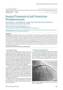
intensive task. However, with the rapid advancement in computer technology and signal processing techniques in the last two decades, automatic detection of arterial pulse waveform features has become more feasible (Oppenheim and Sittig 1995, Karamanoglu 1997). The aim of the present study is to present a method for the automatic detection of LVET from the finger PPG-POW signal and to evaluate the reliability of the measurement by direct comparison with the LVET derived from Doppler aortic flow velocity. The aortic flow derived LVET is believed to closely resemble the true LVET from a physiological viewpoint since it is measured at the ascending aorta. The comparison was made in two ways: first, examination of the correlation between the PPG-POW LVET and the aortic LVET; second, determination of the level of agreement between the two methods using the technique proposed by Bland and Altman (1986). Correlation and agreement were assessed not only on a pooled basis (with all data included), but also on an intra-subject basis. To investigate the performance of the PPG-POW technique for tracking individual change in LVET, the subjects underwent graded head-up tilt, which was capable of simulating a range of volume status and inducing a wide range of LVET (Stafford et al 1970). 2. Methods 2.1. Subject 13 healthy subjects (12 males and 1 female, mean age of 30 years, range 180–44 years) were studied. Prior to the experiment, the subjects were requested to provide information about their physical condition and none reported any symptoms or signs of cardiovascular or respiratory disease. Written informed consent was obtained from all participants, and the study was approved by the Human Research Ethics Advisory (HREA) Panel of the University of New South Wales. 2.2. Measurement devices and systems The PPG-POW signal was measured from the tip of the right index finger using a reflection mode infrared finger probe (ADInstruments, Sydney, Australia). The signal was recorded and digitized at a sampling rate of 1000 Hz using the Powerlab data acquisition system (ADInstruments, Sydney, Australia). Doppler flow velocity was measured at the ascending aorta using the USCOM ultrasonic cardiac output monitor (USCOM, Sydney, Australia). Previous studies have demonstrated the reliability of the device for the noninvasive monitoring of cardiac output (Critchley et al 2005, Knobloch et al 2005, Tan et al 2005). A clear advantage of the USCOM device is that one can learn how to use the device in a short period of training and its use does not demand the degree of expertise as required for echocardiography. The ultrasound measurements were performed by the same trained investigator throughout the study to maintain consistency. The USCOM device uses a handheld piezoelectric 2.2 MHz continuous wave (CW) Doppler ultrasound probe to insonate the aortic flow. As recommended by the manufacturer, the probe could be placed either at the suprasternal notch or at the supraclavicular fossa area (lateral to the sternocleidomastoid muscle) for the measurement of aortic flow. During each subject trial, measurement attempts were made at both positions, and the probe position that consistently produced high quality images at all the tilt angles was chosen as the preferred probe position. The USCOM device employed in the current study was a modified version that provided the capability of transmitting the digital images of aortic flow velocity profiles to a computer for further processing.
4
G S H Chan et al
Figure 1. Image of aortic flow velocity profiles from a subject (top) and the extracted aortic flow velocity waveform (bottom).
2.3. Measurement protocol The subjects were advised not to eat for at least 2 h prior to the study, and any meal needed to be free of alcohol and caffeine beverages. They were also asked not to undergo intensive exercise within 12 h before the study. All measurements were made in a quiet dimly lit room at an ambient temperature of approximately 24 ◦ C. The subject initially rested in a supine position on the tilt table for a period of 20 min. The subject’s feet were supported by a footboard and straps were applied at the levels of waist and knees to stabilize the body during head-up tilting. Measurements were made at each of the following tilt angles in incremental order: 0◦ , 10◦ , 20◦ , 30◦ , 40◦ , 50◦ , 60◦ and 80◦ . At each tilt angle, PPG-POW was recorded for a period of 15 s, immediately followed by 15 s of Doppler flow measurement using the USCOM device. A 15 s measurement period is sufficient to cover at least one respiratory cycle allowing the influence of respiratory phase on the measurements to be minimized by averaging. Once measurements at the current tilt angle were completed, the subject was tilted to the next angle. After each tilt, and before the next phase of measurement started, a 1.5 min adaptation period allowed the measured cardiovascular variables to settle at a stable level, which generally takes up to 30 s (Toska and Walloe 2002). Measurements were made with the subject breathing spontaneously. Finger PPG-POW signals were acquired with the subject’s forearm supported by an armrest, which was adjusted to a comfortable level close to the level of the heart. 2.4. LVET measurement from aortic flow 2.4.1. Extraction of the aortic flow waveform. The processing of the aortic flow velocity profiles was carried out in Matlab (the MathWorks Inc., Natick, USA). A program was written to track the instantaneous peak flow velocity values from the spatial velocity spectra based on the threshold method (Evans and McDicken 2000). Figure 1 illustrates the aortic flow waveform extracted from a given aortic flow velocity profile. A threshold peak detection process was carried out to identify each pulse from the aortic flow waveform. To provide a
Automatic detection of left ventricular ejection time from a photoplethysmographic pulse oximetry waveform
5
sufficient time resolution for LVET measurement, the aortic flow waveform was interpolated to a sampling rate of 1000 Hz by cubic spline interpolation. 2.4.2. LVET detection. In agreement with previous studies (Bennett et al 1984, Sabbah et al 1986), the aortic flow waveforms obtained in the present study showed a clear dicrotic notch before the diastolic component that could be used to signify the end of ventricular ejection (see figure 1). To facilitate feature detection, an average aortic flow waveform was first created from the pulses registered during the 15 s recording period. A semi-automated program was written to identify the onset and the end of systolic ejection from the average aortic flow waveform. The measurement of LVET from the aortic flow waveform was carried out well before the processing of PPG-POW began; thus any possible bias introduced by prior knowledge of the PPG-POW measurements could be ruled out. 2.5. LVET measurement from PPG-POW 2.5.1. Pre-processing and pulse detection. A series of pre-processing steps were required to facilitate the detection of LVET from PPG-POW. All computations were implemented in Matlab. There were three main stages of pre-processing: (1) Lowpass filtering—an eighth-order Butterworth lowpass filter with a 3 dB point at 18 Hz was designed to remove high frequency noise from the PPG-POW signal. Zero-phase filtering was implemented, which involved filtering the signal in both forward and backward directions, to eliminate phase distortion. (2) Baseline removal—the baseline of the PPG-POW signal was approximated by moving averaging with a 2 s window (3 dB point at 0.23 Hz), and this baseline component was subsequently subtracted from the PPG-POW signal. (3) Differentiation—a five-point digital differentiator was designed to differentiate the PPGPOW signal and obtain the first derivative (d1), the second derivative (d2), the third derivative (d3) and the fourth derivative (d4) of PPG-POW, namely d1PPG-POW, d2PPGPOW, d3PPG-POW and d4PPG-POW. The d3PPG-POW and the d4PPG-POW were smoothed by moving averaging with a 31.3 ms window (3 dB point at 14 Hz). Although moving average filtering is essentially lowpass filtering, it has a very gradual roll-off, and therefore can minimize distortion of the waveform morphology. A threshold detection algorithm was implemented for detecting the systolic peaks in d1PPG-POW, d2PPG-POW and d3PPG-POW (d1 sys pk, d2 sys pk, d3 sys pk). All the data traces were free of artefacts and therefore artefact rejection was not necessary. 2.5.2. PPG-POW-based LVET detection method. The physiological basis for the detection of LVET from PPG-POW was the close association between the first derivative of PPG-POW (d1PPG-POW) and the arterial flow waveform (Cook 2001, Wisely and Cook 2001). The previous study involving the ear densitogram (Quarry-Pigott et al 1973) suggested that the onset of systolic ejection could be recognized as the point of ‘rapid change from thick to thin’ (in relation to the thickness of the line printed on the graph paper) at the foot of the ear first derivative pulse. In mathematical terms, this point would be the maximum acceleration with respect to d1PPG-POW, or in other words, the systolic peak of d3PPG-POW (d3 sys pk). The study also suggested that the end of systolic ejection would appear as a nadir (or negative trough) in the ear first derivative waveform. However, based on observations of the finger d1PPG-POW recorded from a number of subjects in a pilot study, it seemed that the end of systole might not always appear as a true notch (local minimum) (see figures 2(b) and (c)).
6
G S H Chan et al
(a)
(b) Figure 2. Illustrations of feature point detection from d1PPG-POW based on the proposed method. The upper and the lower traces correspond to d1PPG-POW and its associated d3PPG-POW. The upward triangle and the circle correspond to the onset and the end point of systolic ejection respectively, which are detected based on the proposed method. The double head arrows between the dotted lines indicate the aortic flow derived LVET. Note the close agreement between the aortic LVET and the time interval between the two feature points located from the derivatives of PPG-POW. (a) The end of ejection appears as a true notch, although it does not correspond to the lowest point in the cardiac cycle. (b) The end of ejection appears at the foot of the diastolic wave but does not correspond to the nadir. (c) The end of ejection appears as an inflection point instead of a true notch.
A more reliable method to identify the end of systolic ejection would be to detect a local maximum in the second derivative of d1PPG-POW, which corresponded to the diastolic peak of d3PPG-POW (d3 dia pk). As illustrated in figure 2, the use of feature points in d3PPG-POW was found to provide an estimate of LVET that was very close to the aortic measurement.
Automatic detection of left ventricular ejection time from a photoplethysmographic pulse oximetry waveform
7
(c) Figure 2. (Continued.)
The observations made in the pilot study regarding the relationship between systolic ejection and the morphology of the PPG-POW derivatives have facilitated the development of a fully automated method to estimate LVET from PPG-POW. A typical problem encountered in high-order derivative analysis is the noise, partly contributed by wave reflections (Iketani et al 2000). To effectively identify the end of systole in the presence of random noise and wave reflections, the proposed LVET detection method consisted of two main parts: the first was to compute a reference value of LVET from an average waveform of d3PPG-POW and the second was to use the reference value to guide beat-to-beat detection of LVET. Estimation of LVET from the average d3PPG-POW. Average waveforms of the PPG-POW derivatives were constructed from the pulses registered during the 15 s recording after further lowpass filtering (zero-phase filtering using an eighth-order Butterworth lowpass filter with a 3 dB point at 14 Hz). The reference point for waveform averaging was the d2 sys pk. To minimize the influence of noise spikes on feature detection, the average waveforms were smoothed by moving averaging with a 25 ms window (3 dB point at 18 Hz). As shown in figure 3, the general form of an average d3PPG-POW consisted of a systolic peak associated with the onset of ejection (d3 sys pk), one or more peaks produced by wave reflection, and a diastolic peak associated with the end of systolic ejection (d3 dia pk). The d3 dia pk was typically followed by a sharp falling edge corresponding to a deep trough in d4 (d4 dia tr) and a positive to negative zero cross corresponding to a local maximum in d2 (d2 dia pk). To detect the d4 dia tr, a trapezium window was constructed based on the expected physiological range of LVET and multiplied with the negated average d4PPG-POW waveform. The maximum point within the window was identified as the d4 dia tr. The negative local minimum in d3 subsequent to the d4 dia tr would be identified as the d3 dia tr, whereas the positive to negative zero cross before the d3 dia tr would correspond to the d2 dia pk. The d3 dia pk would typically be selected as the d3 local maximum/inflection point before the d2 dia pk, or in some cases, the d3 local maximum/inflection point before the d4 dia tr if that was much higher than the former. A reference value of LVET (LVET ref) was computed from the
8
G S H Chan et al
Figure 3. Detection of the onset and the end of systolic ejection and estimation of reference LVET (LVET ref) from the average d3PPG-POW. The upward triangle, square, diamond and downward triangle correspond to d3 sys pk, d3 dia pk, d4 dia tr and d3 dia tr respectively. The dotted line indicates the trapezium window for detecting the d4 dia tr.
average waveform as the time interval between the d3 sys pk and the d3 dia pk, as illustrated in figure 3. Beat-to-beat LVET detection. The proposed beat-to-beat LVET detection algorithm involved the identification of the two d3 local maxima from each cardiac period which provided a value of LVET that was closest to LVET ref. The initial selection of d3 dia pk was based on the assessment of the sign and the magnitude of the two selected d3 local maxima and their following d2 local maxima as well as the closeness of their corresponding LVET values with LVET ref, and the final selection was based on the closeness of their LVET values with the moving average computed from the initial selections. Once the d3 dia pk was located, three estimates of LVET were computed: the first one was based on the time interval between the d3 sys pk and the d3 dia pk; the second one was based on the time interval between the d3 sys pk and the d1 local minimum (d1 notch) occurring immediately after the d3 dia pk; the third one was based on the time interval between the d2 sys pk and the d2 dia pk. The final LVET would be computed as the average of the three estimates, unless the d2 dia pk was negative or the d1 notch occurred much later than the d3 dia pk, in which case the final LVET was obtained solely based on the first estimate. The beat-to-beat detection of the onset and the end of systolic ejection from d1PPG-POW using the proposed method are illustrated in figure 2. Since the locations of the characteristic points were always identified automatically based on well-defined waveform features without involving any manual adjustment, the results obtained were completely free of potential measurement bias introduced by human assessment. 2.6. Data analysis The LVET measurements derived from the average aortic flow waveforms were compared against the mean values of the beat-to-beat LVET measurements obtained from PPG-POW over the 15 s recording period. Two methods were employed for comparison. The first method
Automatic detection of left ventricular ejection time from a photoplethysmographic pulse oximetry waveform
9
Figure 4. The regression plot of the aortic LVET against the PPG-POW LVET using the pooled data from all 13 subjects.
was least-squares linear regression analysis and the computation of correlation coefficient (r). The relationship between the two measurements was considered significant if p < 0.05. The second method utilized the technique proposed by Bland and Altman (1986). The differences between each pair of measurements were plotted against the average of the pair. Bias (mean difference) and limits of agreement (mean difference ±2 SD) were used to assess the level of agreement between the two measurements. Percentage error was computed according to a previous recommendation (Critchley and Critchley 1999) as (2 SD/mean LVET) × 100%. A comparison was made both on the pooled data (including all measurements from the 13 subjects) and on an intra-subject basis (using the data set collected from an individual subject at the eight different tilt angles). The range (minimum and maximum), group average and SD were computed from the intra-subject biases of the 13 subjects.
3. Results The regression plot of the PPG-POW derived LVET against the Doppler aortic flow derived LVET using the pooled data from all 13 subjects is shown in figure 4. There was a strong and statistically significant linear correlation between the two measurements (r = 0.897, p < 0.0001). The Bland–Altman plot comparing the two measurements is shown in figure 5. The overall bias between the measurements was −14 ms (SD 14 ms), and the limits of agreement were −41 ms and 13 ms. The overall percentage error was 9.7%. The results from intra-subject regression analysis are summarized in table 1. The correlation coefficients (r) were high, ranging from 0.860 to 0.996, with a group average of 0.945 (SD 0.043). All the regression relationships were statistically significant (p < 0.05). The regression plots for the subjects with the highest and the lowest correlation are shown in figure 6. The intra-subject bias ranged from −42 ms to −2 ms, with a group average of −14 ms (SD 10 ms). The intra-subject percentage error ranged from 2.8% to 13.8%, with a group average of 6.5% (SD 2.8%).
10
G S H Chan et al
Figure 5. The Bland–Altman plot of the PPG-POW LVET and aortic LVET using the pooled data from all 13 subjects. The dotted lines on the top, middle and bottom correspond to mean difference +2 SD, mean difference, and mean difference −2 SD. Table 1. Results from intra-subject regression and bias analysis: r = correlation coefficient, p = p-value of regression, m = slope of regression line, c = intercept of regression line (in ms), Bias = mean difference (in ms), SD = standard deviation of difference (in ms), Upper = Bias + 2 SD (in ms), Lower = Bias − 2 SD (in ms), % error = percentage error. The two rows at the bottom of the table correspond to the group averages ± standard deviations (Mean ± Std Dev). Subject
r
p
m
c
1 2 3 4 5 6 7 8 9 10 11 12 13
0.954 0.928 0.916 0.995 0.952 0.962 0.953 0.966 0.861 0.977 0.977 0.979 0.860
0.0002 0.0009 0.0014 0.0000 0.0003 0.0001 0.0002 0.0001 0.0060 0.0000 0.0000 0.0000 0.0061
1.08 1.00 0.78 0.93 0.91 0.60 0.96 0.83 1.12 0.87 1.12 0.79 0.96
20 5 81 34 34 127 23 53 −31 40 −16 69 21
Mean 0.945 0.0012 0.92 ±Std Dev ±0.043 ±0.0022 ±0.15
35 ±41
Bias
SD
Upper
Lower
% error
−42 −7 −20 −16 −11 −22 −10 −9 −2 −5 −15 −8 −11
10 7 10 4 6 13 7 8 19 10 10 6 9
−23 8 −1 −8 1 3 4 7 36 15 4 3 8
−61 −21 −40 −23 −23 −47 −23 −26 −40 −25 −35 −19 −29
6.7 4.8 6.7 2.8 4.5 9.2 4.3 6.3 13.8 7.8 7.4 3.8 6.9
−14 ±10
9 ±4
4 ±13
−32 ±12
6.5 ±2.8
4. Discussion Results from the present study highlight the potential value of the finger PPG-POW for noninvasive and continuous LVET monitoring. The correlation between the PPG-POW derived LVET and the aortic flow derived LVET was high and significant (r = 0.897,
Automatic detection of left ventricular ejection time from a photoplethysmographic pulse oximetry waveform
11
(a)
(b)
Figure 6. The individual regression plots of the aortic LVET against the PPG-POW LVET for the subjects with (a) the highest correlation and (b) the lowest correlation.
p < 0.05). The overall bias was −14 ms, and the percentage error was smaller than 10%. Such results were promising given that the LVET was monitored at a peripheral site far away from the heart. However, since the slope of the overall regression line was not unity and a seemingly subject-dependent negative bias was present (which ranged from −42 ms to −2 ms), the PPGPOW-based technique would not be suitable for clinical evaluation of LVET when absolute accuracy was demanded. The underestimation of LVET by peripheral measurement could be attributed to the dependence of pulse transmission time on pressure (Hegrenaes 1983). Higher intravascular pressure at the time of the aortic valve closure (end systolic pressure) compared with the onset of upstroke (end diastolic pressure of the previous beat) will lead to faster
12
G S H Chan et al
transmission of the diastolic wavefront compared with the systolic wavefront, and as a result causing the peripheral LVET to be shorter than the central LVET. Although absolute accuracy may not be achievable, the PPG-POW-based technique can still be useful for tracking the directional change in LVET of an individual. This was supported by the strong intra-subject correlation between the PPG-POW LVET and the aortic LVET (r = 0.945 ± 0.043, mean ± SD). The ability to identify a decrease in LVET associated with a decrease in preload can be very valuable for the clinicians to detect progressive hypovolaemia or blood loss at an early stage (Geeraerts et al 2004). There is a clear benefit of using finger pulse oximetry for continuous beat-to-beat monitoring of LVET variation—it is totally noninvasive and causes minimal discomfort to the patients, and besides, it has been a routine patient monitoring technique for some years. Moreover, the finger PPG can be acquired with relatively low cost and miniature optical sensors, and therefore is suitable for implementation on personal health care devices for mobile physiological monitoring (Asada et al 2003). An important contribution of the present study is the demonstration of a fully automated algorithm for the beat-to-beat detection of LVET from PPG-POW. The design of the algorithm was inspired by the approach suggested by Oppenheim and Sittig (1995), who proposed a hybrid algorithm combining signal processing and rule-based decision making for accurate and reliable detection of the waveform feature which indicated the end of systolic ejection. A new element introduced by the current study is the use of a reference value derived from an average waveform to guide beat-to-beat detection. Detecting beat-to-beat features directly from the PPG-POW derivatives is very difficult because of the random noise and wave reflections. Simply reducing the bandwidth of PPG-POW by lowpass filtering (e.g. to below 10 Hz) may not be the best option since the high-order derivatives consist of high frequency components that may span up to 18 Hz. The solution proposed by the present study is to first obtain a reference value of LVET from the average waveform and then use it to guide beat-to-beat detection from the 18 Hz bandwidth PPG-POW signal. With this approach, LVET could be estimated beat-by-beat with minimal loss in information caused by filtering or waveform averaging. A limitation of the present study was the relatively small sample size. Since the focus of the present study was to investigate the feasibility of measuring intra-subject change in LVET using PPG-POW, it was sensible to have a wide range of tilt angles to represent a wide range of intra-subject variation rather than having a large sample size but at only one posture. Future work should involve testing the algorithm on a wider population, including different age groups, and on patients who suffer from cardiovascular diseases. It was anticipated that some pathological conditions such as aortic valve disease might cause difficulties in LVET estimation. Moreover, the finger PPG-POW signal is known to be susceptible to the influences of contact force (Teng and Zhang 2004) and local vascular tone (Avolio 2002). Whether such influences would affect the accuracy of the measurement is yet to be addressed. Clearly, for the proposed method to be utilized in clinical monitoring, more extensive tests are required to explore its limitations and to optimize its performance.
5. Conclusion The present study has presented a novel algorithm for the automatic beat-to-beat detection of left ventricular ejection time (LVET) from the finger photoplethysmographic pulse oximetry waveform (PPG-POW), based on a combination of derivative analysis, waveform averaging and rule-based logic. The performance of the algorithm was evaluated on 13 healthy subjects during graded head-up tilt. Overall, the correlation between the PPG-POW derived LVET and
Automatic detection of left ventricular ejection time from a photoplethysmographic pulse oximetry waveform
13
the aortic flow derived LVET was high and significant, but a negative bias existed between the two measurements. Although the results from the PPG-POW-based technique would not be sufficient to satisfy the requirement for clinical evaluation of LVET when absolute accuracy was demanded, the strong correlation between the PPG-POW LVET and the aortic LVET on an intra-subject basis would support the application of PPG-POW to detect the directional change in LVET of an individual. This could be very useful for the early identification of progressive hypovolaemia or blood loss. The present study has demonstrated a promising approach to extract potentially useful information from a noninvasive, easy-to-obtain signal that could be readily acquired either from existing patient monitoring equipment or from inexpensive instrumentation. More extensive investigation is necessary to evaluate the applicability of the present approach in clinical care monitoring.
References Asada H H, Shaltis P, Reisner A, Rhee S and Hutchinson R C 2003 Mobile monitoring with wearable photoplethysmographic biosensors IEEE Eng. Med. Biol. Mag. 22 28–40 Avolio A 2002 The finger volume pulse and assessment of arterial properties J. Hypertens. 20 2341–3 Bennett E D, Barclay S A, Davis A L, Mannering D and Mehta N 1984 Ascending aortic blood velocity and acceleration using Doppler ultrasound in the assessment of left ventricular function Cardiovasc. Res. 18 632–8 Bland J M and Altman D G 1986 Statistical methods for assessing agreement between two methods of clinical measurement Lancet 1 307–10 Cook L B 2001 Extracting arterial flow waveforms from pulse oximeter waveforms apparatus Anaesthesia 56 551–5 Critchley L A and Critchley J A 1999 A meta-analysis of studies using bias and precision statistics to compare cardiac output measurement techniques J. Clin. Monit. Comput. 15 85–91 Critchley L A, Peng Z Y, Fok B S, Lee A and Phillips R A 2005 Testing the reliability of a new ultrasonic cardiac output monitor, the USCOM, by using aortic flowprobes in anesthetized dogs Anesth. Analg. 100 748–53 Evans D H and McDicken W N (eds) 2000 Doppler Ultrasound: Physics, Instrumentation and Clinical Applications 2nd edn (Chichester: Wiley) Geeraerts T, Albaladejo P, Declere A D, Duranteau J, Sales J P and Benhamou D 2004 Decrease in left ventricular ejection time on digital arterial waveform during simulated hypovolemia in normal humans J. Trauma-Inj. Infect. Crit. Care 56 845–9 Hegrenaes L 1983 Left ventricular systolic time intervals. A comparison between the conventional carotid pulse curve method and the Doppler ultrasound method Eur. Heart J. 4 313–9 Iketani Y, Iketani T, Takazawa K and Murata M 2000 Second derivative of photoplethysmogram in children and young people Japan. Circ. J. 64 110–6 Karamanoglu M 1997 A system for analysis of arterial blood pressure waveforms in humans Comput. Biomed. Res. 30 244–55 Knobloch K, Lichtenberg A, Winterhalter M, Rossner D, Pichlmaier M and Phillips R 2005 Non-invasive cardiac output determination by two-dimensional independent Doppler during and after cardiac surgery Ann. Thorac. Surg. 80 1479–83 Lewis R P, Rittogers S E, Froester W F and Boudoulas H 1977 A critical review of the systolic time intervals Circulation 56 146–58 Middleton P M and Henry J A 2000 Pulse oximetry: evolution and directions Int. J. Clin. Pract. 54 438–44 Murray W B and Foster P A 1996 The peripheral pulse wave: information overlooked J. Clin. Monit. 12 365–77 Nichols W W and O’Rourke M F (eds) 1998 McDonald’s Blood Flow in Arteries: Theoretical, Experimental, and Clinical Principles 4th edn (London/New York: Arnold/Oxford University Press) Oppenheim M I and Sittig D F 1995 An innovative dicrotic notch detection algorithm which combines rule-based logic with digital signal processing techniques Comput. Biomed. Res. 28 154–70 Quarry-Pigott V, Chirife R and Spodick D H 1973 Ejection time by ear densitogram and its derivative. Clinical and physiologic applications Circulation 48 239–46 Sabbah H N, Khaja F, Brymer J F, McFarland T M, Albert D E, Snyder J E, Goldstein S and Stein P D 1986 Noninvasive evaluation of left ventricular performance based on peak aortic blood acceleration measured with a continuous-wave Doppler velocity meter Circulation 74 323–9 Stafford R W, Harris W S and Weissler A M 1970 Left ventricular systolic time intervals as indices of postural circulatory stress in man Circulation 41 485–92
14
G S H Chan et al
Tan H L, Pinder M, Parsons R, Roberts B and van Heerden P V 2005 Clinical evaluation of USCOM ultrasonic cardiac output monitor in cardiac surgical patients in intensive care unit Br. J. Anaesth. 94 287–91 Teng X F and Zhang Y T 2004 The effect of contacting force on photoplethysmographic signals Physiol. Meas. 25 1323–35 Toska K and Walloe L 2002 Dynamic time course of hemodynamic responses after passive head-up tilt and tilt back to supine position J. Appl. Physiol. 92 1671–6 Weissler A M, Harris W S and Schoenfeld C D 1968 Systolic time intervals in heart failure in man Circulation 37 149–59 Wisely N A and Cook L B 2001 Arterial flow waveforms from pulse oximetry compared with measured Doppler flow waveforms apparatus Anaesthesia 56 556–61
Endnotes (1) Author: Please be aware that the colour figures in this article will only appear in colour in the Web version. If you require colour in the printed journal and have not previously arranged it, please contact the Production Editor now. Reference linking to the original articles References with a volume and page number in blue have a clickable link to the original article created from data deposited by its publisher at CrossRef. Any anomalously unlinked references should be checked for accuracy. Pale purple is used for links to e-prints at arXiv.