1881 Journal of Food Protection, Vol. 65, No. 12, 2002, Pages 1881–1887
Bacterial Contamination of Cucumber Fruit through Adhesion†,‡ LAURA D. REINA, HENRY P. FLEMING,*
AND
FREDERICK BREIDT, JR.
U.S. Department of Agriculture, Agricultural Research Service, and North Carolina Agricultural Research Service, Department of Food Science, North Carolina State University, Raleigh, North Carolina 27695-7624 , USA MS 01-472: Received 13 December 2001/Accepted 25 June 2002
ABSTRACT In this study, the adhesion of bacteria to fresh cucumber surfaces in aqueous suspension was shown to be dependent on time of incubation, inoculum species and concentration, and temperature. The adhesion of bacteria to the fruit in wash water was less extensive at lower temperatures and shorter exposure times. Various species of bacteria were adsorbed to cucumber surfaces in the following relative order: Salmonella Typhimurium . Staphylococcus aureus . Lactobacillus plantarum . Listeria monocytogenes. Cells were adsorbed at all temperatures tested (5, 15, 25, and 358C) at levels that depended on incubation time, but the numbers of cells adsorbed were larger at higher incubation temperatures. Levels of adhesion of bacteria to dewaxed fruit were higher for L. monocytogenes and lower for Salmonella Typhimurium, L. plantarum, and S. aureus than were levels of adhesion to waxed fruit.
In the past decade, outbreaks of human illness associated with the consumption of raw vegetables and fruits have become more frequent in the United States (4, 6, 7). Fruits and vegetables are often in contact with soil, insects, and animals during growing and harvesting in the eld (26). Many different kinds of microorganisms have been found on fresh produce, and the numbers of these microorganisms can range from 103 to 109 CFU/g (34). The agricultural practices and hygienic conditions used during harvesting, processing, packaging, transport, and storage in uence the initial microbial population (32, 34). Fruits and vegetables are cleaned, cooled, and conveyed in water and are treated with aqueous formulations of chemicals after harvesting to remove foreign matter and microorganisms (13). The degree to which washing reduces the microbial load depends on the produce in question and the procedure used. Recycling of the wash water, a common practice in washing and hydrocooling, can lead to crosscontamination (5). Microorganisms in the water may contaminate fruit through lenticels, stomata, and injuries incurred during harvesting (28). Warmed tomatoes immersed in cool water containing bacteria were found to absorb water and bacteria. When the tomatoes immersed in water were at the same temperature as the water, they did not absorb water, but their surfaces were contaminated with bacteria (2, 3). Knowledge about microbial contamination of fruits and * Author for correspondence . Tel: 919-515-2979 ; Fax: 919-856-4361 ; E-mail: h
[email protected] . † Mention of a trademark or proprietary product does not constitute a guarantee or warranty of the product by the U.S. Department of Agriculture or the North Carolina Agricultural Research Service, nor does it imply approva l to the exclusion of other products that may be suitable. ‡ Paper number FSR01-40 of the Journal Series of the Department of Food Science, North Carolina State University, Releigh, N.C.
vegetables will be helpful in determining ways to prevent or reduce the establishment of pathogens on vegetable tissues, thus reducing the risk of potential illness due to the consumption of contaminated fruits and their products. Such knowledge will also be helpful in determining ways to reduce the risk of cross-contamination during postharvest handling (18). A better understanding of the relative importance of bacteria and of the factors involved in the attachment (adhesion) of bacteria could be useful in designing improved methods for the washing and hydrocooling of produce. The objectives of this study were to determine the relative levels of adhesion of selected species of bacteria to fresh cucumber surfaces and to determine how adhesion might be related to bacterial hydrophobicity, contact time, and temperature. MATERIALS AND METHODS Cucumbers. Size 2B (3.43 to 3.75 cm in diameter) and 3B (4.37 to 5 cm in diameter) Calypso cultivar cucumbers were obtained from Mount Olive Pickle Company. Pickling cucumbers of an unknown cultivar were obtained from a local supermarket. All fruits were in good condition and free of mechanical damage. Fruits were washed with cold water and stored at 68C for up to 3 days before they were used. None of the cucumbers used in this study were arti cially waxed. Bacterial cultures. Bacteria (Table 1) were stored at 2828C in media containing 15% glycerol (Staphylococcus aureus, Salmonella Typhimurium, and Listeria monocytogenes were stored in Trypticase soy broth, and Lactobacillus plantarum was stored in deMan Rogosa Sharpe [MRS] broth). Chloramphenicol-resistant S. aureus and L. plantarum were obtained by electroporation with pGk12 as described by Breidt and Fleming (6). L. monocytogenes and Salmonella Typhimurium were obtained from the culture collection of Dr. P. M. Foegeding (Department of Food Science, North Carolina State University).
1882
REINA ET AL.
J. Food Prot., Vol. 65, No. 12
TABLE 1. Relative hydrophobicities of bacteria Relative hydrophobicit y (%)b
Bacterial species
Straina
Bacillus subtilis Listeria monocytogenesd Lactobacillus plantarum L. plantarumd Staphylococcus aureusd Salmonella Typhimurium d Enterobacter aerogenesd E. aerogenes
B151 (ATCC 19221) B88 (NCK 157 cm1 ) LA70 (ATCC 14917) LA219 (MOP 3 cm1 ) B195 (153 CPS cm1 ) B38 (ATCC 14028) B146 (ATCC 13048) B148 (ATCC 29940)
8 23 35 59 96 63 63 10
(2.1) (5.4) (1.3) (1.6) (1.6) (3.2) (0.5) (0.5)
Reference c
E D C B A B B E
32 22 — — 27 25 27 —
a
Strain numbers refer to the U.S. Department of Agriculture Food Fermentation Laboratory designations. Averages for two samples; experiments were repeated four times. Standard deviations are shown in parentheses. Values with different letters are signi cantly different (P , 0.05) by Duncan’s multiple-range test. c Studies reporting results similar to ours regarding hydrophobicity. d The time course for adhesion of this species at 4 to 58C was determined. b
Hydrophobicity determination. The microbial-adherence-to-hydrocarbons method was used to determine bacterial cell hydrophobicity (11). Bacteria were grown overnight in the appropriate medium to an optical density (OD) of 1 to 1.2. Then, cells were washed twice with 100 mM sodium phosphate (pH 6.8) by centrifugation at 709 3 g for 10 min each time. The cells were diluted to an OD 440 of 0.8 to 1.0. The resulting turbid, aqueous suspension of washed microbial cells (3 ml) was mixed by vortexing in the presence of 1 ml of hexadecane and was then incubated for 15 min at 358C in a water bath. After the suspension had been mixed, the two phases were allowed to separate. The percentage of adhering cells was determined by the decrease in absorbance of the original cell suspension. The relative hydrophobicity (%) of the strain was determined as [(initial A 440 2 nal A440 ) 3 100]/initial A440 . The integrity of the cells was con rmed by phase-contrast microscopy. Lysis or clumping of the cells due to hexadecane, which might have caused a decrease in absorbance, was ruled out. Bacillus subtilis was included in each test as a control. We found the hydrophobicity of this bacterium to be consistent with that reported by Wyncek et al. (33). Inoculum preparation. S. aureus and L. monocytogenes were grown overnight in Trypticase soy broth (BBL Microbiology Systems, Cockeysville, Md.) containing 5 mg of chloramphenicol per ml. L. plantarum MOP3 (LA 219) was grown in MRS broth (Difco Laboratories, Detroit, Mich.) with 5 mg of chloramphenicol per ml. Salmonella Typhimurium was grown in Trypticase soy broth (BBL). Cells were harvested by centrifugation (709 3 g for 10 min) and washed twice with distilled water. Cells were resuspended in water to produce inocula containing approximately 1 3 106 or 1 3 108 CFU/ml. Inoculum levels of 103, 104, 106, and 108 CFU/ml were studied. Adhesion studies. Cucumbers (200 g, three to four fruits) were equilibrated at ve different air temperatures (4, 10, 20, 30, and 408C) and then immersed for 30 or 60 min in water containing approximately 1 3 108 CFU/ml. The cucumbers were held under water with a plastic mesh. The incubation temperatures were 5, 10, 15, 25, and 358C. After immersion for the designated times, the cucumbers were washed twice with 23 sterile distilled water (at the temperature of the inoculum) on a platform shaker (gyratory shaker model G2, New Brunswick Scienti c Co., Inc., Edison, N.J.) rotating at 100 rpm for 1 min. Bacteria that were not
removed by washing were considered to have adsorbed to the surfaces of the cucumbers. Samples of these fruits were obtained for scanning electron microscopy (SEM) and for the determination of bacterial cell counts. SEM. Samples of the fruit were obtained for SEM by dissecting sections (5 mm2 by 1 mm thick) from three different skin areas (the blossom end, the stem end, and the middle). Samples were prepared for SEM by general procedures described previously (9). The samples were xed with 3% glutaraldehyde in 0.05 M potassium phosphate buffer, dried by critical point drying with CO2 (Tousimis Research Corp., Rockville, Md.), and coated with a gold-palladium alloy (80:20) in a Hummer V (Anatech LTD, Spring eld, Va.). Samples were observed at various magni cation levels at 15 kV with a Philips 505T SEM (Philips, Eindhoven) operating in the secondary electron mode. Bacterial cell counts. Cucumbers (200 6 5 g) were blended in a Waring blender for 2 min at high speed with 400 g of sterile saline (0.85%). The slurry was syringe ltered with Whatman no. 4 lter paper to remove large particles. Dilutions were made in sterile saline solution and plated with a spiral plater Model D (Spiral Systems, Inc., Cincinnati, Ohio). Samples were plated on modi ed MRS agar containing 0.02% sodium azide and 5 mg of chloramphenicol per ml and modi ed Oxford agar containing 10 mg of colistin sulfate per liter plus 20 mg of moxalactan per liter and 5 mg of chloramphenicol per ml; S110 with chloramphenicol was used for S. aureus, and salmonella-shigella agar was used for Salmonella Typhimurium. Dewaxing procedure. Cucumbers were dewaxed by the procedure of Silva Fernandez et al. (29). Cucumbers (150 g) were immersed in beakers containing hexane (300 ml) and placed on a rotary shaker for 1 min at 100 rpm. This procedure was carried out twice. Cucumbers were allowed to dry under the hood and were washed with distilled water. Adhesion studies were carried out as described above. Statistical analysis. Linear regression analysis and analysis of variance of the data were carried out with SAS software (version 6, SAS Institute, Cary, N.C.).
RESULTS AND DISCUSSION The relative hydrophobicities of the bacteria studied ranged from 8 to 96%. There were considerable differences
J. Food Prot., Vol. 65, No. 12
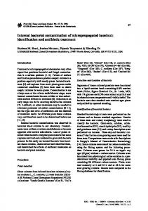
FIGURE 1. Effect of time of incubation on the adhesion of bacteria to cucumber surfaces in water at 58C. Numbers are the averages of duplicates obtained by spread plating.
in hydrophobicity, even between the different strains of Enterobacter aerogenes and L. plantarum that were tested (Table 1). Hydrophobicity was independent of the Gram stain status or pathogenicity of bacteria. Bacterial surface hydrophobicity has been found to vary depending on a variety of factors, including culture age, growth medium, pH, and subculturing techniques (15, 25, 27). In the present study, cultures were of the same age. Differences in hydrophobic ity could be species speci c. According to our results, the hydrophobicities of S. aureus, Salmonella Typhimurium, E. aerogenes 13048, and L. plantarum MOP3 were higher than those of L. plantarum ATCC 14917, L. monocytogenes, and E. aerogenes 29940. It is not known whether the results obtained here can be considered typical of these particular species. However, the fact that the two organisms showing the highest hydrophobicities were pathogenic indicates the potential for pathogens to adhere well to fruit and vegetable surfaces. Lachica (20) reported that 6 of 11 pathogenic bacteria that affect the urinary, digestive, and respiratory tracts were shown to have high cell surface hydrophobicities, which were associated with the binding of bacteria to the host cell. The time and temperature of exposure of whole cucumbers in water to bacteria signi cantly in uenced the adhesion of the bacterial species tested to cucumber surfaces. In preliminary tests, the six species of bacteria listed in Table 1 adhered to cucumbers at undetectable levels (,20 CFU/g) when incubated in water at 4 to 58C (data not shown). However, when the levels of inoculum were higher and time of contact with water was extended to 60 min, bacteria could be enumerated (Fig. 1). The degree of adsorption of bacteria to the fruit surface was somewhat related to the hydrophobicity of the strain. For instance, L. monocytogenes, the least hydrophobic bacterium tested, was the least adsorbed (Fig. 2). Similar results were obtained for the adhesion of L. monocytogenes to the surfaces of cantaloupes (31). However, S. aureus, the most hydrophobi c bacterium tested (96% hydrophobic), was less adsorbed than Salmonella Typhi-
BACTERIAL ADHESION TO CUCUMBERS
1883
FIGURE 2. Effect of temperature of incubation on the adhesion of bacteria to cucumber surfaces in water. The incubation time was 60 min. Points represent the means of two replicates.
murium, which was 60% hydrophobic . Perhaps other factors, such as the presence of agella, could have contributed to the adhesion of bacteria (23). Flagella have been suggested to play a role in bacterial adhesion and colonization, because agellated bacteria presumably should reach the surface of attachment faster (8, 10, 16, 17). The attachment of bacteria to surfaces has been reported to be a two-phase process. The cells rst become associated with the surface through loose, reversible sorption, and then a time-dependent irreversible type of lm attachment develops through the formation of viscous polymers (14). In this study, we considered only attachment of bacteria to the cucumber surface due to reversible sorption, which could be mediated by hydrophobici ty, agella, London–van der Waals forces, etc. The effect of incubation temperature on the adhesion of four bacterial species is illustrated in Figure 2. The overall slopes of data shown in the gure indicate that the effect of temperature on the adhesion of Salmonella Typhimurium was signi cantly (P , 0.05) stronger than its effects on the adhesion of the other three species. Differences among the other three species were not signi cant (P . 0.05). However, within each species, there was a signi cant (P , 0.05) increase in the degree of adhesion as the temperature was increased from 5 to 358C. Cucumbers were treated with hexane to remove wax and to change the surface characteristics of the fruit (Fig. 3A and 3B). The relative adhesion by L. monocytogenes was more extensive for the dewaxed fruit (Table 2). Counts of L. plantarum, S. aureus, and Salmonella Typhimurium were lower on the dewaxed fruit. Salmonella Typhimurium was found to be 60% hydrophobic , and S. aureus was found to be 96% hydrophobic . If the attachment of Salmonella and Staphylococcus to cucumber surfaces is due to hydrophobi c interactions (12), then the removal of wax from the cucumbers, resulting in a reduction of these hydrophobi c interactions, facilitates the removal of the bacteria during washing. The results of an analysis of variance for the data shown in Table
1884
REINA ET AL.
J. Food Prot., Vol. 65, No. 12
FIGURE 3. Surfaces of pickling cucumbers. (A) Calypso variety with wax. (B) Calypso variety without wax. (C) Calypso variety with trichome intact. (D) Calypso variety with trichome partially broken. (E) Calypso variety with stomate open. Bar 5 10 mm in each of the scanning micrographs. Magni cation of pictures without dimensional bars (A, C, and E), 32,400.
2 indicate a statistically signi cant difference (P , 0.05) between the number of bacteria adsorbed to the waxed fruit and the number adsorbed to the dewaxed fruit. However, with regard to adsorption to dewaxed fruit, only L. plantarum was signi cantly different (P , 0.05) among the four species tested. From these results, we can conclude that bacteria will be adsorbed to the surfaces of either waxed or
dewaxed cucumbers, as long as there is enough time to allow surface interactions between bacteria and the cucumber surface. Once bacteria are attached to the surface, it is very dif cult to remove them, as demonstrated by Ukuku and Fett (31). Depending on the storage temperature of the fruit, bacteria will grow or will form extracellular polymers, as was the case for Pseudomonas attached to meat surfaces at 208C
J. Food Prot., Vol. 65, No. 12
BACTERIAL ADHESION TO CUCUMBERS
1885
FIGURE 4. Adhesion of bacteria to the surfaces of pickling cucumbers (Calypso variety) with wax. (A) S. aureus. (B) L. plantarum. (C) L. monocytogenes. (D) Salmonella Typhimurium. (E) E. aerogenes ATCC 13048. Bar 5 10 mm.
(14). Bacteria are more easily removed when they are not allowed to adsorb to the surface via extracellular polymers. The removal of attached bacteria from the surfaces of cantaloupes upon washing after 12 days of storage at refrigeration temperatures was more dif cult (31). Figure 3 shows the surfaces of fresh cucumbers and illustrates features that may be involved in the adhesion and physical entrapment of microorganisms. The presence (Fig.
3A) or absence (Fig. 3B) of wax may in uence the types of microorganisms that adhere to the surface. Trichomes (Fig. 3C and 3D) and stomata (Fig. 3E) can serve to physically entrap microorganisms. SEMs for the attachment of different bacteria are shown in Figure 4. Bacteria were adsorbed to the surface of the cucumber whether or not the fruit was treated with hexane (Fig. 4). Clusters of bacteria were observed with
1886
REINA ET AL.
J. Food Prot., Vol. 65, No. 12
TABLE 2. Effects of epicuticular wax on the adhesion of bacteria to the surfaces of cucumbers in water at 208C for 60 mina RA for fruit with wax
Bacterial species
Listeria monocytogenes Lactobacillus plantarumb Staphylococcus aureus Salmonella Typhimurium
0.3 0.2 13.6 14.6
(0.3) B (0.01) B (3.2) A (3.1) A
RA for dewaxed fruit
1.85 0.02 1.4 1.0
(1.7) B (0.01) C (0.03) B (0.8) B
3. 4.
5.
6.
a
Relative adhesion (RA) 5 (CFU/g of fruit/CFU/ml of inoculum) 3 100. Values are the means for two replicates. Standard deviations are shown in parentheses. Means with different letters are signi cantly different (P # 0.05) by Tukey’s multiple-range test. b LA219.
7.
8.
9.
SEM (Fig. 4A, 4D, and 4E). Figure 4B and 4C shows evenly distributed bacteria. This clustering of bacteria could be due to the adhesion of bacteria to cucumber surfaces, as well as interbacterial adhesion (19). When a cell is adsorbed to a surface, other parts of the cell are free to adhere to other surfaces, which may explain why cells tend to appear in clusters under SEM. Figure 4E shows bacteria clustering around a hole of unknown origin (possibly caused by an insect sting or another type of injury). This hole might have allowed the leakage of nutrients from the fruit, thus attracting bacteria. In one study, cells of Escherichia coli O157:H7 were found to attach preferentially to cut edges of lettuce over the intact leaf surface (30). In another study, when tomatoes were inoculated with bacteria, the largest numbers of bacteria were recovered from surface scars, blossom scars, and stem scars (21). Annous et al. (1) found that E. coli attached mainly to the calyx and stem scars of arti cially contaminated Golden Delicious apples. The adhesion of bacteria to cucumber surfaces was found to depend on temperature, species, characteristics of the cucumber surface, and time of exposure. Bacterial adhesion to meat surfaces has been found to be a time-dependent process, and once attached, bacteria have been found to be dif cult to remove (14, 24). Our results indicate that low water temperatures and short periods of contact with the fruit will reduce the potential for bacterial adhesion during the postharvest washing and hydrocooling of cucumbers. In addition, treatment of the water with chemicals to keep the microbial load low is fundamental to the avoidance of contamination of the produce. ACKNOWLEDGMENT This investigation was supported in part by a research grant from Pickle Packers International, Inc., St. Charles, Ill.
10.
11.
12.
13.
14.
15.
16.
17.
18.
19. 20.
21.
REFERENCES 1.
2.
Annous, B. A., G. Sapers, A. Mattrazzo, and D. Riordan. 2001. Ef cacy of washing with a commercial ated brush water, using conventional and experimental washing agents, in reducing populations of Escherichia coli on arti cially inoculated apples. J. Food Prot. 64:159 –163. Bartz, J. A. 1982. In ltration of tomatoes immersed at different tem-
22.
23.
peratures to different depths in suspensions of Erwinia carotovora sp. carotovora . Plant Dis. 66:302–306. Bartz, J. A., and R. K. Showalter. 1981. In ltration of tomatoes by aqueous suspensions . Phytopathology 71:515–518. Beuchat, L. R. 1998. Surface decontaminatio n of fruits and vegetables eaten raw: a review. WHO/FSF/FOS/98.2. World Health Organization, Geneva. Brackett, R. E. 1993. Microbial quality, p. 134 –137. In R. L. Shew et and S. E. Prussia (ed.), Postharvest handling. A systems approach. Academic Press, New York. Breidt, F., and H. P. Fleming. 1992. Competitive growth of genetically marked malolactic de cient Lactobacillus plantarum in cucumber fermentations. Appl. Environ. Microbiol. 58:3845–3849. Burnett, S. L., and L. R. Beuchat. 2000. Human pathogens associated with raw produce and unpasteurized juices, and dif culties in decontamination. J. Ind. Microbiol. Biotechnol. 25:281–287. Butler, J. L., J. C. Stewart, C. Vanderzant, Z. L. Carpenter, and G. C. Smith. 1979. Attachment of microorganisms to pork skin and surfaces of beef and lamb carcasses. J. Food Prot. 42:401–406. Corey, K. A., D. V. Schlimme, and N. A. Channey. 1988. Changes in epicuticular wax on watermelon fruits during ripening. HortScience 23:730 –731. Dickson, J. S., and E. K. Daniels. 1991. Attachment of Salmonella typhimurium and Listeria monocytogene s to glass as affected by surface lm thickness, cell density, and bacterial motility. J. Ind. Microbiol. 8:281–284. Doyle, R. J., and M. Rosenberg. 1990. Microbial cell surface hydrophobicity: history, measuremen t and signi cance, p. 1–37. In R. J. Doyle and M. Rosenberg (ed.), Microbial surface hydrophobicity . American Society for Microbiology, Washington, D.C. Duncan-Hewitt, W. C. 1990. Nature of the hydrophobi c effect, p. 39–73. In R. J. Doyle and M. Rosenberg (ed.), Microbial surface hydrophobicity . American Society for Microbiology, Washington, D.C. Eckert, J. W. 1975. Postharvest diseases of fresh fruits and vegetables—etiology and control, p. 81–89. In N. F. Haard and D. K. Salunkhg (ed.), Symposium: postharvest biology and handling of fruits and vegetables. AVI Publishing, Westport, Conn. Firstenberg-Eden, R., S. Notermans, F. Thiel, S. Henstra, and E. H. Kamplemache r. 1979. Scanning electron microscopi c investigations into attachment of bacteria to teats of cows. J. Food Prot. 44:602– 607. Gilbert, P., E. Evans, I. G. Duguid, and M. R. Brown. 1991. Surface characteristics and adhesion of E. coli and Staphylococcu s epidermidis. J. Appl. Bacteriol. 71:72–77. Herald, P., and E. Zottola. 1988. Attachment of Listeria monocytogenes to stainless steel surfaces at various temperatures and pH values. J. Food Sci. 53:1549 –1552. Husmark, U., and U. Ronner. 1990. Forces involved in adhesion of Bacillus cereus spores to solid surfaces under different environmental conditions. J. Appl. Bacteriol. 69:557–562. Janisiewicz, W., W. S. Conway, and B. Leverentz. 1999. Biological control of postharvest decays of apple can prevent growth of E. coli O157H7 in apple wounds. J. Food Prot. 62:1372–1375. Jones, G. W., and R. E. Isaacson. 1982. Proteinaceous bacterial adhesions and their receptors. Crit. Rev. Microbiol. 10:229 –260. Lachica, V. 1990. Signi cance of hydrophobicit y in the adhesivenes s of pathogenic Gram-negative bacteria, p. 297–313. In R. J. Doyle and M. Rosenberg (ed.), Microbial surface hydrophobicity . American Society for Microbiology, Washington, D.C. Lukasik, J., M. Bradley, T. Scott, W.-Y. Hsu, S. Farrah, and M. Tamplin. 2001. Elution, detection, and quanti cation of Polio I, bacteriophages, Salmonella montevideo, and Escherichia coli O157H7 from seeded strawberries and tomatoes. J. Food Prot. 63:292–297. Mafu, A. A., D. Roy, J. Goulet, and L. Savoi. 1991. Characterization of physicochemica l forces involved in adhesion of Listeria monocytogenes to surfaces. Appl. Environ. Microbiol. 7:1969 –1973. Meadows, P. S. 1971. The attachment of bacteria to solid surfaces. Arch. Microbiol. 75:374 –381.
J. Food Prot., Vol. 65, No. 12
24. 25. 26.
27.
28.
29.
Notermans, S., R. Firstenberg-Eden, and M. V. Schothorst. 1979. Attachment of bacteria to teats of cows. J. Food Prot. 42:228–232. Olsson, J., and G. Westergren. 1972. Hydrophobi c surface properties of oral streptococci. FEMS Microbiol. Lett. 15:319 –323. Pao, S., C. L. Davis, and M. E. Parish. 2001. Microscopic observation and processing validation of fruit sanitizing treatments for enhanced microbiological safety of fresh orange juice. J. Food Prot. 64:310 –314. Rosenberg, M., D. Gutnick, and E. Rosenberg. 1980. Adherence of bacteria to hydrocarbons : a simple method for measuring cell-surface hydrophobicity. FEMS Microbiol. Lett. 9:29–33. Sharon, E., Y. Bashan, Y. Okon, and Y. Henis. 1981. Presymptomatic multiplication of Xanthomona s campestris pv. Vesicatoria on the surface of pepper leaves. Can. J. Bot. 60:1041–1045. Silva Fernandez, A. M., E. A. Baker, and J. T. Martin. 1964. Studies
BACTERIAL ADHESION TO CUCUMBERS
30.
31.
32.
33.
34.
1887
on plant cuticule. VI. The isolation and fractionation of cuticular waxes. Ann. Appl. Biol. 53:43–58. Takeuchi, K., and J. Frank. 2001. Expression of red-shifted green uorescent protein by Escherichia coli O157H7 as a marker for the detection of cells on fresh product . J. Food Prot. 64:298–304. Ukuku, D. O., and W. Fett. 2001. Surface characteristics and adhesion of Salmonella, Listeria monocytogenes , and Escherichia coli on cantaloupe surfaces. IFSM meeting, Minneapolis. Vescovo, M., C. Orsi, G. Scolari, and S. Torriani. 1995. Inhibitor y effect of selected lactic acid bacteria on micro ora associated with ready-to-use vegetables. Lett. Appl. Microbiol. 21:121–125. Wyncek, K., A. Kaples, and P. M. Foegeding. 1990. Hydrophobicit y of Bacillus and Clostridium spores. Appl. Environ. Microbiol. 9: 2600–2605. Zagory, D. 1999. Effects of post-processin g handling and packaging on microbial populations. Postharvest Biol. Technol. 15:313–321.