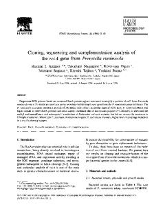
. (2007), 17(10), 1607–1615
J. Microbiol. Biotechnol
Bacterial β-Lactamase Fragment Complementation Strategy Can Be Used as a Method for Identifying Interacting Protein Pairs PARK, JONG-HWA, JUNG HO BACK, SOO HYUN HAHM, HYE-YOUNG SHIM, MIN JU PARK, SUNG IL KO, AND YE SUN HAN* Department of Advanced Technology Fusion and Bio/Molecular Informatics Center, Konkuk University, Seoul 143-701, Korea
Received: February 8, 2007
Accepted: May 5, 2007
Abstract We investigated the applicability of the TEM-1 β-
lactamase fragment complementation (BFC) system to develop a strategy for the screening of protein-protein interactions in bacteria. A BFC system containing a human Fas-associated death domain (hFADD) and human Fas death domain (hFasDD) was generated. The hFADD-hFasDD interaction was verified by cell survivability in ampicillin-containing medium and the colorimetric change of nitrocefin. It was also confirmed by His pull-down assay using cell lysates obtained in selection steps. A coiled-coil helix coiled-coil domain-containing protein 5 (CHCH5) was identified as an interacting protein of human uracil DNA glycosylase (hUNG) from the bacterial BFC cDNA library strategy. The interaction between hUNG and CHCH5 was further confirmed with immunoprecipitation using a mammalian expression system. CHCH5 enhanced the DNA glycosylase activity of hUNG to remove uracil from DNA duplexes containing a U/G mismatch pair. These results suggest that the bacterial BFC cDNA library strategy can be effectively used to identify interacting protein pairs. Keywords: β-Lactamase fragment complementation, hFADD, hFasDD, nitrocefin, cDNA library, human uracil DNA glycosylase, coiled-coil helix coiled-coil domain-containing protein 5
The need to develop strategies to investigate proteinprotein interactions, especially proteins associated with pathologic cellular processes caused by abnormal proteinprotein interactions, has been progressively increased. To understand these protein interactions, several techniques including the yeast two-hybrid (YTH) system, fluorescence resonance energy transfer (FRET) system, and protein fragment complementation assay (PCA) have been developed [3, 14, 22]. Although lots of works related to protein-protein *Corresponding author
Phone: 82-2-2049-6050; Fax: 82-2-452-5558; E-mail:
[email protected]
interactions have been achieved by fusion protein-based assays such as YTH, it is not applicable to studies of the kinetics of protein-protein interaction, and it is hard to detect the interaction of proteins such as cell surface receptors. A method based on FRET is thought to be one of most accurate methods used to monitor dynamic interactions. However, it is also hard to detect a protein interaction because changes in fluorescence assayed by FRET are small. The PCA system has been suggested to be a more useful tool than other methods to study protein-protein interactions [2, 14]. PCA systems, based on interaction-driven reconstitution of the activity of a reporter protein that is genetically split into two fragments and produced as separate members of a protein-protein interaction pair, have been developed as suitable methods to monitor inducible protein interactions [14, 23]. PCA systems have been successfully used for investigations of protein-protein interactions [4, 24, 25, 27]. Several enzymatic or fluorescent reporter proteins including mouse dihydrofolate reductase (mDHFR), green fluorescent protein (GFP), luciferase, and TEM-1 β-lactamase have been used for PCA applications in prokaryotes or eukaryotes [5, 14, 20]. TEM-1 β-lactamase is an attractive reporter enzyme in PCA application because of the fact that it is small, monomeric, and nontoxic [8, 28]. In addition, β-lactamase is easily expressed in many hosts and there are no orthologs to exist in mammalian cells, which makes this system applicable to both prokaryotic and eukaryotic cells [15]. In screening work to identify an interacting protein with a target protein using bacterial cells, the PCA system can be used as a useful tool, because the interaction between target and screening proteins can be confirmed by a direct pulldown assay on a selected cell lysate. However, the use of PCA for screening of interacting proteins in bacterial cells has only been reported in a few studies. A PCA system based on mDHFR has been effectively used to identify interacting pairs of heterodimerizing polypeptides by a leucine zipper library-versus-library strategy [21]. However, a TEM-1 BFC
1608
PARK
et al.
cDNA library strategy for identifying interacting protein pairs is not yet fully developed. Human uracil DNA glycosylase (hUNG) is an enzyme responsible for excising uracil residues from DNA by cleaving the N-glycosylic bond between the uracil base and the deoxyribose phosphate backbone DNA [12, 16]. Two hUNG proteins, mitochondrial hUNG1 and nuclear hUNG2 [13], have been shown to play a specific role in removing misincorporated uracil in a post-replicative process and in removing deaminated cytosine residues [11, 19]. In this report, we investigated the applicability of the TEM-1 BFC system in identifying interacting proteins in cells. To accomplish this, we constructed the bait and prey plasmids of the BFC system containing a human Fas-associated death domain (hFADD) and human Fas death domain (hFasDD), which have been known to interact with each other [6], and analyzed β-lactamase activities by the complementation resulting from the hFADD-hFasDD interaction. In addition, hUNG interacting proteins were screened from pretransformed cells using cDNA library plasmids of the BFC system, and their functions on hUNG glycosylase activity were further investigated. Escherichia coli
E. coli
MATERIALS AND METHODS Cell Lines
strain BL21(DE3) was used as a host for the cloning and BFC studies. Human embryonic kidney Hek293 cells were cultured in Dulbecco’s modified Eagle’s medium (DMEM) supplemented with 10% fetal bovine serum (FBS) and 1% penicillin-streptomycin solution. Escherichia coli
Construction of Plasmids
Two gene fragments corresponding to residues 1-197 (α-197) and 198-290 (ω-198) of TEM-1 β-lactamase contained in the pQE30 plasmid (Qiagen) were separately amplified using PCR. The α-197 fragment was amplified as a form containing NGR tripeptides and the (GGGGS)3 linker at the C-terminal region and subcloned into a pET28a plasmid. Subsequently, a gene fragment encoding residues 218-335 of hFasDD (GenBank Accession No. NM000043) and His6 epitope was inserted into the Cterminal region of α-197-linker-NGR. Furthermore, PCR was performed to generate a M182T mutant gene of the α-197 fragment. Finally, a pSBLNL-hFasDD-His vector (Fig. 1B) was generated by replacing this PCR fragment between the NcoI and SfcI restriction sites of pET-28a-a-197-NGRLinker-hFasDD-His. The ω-198 fragment containing the (GGGGS)3 linker at the C-terminal region was amplified by PCR and subcloned into a pET-28a plasmid. A PCR fragment encoding the c-myc epitope and residues 1-208 of hFADD (GenBank Accession No. BC000334) was inserted into the N-terminal region of linker-ω-198. Then,
Fig. 1. Bait and prey plasmids constructions.
A. Bait plasmid, pSLBLC-c-myc-hFADD: c-myc-hFADD was subcloned
as N-terminally fused to a C-terminal domain of β-lactamase (ω-198) connected via a linker region [(GGGGS)3]. B. Prey plasmid, pSBLNLhFasDD-His: hFasDD corresponding to a cognate binding partner for hFADD was subcloned as a C-terminal fusion to an N-terminal fragment of β-lactamase (α-197) containing the stabilizing substitution M182T. The connecting linker (GGGGS)3 was extended by NGR tripeptides to promote BFC. Both plasmids contain a signal sequence (SS) of β-lactamase (residues 1-23) for the periplasmic targeting. All fusion proteins were expressed via the regulation of the T7 promoter and lac operator.
a signal sequence coding region (residues 1-23) of βlactamase between NcoI and BamHI sites was cloned. Finally, a fragment containing a T7 promoter, a lac operator, a signal sequence of β-lactamase, c-myc-hFADD, and the (GGGGS)3 linker-ω-198 coding region was cloned between the BamHI and SalI sites of the pLysS plasmid (Stratagene) to generate the final pSLBLC-c-myc-hFADD vector (Fig. 1A). A gene fragment that corresponds to residues 78-304 without the mitochondrial targeting sequence of hUNG1 (GenBank Accession No. NM003362) was amplified and cloned between the SfiI and SalI sites of the pSLBLC-c-myc vector, in which hFADD was deleted by SfiI and SalI reaction (pSLBLC-c-myc-hUNG). All constructs were confirmed by DNA sequence analyses. β-Lactamase Activity Assay
A nitrocefin disk (Cefinase; Clontech) was used to determine β-lactamase activity produced by BFC from transformed cells. Equal amounts of overnightcultured cells were applied onto a nitrocefin disk using a pipette and incubated for 30 min at room temperature. The presence of β-lactamase activity was detected by observing the nitrocefin color change from yellow to red. These cells were also applied on to a LB agar medium containing IPTG and different formulations of antibiotics. After 16 h incubation, cell survivals in medium containing ampicillin were monitored to determine β-lactamase activity. E. coli
His Pull-down Assay
Fusion protein expressions from cells were induced using 0.2 mM IPTG at A600=0.6-0.8 for 12 h at 18oC. Cells E. coli
BACTERIAL BFC FOR IDENTIFYING INTERACTING PROTEIN PAIRS
were harvested and lysed by sonication in a buffer solution (50 mM NaH2PO4, 300 mM NaCl, 10 mM imidazole, pH 8.0) with 10 µg/ml PMSF. After centrifugation, supernatants were incubated for 3 h at 4oC with an equal amount of NiNTA beads (Qiagen). The beads were washed five times with ice-cold buffer solution, and then resuspended in 200 µl of elution buffer (50 mM NaH2PO4, 300 mM NaCl, 250 mM imidazole, pH 8.0). After incubation for 10 min and centrifugation, supernatants were collected and analyzed by Western blot analysis.
Transient Expression of hUNG and CHCH5
To express tagged hUNG (c-myc-hUNG) and CHCH5 (flag-CHCH5), pCMV Tag3A-hUNG and pCMV Tag2CCHCH5 plasmids were transiently transfected, respectively, into 80%-90% confluent Hek293 cells using Transfectin lipid reagent (Bio-Rad). After thirty-six hours of incubation, cells were collected and lysed using 1 ml of lysis buffer [50 mM Tris-HCl (pH 8.0), 100 mM NaCl, 5 mM EDTA, 1 mM NaF, 1 mM Na3VO4, 1% Nonidet P-40, 10 µg/ml PMSF, protease inhibitor cocktail (Sigma)] for 1 h at 4oC with occasional vortexing. After centrifugation, supernatants were collected and used in the Western blot analysis and/or immunoprecipitation experiment.
Immunoprecipitation
Total cell lysates were collected from Hek293 cells transfected with different sets of plasmids and were cleared with 30 µl of protein G-Sepharose beads (Santa Cruz Biotech.) to remove nonspecific proteins. After 3 h incubation with 2 µg of anti-c-myc antibody at 4oC with gentle rotation, residual antibody and bound proteins were pulled down with 30 µl of protein G-Sepharose beads at 4oC for 3 h. Protein-bead complexes were precipitated and washed five times with washing buffer (1:1 mixture of lysis buffer and PBS) and eluted with 2× SDS-PAGE gel loading buffer.
Partial Purification of hUNG and CHCH5 Using Immunoaffinity Chromatography
hUNG or CHCH5 proteins tagged with c-myc or flag epitope were partially purified by immunoaffinity batch chromatography using anti-c-myc or anti-flag antibodies covalently attached to agarose gel by hydrazide linkage (Sigma). Protein extracts were collected from Hek293 cells transfected with pCMV Tag3A-hUNG1 or pCMV Tag2C-CHCH5 plasmids. The protein extracts (1 mg) were mixed with 200 µl of anti-c-myc or anti-flag antibodyagarose conjugate (at ratio 1 mg of antibody/ml of agarose gel) preequilibrated with PBS or TBS buffer. After overnight incubation at 4oC, gels were washed with 20 gel volumes of PBS or TBS solution. c-myc-tagged hUNG or flag-tagged CHCH5 was separated from anti-c-myc or anti-flag antibody-agarose gels using elution buffer (anti-c-
1609
myc antibody-agarose gels: 0.1 M ammonium hydroxide, pH 11; anti-flag antibody-agarose gels: 0.1 M glycine-HCl, pH 3.5) and neutralized with 1 N acetic acid or 1 M TrisHCl (pH 9.0) solution. Eluates were dialyzed using a solution (20 mM Tris-HCl, pH 8.0, 1 mM EDTA), and the protein concentrations were determined using the Bio-Rad protein assay kit and the resulting proteins were stored at -70oC.
Western Blot Analysis
Protein samples resolved using SDS-PAGE were transferred onto PVDF membranes and blocked with 5% nonfat dried milk in TBS-T (TBS with 0.05% Tween 20). The membranes were incubated with anti-c-myc, anti-His, and anti-flag antibodies (Santa Cruz Biotech.), followed by incubation with horseradish peroxidase (HRP)-conjugated secondary antibodies (Santa Cruz Biotech.). The signal was detected using the ECL Western blotting detection reagents (Pierce).
hUNG DNA Glycosylase Activity Assay
DNA glycosylase activity was assayed on a 32 mer duplex substrate of oligonucleotide carrying uracil (U) at position 16 (5'-GGA TCC TCT AGA GTC UAC CTG CAG GCA TGC AA-3') and complement oligonucleotide carrying guanine in the pairing position of uracil (3'-CCT AGG AGA TCT CAG GTG GAC GTC CGT ACG TT-5') (BioSynthesis Inc., U.S.A.). Uracil-containing oligonucleotides were 5'-end labeled with T4 polynucleotide kinase (New England Biolabs.) in the presence of [γ-32P] ATP and purified through a Microspin G-50 column (Amersham Biosciences). Oligonucleotide duplexes were prepared by annealing with 1.5-fold molar excesses of the unlabeled complementary oligonucleotide. The activity assay was assembled in 20 µl volumes with different concentrations of proteins and 1 pmole of radiolabeled 32 mer oligonucleotide duplexes in a reaction buffer containing 20 mM Tris-HCl (pH 7.5), 100 mM NaCl, and 1 mM DTT. After incubating at 37oC for 1 h, reactions were terminated using 2× alkaline loading buffer [0.5 N NaOH, 95% formamide, 10 mM EDTA (pH 8), 0.05% bromophenol blue] and heated at 95oC for 5 min. Reaction mixtures were resolved using electrophoresis on a denaturing 18% polyacrylamide gel containing 7 M urea in 1× TBE buffer (90 mM Tris, 90 mM boric acid, and 2 mM EDTA). The gel was dried and placed on an imaging plate, and DNA cleavage products were quantified using a BAS2000 image analyzer (Fuji, Tokyo, Japan).
RESULTS Bacterial BFC by a Protein-Protein Interaction
Two plasmid vectors, pSLBLC-c-myc-hFADD and pSBLNLhFasDD-His, were designed and constructed to complement
1610
PARK
et al.
an enzymatic activity of β-lactamase by T7 promoterdriven coexpression of an interacting protein pair, hFADD and hFasDD. The α-197 and ω-198 fragments of β-lactamase were fused through a triple pentapeptide (GGGGS) linker to the N-terminal of hFasDD and the C-terminal of hFADD, respectively (Fig. 1). NGR tripeptide and M182T, which has been known to increase the complementation [27], were also introduced in the α-197 fragment. cells transformed with one or two of the above plasmids were obtained by overnight cultivation from medium containing different antibiotics. Expressions of fused hFADD or hFasDD from transformed were induced by IPTG and confirmed using SDS-PAGE followed by Coomassie brilliant blue staining and Western blot analysis. As shown in Fig. 2, fused hFADD and hFasDD E. coli
E. coli
Fig. 3. E. coli clone transformed with both pSLBLC-c-myc-
hFADD and pSBLNL-hFasDD-His plasmids showed β-lactamase activities induced by BFC. A. β-Lactamase activity was analyzed by the colorimetric change method
of nitrocefin. Lanes 1, 2, 3, and 4 represent E. coli cell transformed with pSLBLC-c-myc-hFADD, pSBLNL-hFasDD-His, pSLBLC-c-myc-hFADD/ pSBLNL-hFasDD-His, and pQE30, respectively. B. β-Lactamase activity was analyzed by the survivability on media containing different formulations of antibiotics. LBK, LBC, LBKCI, and LBKCAI represent the LB medium containing kanamycin, chloramphenicol, kanamycin/chloramphenicol/IPTG, and kanamycin/chloramphenicol/ampicillin/IPTG, respectively.
proteins were expressed as expected band sizes, ~40 and 32 kDa, respectively. transformant obtained from cotransfection of both plasmids expressed both proteins (Figs. 2A, 2B lane 3). BFC from cotransfected with both plasmids was confirmed by observing β-lactamase activity using the nitrocefin color change assay and measuring cell growth in an ampicillin-containing medium (Fig. 3). cotransformed with both plasmids changed the yellow color of nitrocefin to red. This color change was also detected in transformed with the normal TEM-1 βlactamase-producing plasmid (pQE30). transformants were cultivated for 16 h in medium containing IPTG and different formulations of antibiotics; cotransfected with both plasmids was able to grow in medium containing 10 µg/ml ampicillin. However, transformed cells with a single plasmid did not grow in ampicillin-containing medium. E. coli
E. coli
E.
coli
Fig. 2. Fusion proteins were expressed in E. coli cells
transformed with pSLBLC-c-myc-hFADD or pSBLNL-hFasDDHis plasmids and determined by 12% SDS-PAGE followed by Coomassie brilliant blue staining (A) or Western blot analysis (B). Arrows indicate fusion proteins bands. Lanes 1, cells transformed with pSLBLC-c-myc-hFADD; 2, cells transformed with pSBLNL-hFasDD-His; 3, cells transformed with pSLBLC-c-myc-hFADD and pSBLNL-hFasDDHis.
E. coli
E. coli
E. coli
BACTERIAL BFC FOR IDENTIFYING INTERACTING PROTEIN PAIRS
1611
down proteins were analyzed using Western blot analysis. His-tag fused α-197-hFasDD was observed from pulleddown eluates of transformed with pSBLNLhFasDD-His or pSLBLC-c-myc-hFADD/pSBLNL-hFasDDHis plasmids (Fig. 4, lanes 2 & 3). c-myc-hFADD-ω-198 pulled-down by the interaction between hFADD and hFasDD was only detected in transformed with both plasmids (Fig. 4, lane 3). E.
coli
E. coli
Screening of hUNG Interacting Proteins Using Bacterial BFC
To screen hUNG interacting proteins using the BFC system, the pSLBLC-c-myc-hUNG plasmid was transformed to pretransformed cells with pSBLNL-cDNA libraries through the electroporation method. cDNA libraries were generated from mRNA, which was purified from DNA-damaged Hek293 cells, using the SuperScript Choice system (Invitrogen) and ligated to EcoRI- and XhoI-digested pSBLNL. Colonies showing β-lactamase activities through physical interaction between hUNG and proteins encoded from cDNA libraries were selected in ampicillin-containing medium Plasmid DNAs were propagated from these colonies and transformed into cells. After overnight incubation in kanamycin-containing medium, plasmid DNA containing cDNA was purified and the sequence information was analyzed. Furthermore, BFC from the interactions between hUNG and proteins encoded in cDNA libraries were confirmed by a β-lactamase activityderived nitrocefin disk color change assay (data not shown). E. coli
Fig. 4. The interaction of hFADD and hFasDD was directly
confirmed by a His pull-down assay using E. coli cell lysates transformed with pSLBLC-c-myc-hFADD and pSBLNL-hFasDDHis. Soluble protein fractions of E. coli cells were collected and mixed with NiNTA beads. After 3 h incubation and elution of bound proteins, the presence of hFADD and hFasDD was analyzed by Western blot analysis using anti-c-myc or anti-His antibodies. Lanes 1, 2, and 3 represent E. coli cell transformed with pSLBLC-c-myc-hFADD, pSBLNL-hFasDD-His, and pSLBLC-c-myc-hFADD/pSBLNL-hFasDD-His, respectively.
E.
coli
.
E.
Interaction Confirmation Using His Pull-down Assay
Interaction between fusion proteins, c-myc-hFADD-ω-198 and α-197-hFasDD-His, was confirmed by the His pull-down assay from transformed cells. Soluble fractions of proteins were incubated with Ni-NTA beads and pulledE. coli
coli
Fig. 5. Sequence information of CHCH5 (A) and amino acid alignment of CHCH5 domain (B).
AAH04498, Homo sapience CHCH5; CAD33933, Mus musculus CHCH5; AAF48701, Drosophila melanogaster CHCH5; QO9254, hypothetical protein C16-C10.11; P42029, NADH dehydrogenase I alpha subcomplex subunit 8; CAD27307, Asperigillus fumigatus CHCH5; CAA92368, Saccharomyces cerevisiae CHCH5.
1612
PARK
et al.
Several clones showing positive β-lactamase activities encoding a coiled-coil helix coiled-coil protein 5 (CHCH5). Alignment of this protein with related members showed the presence of a conserved [coiled coil 1]-[coiled coil 2][helix 2] domain (CHCH domain) (Fig. 5).
Interaction Confirmation of hUNG1 and CHCH5 Using Immunoprecipitation
A gene fragment corresponding to the amino acid coding region of CHCH5 (GenBank Accession No. BC004498) was amplified by PCR and subcloned to a mammalian expression vector, pCMV Tag 2C. The hUNG gene of pSLBLC-c-myc-hUNG was also subcloned to pCMV Tag3A. Transiently expressed hUNG and CHCH5 from Hek293 cells were detected as ~30 and 17 kDa, respectively, which corresponded to the theoretically calculated values (Fig. 6A). Different sets of plasmids were transfected to Hek293 cells. After thirty-six hour incubation, cells were collected and immunoprecipitated as described in the Materials and Methods section. CHCH5 that was precipitated by the physical interaction with hUNG in immunoprecipitation using anti-c-myc antibody was detected in Western blot analysis using anti-flag antibody (Fig. 6B)
Immunoaffinity Purification and hUNG Activity Assay
hUNG tagged with c-myc or CHCH5 tagged with flag epitope were purified by immunoaffinity batch chromatography using anti-c-myc or anti-flag antibodies with conjugated agarose gels. The presence of hUNG or CHCH5 proteins in the purified fractions were confirmed by silver staining and Western blot analysis (data not shown). Most of the proteins purified from cell lysates that were transfected with pCMV Tag3A-c-myc-hUNG or pCMV Tag2C-flagCHCH5 were c-myc-hUNG or flag-CHCH5. The DNA glycosylase activity of hUNG was examined using a 32
Fig. 6. CHCH5 physically interacts with hUNG.
A. Hek293 cells were transfected with pCMV Tag3A-hUNG or pCMV
Tag2C-CHCH5. Cell lysates were collected and the expression of hUNG and CHCH5 was confirmed by Western blot analysis using anti-c-myc or anti-flag antibodies. B. Total cell lysates were collected from Hek293 cells transfected with different sets of plasmids. After coimmunoprecipitation using anti-c-myc antibody, elution samples were resolved by 12% polyacrylamide gel and analyzed by Western blot analysis using anti-flag or anti-c-myc antibodies.
Fig. 7. hUNG purified by immunoaffinity batch chromatography showed the DNA glycosylase activity, but not CHCH5.
A. Purified hUNG fractions (50, 100, 200 ng) were reacted with 1 pmole of 5'-end labeled U/G mismatch 32 mer duplexes for 1 h at 37oC. The cleavage
products were analyzed by 18% denaturing polyacrylamide gel electrophoresis and a BAS2000 image analyzer. A protein fraction (200 ng) purified from Hek293 cells transfected with pCMV tag3A by the same methods as hUNG purification was reacted as a negative control (lane 3). B. Purified CHCH5 fractions (20, 50, 100 ng) were reacted and analyzed as described above. The hUNG fraction (200 ng) was reacted as a positive control (lane 5).
BACTERIAL BFC FOR IDENTIFYING INTERACTING PROTEIN PAIRS
1613
Fig. 8. CHCH5 enhanced the DNA glycosylase activity of hUNG.
A. Purified hUNG (100 ng) and CHCH5 (20, 50 ng) were reacted with 1 pmole of 5'-end labeled U/G mismatch 32 mer duplexes for 1 h at 37oC. The cleavage products were analyzed by 18% denaturing polyacrylamide gel electrophoresis and a BAS2000 image analyzer. Protein fractions purified from Hek293 cells transfected with pCMV tag3A or pCMV Tag2C by the same methods as hUNG or CHCH5 purification were reacted as a negative control (lane 3). B. Cleavage products of two independent assays were quantified using a BAS2000 image analyzer. An average content of hUNG fraction-treated cleavage product was estimated as 100.
mer oligonucleotide duplex containing the U/G pair at position 16. Proteins purified from Hek293 cell lysates transfected with pCMV Tag3A were used as a control. Incubation of a 32 mer oligonucleotide duplex with hUNG resulted in cleaved 15 mer oligonucleotides, suggesting the anticipated glycosylase activity of hUNG (Fig. 7A). When concentrations of hUNG were increased, the amount of cleaved oligonucleotides also increased in a dose-dependent manner. This effect was not detected in the reaction mixture of proteins purified from cell lysates transfected with pCMV Tag3A. Incubation of CHCH5 with one pmole of radiolabeled 32 mer oligonucleotide duplexes did not show any cleaved 15 mer oligonucleotides products (Fig. 7B). However, the addition of CHCH5 increased the glycosylase activities of hUNG when 20 and 50 ng of CHCH5 proteins were mixed with 100 ng of hUNG and incubated with 1 pmole of radiolabeled 32 mer oligonucleotide duplexes for 1 h at 37oC (Fig. 8A, lanes 4 and 5). Protein mixtures purified from cell lysates transfected with pCMV Tag3A or pCMV Tag2C, did not show any presence of cleaved oligonucleotides (Fig. 8A, lanes 2 and 6).
DISCUSSION Herein, we report the potential applicability of a bacterial BFC system for identifying interacting proteins using a cDNA library fused to the complementary reporter fragment. As shown in Fig. 3, showing BFC could be selected from observing their survivability in ampicillin-containing media and β-lactamase activities-derived colorimetric changes of nitrocefin from yellow to red that are derived E. coli
from the complementation of two fragments (α-197, ω198) through physical interaction between the protein pairs. In mammalian cells, BFC can be detected by the conversion of the membrane-permeable substrate CCF2/ AM (esterified form of CCF2, coumarin cephalosporin fluorescein) into CCF2, which is a nonpermeable FRET substrate containing two different fluorophores connected a permissive cephalosporin β-lactam ring [1, 29]. However, BFC detection using a CCF2/AM substrate can not be achieved in bacterial system owing to the absence of esterase. Recently, a fluorescence detection method using genetically engineered with esterase has been suggested as an interesting alternative or complement to survival-based selection or colorimetric analysis using nitrocefin in protein library application involving the identification of target protein-specific binding proteins BFC [18]. Thus, the use of BFC is expected to provide a more efficient strategy in identifying interacting proteins. Although the YTH system is a powerful method to screen the physically interacting proteins [3, 7], it has a limitation to confirm its results using alternative methods such as pull-down assay or immunoprecipitation. However, the bacterial BFC strategy has an advantage in that physical interaction of screened protein pairs can be confirmed by a direct pull-down assay using cell lysates obtained from screening steps. The physical interaction between hFADD and hFasDD was confirmed by a His pull-down assay using the cell lysate obtained from transformant showing β-lactamase activity (Fig. 4). These results suggest that the BFC strategy may be developed as a screening method to reduce the time and cost to identify interacting proteins. via
E.
coli
via
E. coli
E. coli
1614
PARK
et al.
From BFC, CHCH5 was identified to interact with hUNG. This was achieved by transforming a BFC bait plasmid containing hUNG into cells that were pretransformed with BFC prey plasmids containing cDNA libraries and selected through their survivability from ampicillin-containing medium and nitrocefin color change assay. CHCH5 contains a conserved CHCH domain, and within each helix of the CHCH domain, there are two cysteines presented in a C-X9-C motif (Fig. 5). Although protein families containing this domain, such as Cox19, Cox19p, Mrp10, and NADH-ubiquinone oxidoreductase 19 kDa (NDUFA8) subunit, have been known, the real function of the CHCH5 domain is not yet clear [9, 10, 17, 26]. Physical interaction of hUNG and CHCH5 was further confirmed by immunoprecipitation assay using a mammalian expression system (Fig. 6). To investigate the effect of CHCH5 protein on the DNA glycosylase activity of hUNG, CHCH5 and hUNG were transiently expressed in Hek293 cells and purified by immunoaffinity batch chromatography. DNA glycosylase activities of purified hUNG increased in a dose-dependent manner (Fig. 7A) and dramatically increased by the addition of purified CHCH5 (Fig. 8). The purified fraction of CHCH5 protein did not show any DNA glycosylase activity to remove uracil from the U/G mismatched DNA duplex (Fig. 7B). These results suggest that the enhancement of hUNG DNA glycosylase activities by the presence of CHCH5 protein may be derived from the interaction between hUNG and CHCH5. In the activity assay using hUNG and CHCH5 expressed and purified in cells, DNA glycosylase activities of hUNG were also increased by the presence of CHCH5 (data not shown). CHCH5 expressed in Hek293 cells by the transient transfection method was shown to distribute in nuclear and mitochondrial fractions of cells (data not shown). Since CHCH5 was identified to interact with 78304 amino acid residues of hUNG, which are conserved in hUNG1 and hUNG2, CHCH5 is also expected to be involved in removing uracil residues by hUNG2 in a postreplicative process. Although further investigations are necessary to establish the real mechanism and function of hUNG-CHCH5 interaction, these results strongly suggest that CHCH5 interacts with hUNG and participates in the BER pathway to remove misincorporated uracil residues enhancing hUNG DNA glycosylase activity. In conclusion, we established the BFC strategy and investigated the applicable possibilities to identify interacting proteins from cDNA libraries fused to the complementary β-lactamase fragment. CHCH5 was identified as a candidate to interact with hUNG1 though this strategy, and hUNG DNA glycosylase activity was dramatically increase by CHCH5. These results mean that BFC could be effectively applicable in cDNA library screening strategies to identify interacting proteins with target proteins. E. coli
E.
coli
E. coli
via
E. coli
E. coli
Acknowledgments This work was supported by the National Research Laboratory Program (M10400000046-04J0000-04610) of the Korean government and the Real Time Molecular Imaging Project of the Korea Ministry of Science and Technology and Korea Research Foundation Grant (Grant KRF-2006-005-J03403). We would like to thank Dr. Darrick S.H.L. Kim (CurXcel Corporation) for helpful comments on the manuscripts.
REFERENCES 1. Campbell, R. E. 2004. Realization of beta-lactamase as a versatile fluorogenic reporter. Trends Biotechnol. 22: 208211. 2. Drewes, G. and T. Bouwmeester. 2003. Global approaches to protein-protein interactions. Curr. Opin. Cell Biol. 15: 199205. 3. Fields, S. and O. Song. 1989. A novel genetic system to detect protein-protein interactions. Nature 340: 245-246. 4. Galaneau, A., M. Primeau, L. E. Trudeau, and S. W. Michnick. 2002. Beta-lactamase protein fragment complementation assays as in vivo and in vitro sensors of protein interactions. Nat. Biotechnol. 20: 619-622. 5. Ghosh, I., A. D. Hamilton, and L. Regan. 2000. Antiparallel leucine zipper-directed protein reassembly: Application to the green fluorescent protein. J. Am. Chem. Soc. 122: 5658-5659. 6. Hill, J. M., G. Morisawa, T. Kim, T. Huang, Y. Wei, Y. Wei, and M. H. Werner. 2003. Identification of an expanded binding surface on the FADD death domain responsible for interaction with CD95/Fas. J. Biol. Chem. 279: 1474-1481. 7. Ito, T., K. Tashiro, S. Muta, R. Osawa, T. Chiba, M. Nishizawa, K. Yamamoto, S. Kuhara, and Y. Sakaki. 2000. Toward a protein-protein interaction map of the budding yeast: A comprehensive system to examine two-hybrid interactions in all possible combinations between the yeast proteins. Proc. Natl. Acad. Sci. USA 97: 1143-1147. 8. Jim, Y. T., T. U. Kim, and H. S. Baik. 2006. Characterization of extended spectrum β-lactamase genotype TEM, SHV, and CTX-M producing Klebsiella pneumoniae isolated from clinical specimens in Korea. J. Microbiol. Biotechnol. 16: 889-895. 9. Jin, C., A. M. Myers, and A. Tzagoloff. 1997. Cloning and characterization of MRP10, a yeast gene coding for a mitochondrial ribosomal protein. Curr. Genet. 31: 228-234. 10. Kang, J. W. and Y. J. Kim. 2005. Enzymatic and energetic properties of an aerobic respiratory chain-linked NADH oxidase system in marine bacterium Vibrio natriegens. J. Microbiol. Biotechnol. 15: 1080-1086. 11. Kavil, B., O. Sundheim, M. Kbari, M. Otterlei, H. Nilsen, F. Skorpen, P. A. Aas, L. Hagen, H. E. Krokan, and G. Slupphaug. 2002. hUNG2 is the major repair enzyme for removal of uracil from U:A matches, U:G mismatches and U in single stranded DNA, with hSMUNG1 as a broad specificity backup. J. Biol. Chem. 277: 39926-39936.
BACTERIAL BFC FOR IDENTIFYING INTERACTING PROTEIN PAIRS
12. Krokan, H. E., F. Drablos, and G. Slupphaug. 2002. Uracil in DNA-occurrence, consequences and repair. Oncogene 21: 8935-8948. 13. Krokan, H. E., M. Otterlei, H. Nilsen, B. Kavli, F. Skorpen, S. Anderson, C. Skjelbred, M. Akbari, P. A. Aas, and G. Slupphaug. 2001. Properties and functions of human uracilDNA glycosylase from the UNG gene. Prog. Nucleic Acid Res. Mol. Biol. 68: 365-386. 14. Michnick, S. W. 2003. Protein fragment complementation strategies for biochemical network mapping. Curr. Opin. Biotech. 14: 610-617. 15. Moore, J. T., S. T. Davis, and I. K. Dev. 1997. The development of beta-lactamase as a highly versatile genetic reporter for eukaryotic cells. Anal. Biochem. 247: 203-209. 16. Nilsen, H. and H. E. Krokan. 2001. Base excision repair in a network of defense and tolerance. Carcinogenesis 22: 987998. 17. Nobrega, M. P., S. C. Bandeira, J. Beers, and A. Tzagoloff. 2002. Characterization of COX19, a widely distributed gene required for expression of mitochondrial cytochrome oxidase. J. Biol. Chem. 277: 40206-40211. 18. Nord, O., A. Gustrin, and P. Nygren. 2005. Fluorescent detection of β-lactamase activity in living Escherichia coli cells via esterase supplementation. FEMS Microbiol. Lett. 242: 73-79. 19. Otterlei, M., E. Warbrick, T. A. Nagelhus, T. Haug, G. Slupphaug, M. Akbari, P. A. Aas, K. Steinsbekk, O. Bakke, and H. E. Krokan. 1999. Post-replicative base excision repair in replication foci. EMBO J. 18: 3834-3844. 20. Paulmurugan, R. and S. S. Gambhir. 2003. Monitoring protein-protein interactions using split synthetic Renilla luciferase protein-fragment-assisted complementation. Anal. Chem. 75: 1584-1589. 21. Pelletier, J. N., K. M. Arndt, A. Pluckthun, and S. W. Michnick. 1999. An in vivo library-versus-library selection
1615
of optimized protein-protein interactions. Nat. Biotechnol.
17: 683-690.
22. Pollok, B. A. and R. Heim. 1999. Using GFP in FRET-based applications. Trends Cell Biol. 9: 57-60. 23. Rossi, F. M., B. T. Blakely, and H. M. Blau. 2000. Interaction blues: Protein interactions monitored in live mammalian cells by beta-galactosidase complementation. Trends Cell Biol. 10: 119-122. 24. Remy, I. and S. W. Mitchnick. 2001. Visualization of biochemical networks in living cells. Proc. Natl. Acad. Sci. USA 98: 7678-7683. 25. Spotts, J. M., R. E. Dolmetsch, and M. E. Greenberg. 2002. Time-lapse imaging of a dynamic phosphorylationdependent protein-protein interaction in mammalian cells. Proc. Natl. Acad. Sci. USA 99: 15142-15147. 26. Triepels, R., L. van den Heuvel, J. Loeffen, R. Smeets, F. Trijbels, and J. Smeritink. 1998. The nuclear-encoded human NADH: Ubiquinone oxidoreductase NDUFA8 subunit: cDNA cloning, chromosomal localization, tissue distribution, and mutation detection in complex-I-deficient patients. Hum. Genet. 103: 557-563. 27. Wehrman, T., B. Kleaveland, J. H. Her, R. F. Balint, and H. M. Blau. 2002. Protein-protein interactions monitored in mammalian cells via complementation of beta-lactamase enzyme fragments. Proc. Natl. Acad. Sci. USA 99: 34693474. 28. Weldhangen, G. F., B. H. Kim, C. H. Cho, and S. H. Lee. 2006. Definitive nomenclature of GES/IBC-type extendedspectrum β-lactamases. J. Microbiol. Biotechnol. 16: 18371840. 29. Zlokarnik, G., P. A. Negulescu, T. E. Knapp, L. Mere, N. Burres, L. Feng, M. Whitney, K. Roemer, and R. Y. Tsien. 1998. Quantization of transcription and clonal selection of single living cells with beta-lactamase as reporter. Science 279: 84-88.