JOURNAL OF CLINICAL MICROBIOLOGY, Feb. 1999, p. 438–441 0095-1137/99/$04.0010 Copyright © 1999, American Society for Microbiology. All Rights Reserved.
Vol. 37, No. 2
Baculovirus Expression of Chimeric Hepatitis B Virus Core Particles with Hepatitis E Virus Epitopes and Their Use in a Hepatitis E Immunoassay ANTOINE TOUZE,1 NATHALIE ENOGAT,1 YVES BUISSON,2
AND
PIERRE COURSAGET1*
Institut de Virologie de Tours and Laboratoire des Processus Infectieux et Tumoraux, Faculte´ des Sciences Pharmaceutiques “Philippe Maupas,” 37200 Tours,1 and Laboratoire de Biologie Clinique, Ho ˆpital d’Instruction des Arme´es du Val de Gra ˆce, Paris,2 France Received 26 May 1998/Returned for modification 9 October 1998/Accepted 18 November 1998
Two hepatitis B core proteins bearing the immunodominant region of the hepatitis E virus (HEV) capsid protein, one at the C terminus of hepatitis B virus core antigen (HBcAg) and the other within the HBcAg immunodominant loop, were constructed. Both chimeric proteins exhibited HEV reactivity, but only the first construct retained HBcAg reactivity. The second construct was used to develop an anti-HEV test which is equivalent to a commercial test for the detection of anti-HEV immunoglobulin G (IgG) but is more sensitive for the detection of anti-HEV IgM. gonucleotide primers 4 and 5 and plasmid pCRII-HBcD were used in an inverse PCR experiment. The sequence encoding amino acids 613 to 654 of the HEV ORF2 (10), epitope 3-2, was synthesized by using oligonucleotides 6 to 9 (Table 1) in a recursive PCR experiment according to the protocol described by Prodromou and Pearl (8). The PCR product was cloned at the 39 end (HBcD-HEV-Ct) or in place of the immunodominant loop of the HBcAg (HBcDHEV-i) (Fig. 1). The HBcD gene and the two chimeric genes were cloned into the pBlueBacIII vector (Invitrogen), and recombinant baculoviruses were generated and used to infect insect cells as previously described (11). Four days postinfection, cells were collected by low-speed centrifugation, resuspended in
Cloning and sequencing of the hepatitis E virus (HEV) genome have led to the identification of three open reading frames (ORF), including ORF2 and ORF3, which encode structural components of the viral particle (10, 12). Two epitopes have been localized at the C termini of ORF2 and ORF3 products and designated 3-2 and 4-2, respectively (13). Synthetic peptides located in these regions have been used to detect antibodies in human sera, and the highly reactive ORF3 peptides have been used for HEV diagnosis (5). The lack of reactivity of ORF2 peptides suggests that the corresponding epitopes are not modeled correctly in the 10- to 15-mer peptides used (5). In order to develop a serological test based on the immunodominant region located at the C terminus of the ORF2 protein, amino acids 613 to 654 were fused to a particulate carrier protein. One of the widely acknowledged particulate carriers of antigenic sequences is represented by hepatitis B virus (HBV) core antigen (HBcAg), which is composed of 183amino-acid monomers. The arginine-rich C-terminal stretch of 39 amino acids is responsible for pregenome binding and can be removed without any effect on capsid self-assembly (1, 2). To expose foreign epitopes on the surface, the corresponding sequences can be added to the N or C terminus of C-terminally truncated HBcAg or into the major B-cell antigenic loop predicted in the vicinity of position 80 (2). On the basis of the above, the sequence encoding the first 144 amino acids of the hepatitis B core gene, HBcD, was amplified by PCR from HBV DNA extracted from the serum of a patient with chronic active hepatitis B by using primers 1 and 2 (Table 1), derived from a published HBV sequence (9). For the introduction of HEV sequence at the 39 end of the HBcD sequence, PCR was carried out by using primers 1 and 3 and purified HBV DNA. PCR products were cloned into the pCRII vector (Invitrogen, San Diego, Calif.). In order to delete the major antigenic loop of the HBcAg (amino acids 72 to 89) and to insert the HEV sequence, oli-
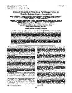
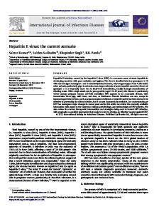
FIG. 1. Schematic representation of the different proteins expressed. The immunodominant loop of HBcAg, epitope c1, is located between amino acids 72 and 89 (■). The C-terminal region of the HBcAg involved in HBV genome binding (p) was deleted in the HBcD protein. HBcD-HEV-Ct is a C-terminal fusion of HBcD with the immunodominant region of HEV ORF2 (epitope 3-2) (s). For the construction of HBcD-HEV-i, the c1 epitope was deleted and replaced by epitope 3-2.
* Corresponding author. Mailing address: Institut de Virologie de Tours and Laboratoire des Processus Infectieux et Tumoraux, Faculte´ des Sciences Pharmaceutiques “Philippe Maupas,” 31 avenue Monge, 37200 Tours, France. Phone: 33 2 47 36 72 56. Fax: 33 2 47 36 71 88. E-mail:
[email protected]. 438
VOL. 37, 1999
NOTES
439
TABLE 1. Sequences of the oligonucleotide primers used for cloning HBV and HEV genes Primer no.
Sequence (59339)a
Positionsb
1 2 3 4 5 6 7 8 9
GGCATGGACATTGATCCTTA GTCTACGGAAGTGTTGATAAG CGTCAAACATCGATAGTCTC GTCATCGATGTCAACACTAATATGGGC ACCATCGATCCAGGTAGCTAGAGTC TTGATCGATGATACCTTGGACTACCCTGCCCGCGCCCATACTTTTGATGAT AAGGCCAAGGGGGCGGCACTCTGGGCAGAAATGATCAAAAGTATGGGCGCGGG AGTGCCGCCCCCTTGGCCTTCAGGGCTGCGCTTTCCCAGTCTACTGTCGCT AGTATCGATCACCTTCATCTTAAGGCGCTGAAGCTCAGCGACAGTAGACTGGAAAGCGC
1900–1919 (upper) 2318–2353 (lower) 2335–2354 (lower) 2158–2184 (upper) 2000–2024 (lower) 6974–7011 (upper) 6998–7041 (lower) 7032–7081 (upper) 7059–7117 (lower)
a b
ClaI restriction sites are boldfaced. In the virus genome considered.
cold phosphate-buffered saline (PBS), and sonicated by three 15-s bursts at 60% maximal power (Vibra-Cell, Strasbourg, France). Cellular lysates were loaded on a CsCl gradient and centrifuged in a Beckman SW28 rotor (for 20 h at 27,000 rpm and 4°C). CsCl gradient fractions were harvested, investigated for density, and then diluted 20-fold in PBS (pH 7.2) to determine seroreactivity by enzyme-linked immunosorbent assay (ELISA). HBcAg reactivity was revealed with an anti-HBc serum (diluted 1:1,000) obtained from a patient suffering from chronic active hepatitis B. HEV reactivity was detected by using a pool of anti-HEV-positive sera (diluted 1:100) obtained from patients with acute sporadic hepatitis E. The ELISA procedure used was as previously described (11). HBcAg reactivity was detected at densities ranging from 1.26
to 1.27 in cells expressing the HBcD gene (Fig. 2). Reactive fractions were pooled and applied to carbon-coated grids, negatively stained with 1.5% uranyl acetate, and observed with a JEOL 1010 electron microscope. Numerous empty core-like particles with an average diameter of 27 nm were observed. These particles were similar to empty HBV nucleocapsids purified from the liver of an HBV carrier (4). Expression of the chimeric HBcD-HEV-Ct protein gave rise to the formation of empty core-like particles with the same overall structure, but more irregular, as that of particles composed of HBcD protein. These particles, with densities ranging from 1.26 to 1.27 in CsCl, exhibit HBcAg- and HEV-positive reactivity (Fig. 2). As described by Borisova et al. with foreign epitopes from HBV (2), the insertion of the 42-amino-acid
FIG. 2. (Top) Detection of HBc ({) and HEV (F) reactivity in CsCl fractions (——, density) obtained from cells infected with baculovirus coding for the HBcD protein (A), the HBcD-HEV-Ct protein (B), or the HBcD-HEV-i protein (C). (Bottom) Particles observed in the CsCl fractions composing the peak of reactivity in the respective preparations. Bars, 50 nm.
440
NOTES
J. CLIN. MICROBIOL.
TABLE 2. Determination of seroreactivities of HBcD, HBcD-HEV-Ct, and HBcD-HEV-i particles by ELISAa
TABLE 3. Detection of anti-HEV IgG and IgM antibodies with a chimeric HEV-HBc recombinant protein (HBcD-HEV-i) in infected patients
No. (%) of sera reactive with:
Serum (no. tested)
HBcD
HBcD-HEV-Ct
HBcD-HEV-i
HEV1 anti-HBc2 (24) Anti-HBc1 anti-HEV2 (10)
0 10 (100)
24 (100) 10 (100)
24 (100) 0
a
HBc and HEV reactivities were tested with sera from patients with sporadic acute hepatitis E (n 5 24) or chronic hepatitis B (n 5 10).
HEV sequence at the C terminus of the HBcD protein did not abolish the formation of core-like particles. The expression of the chimeric HBcD-HEV-i protein gave rise to the formation of smaller particles resembling capsomeres, with an average diameter of 12 nm. These particles had densities ranging from 1.25 to 1.30 in CsCl and retained HEV reactivity only (Fig. 2). These structures are likely to be capsomere-like particles constituted by the assembly of six dimers of the HBc protein, as described by Bringas (3), during partial dissociation of recombinant core-like particles. The fact that the HBcD-HEV-i protein did not self-assemble into core-like particles but only into capsomere-like particles might be the result of conformational constraints introduced by the foreign HEV sequence used. The constraints might be due to excessive length or to the electric charge of the sequence introduced (1). The results obtained indicate that the immunodominant epitope is modeled correctly and exposed to immunoglobulin G (IgG) in both chimeric proteins. According to Crowther et al. (6), foreign sequences inserted into the immunodominant loop of HBcAg are external and maximally exposed at the tip of a protruding domain, whereas sequences fused to the C terminus can emerge through the holes in the capsid and thus become exposed on the surface. The HBV and HEV reactivities of the three recombinant particles are summarized in Table 2. The two chimeric recombinant proteins containing HEV epitopes were both reactive with sera from 24 HEV-infected patients. However, the prevalence of anti-HBc antibodies, which is as high as 50 to 90% in areas where HEV infection is endemic, especially in Southeast Asia and Africa, means that these particles could not be used for the diagnosis of HEV infection. To investigate the diagnostic potential of the HBcD-HEV-i particles, we used a panel of 99 sera from 46 patients with confirmed acute hepatitis type E living in areas where HEV infection is endemic. These serum samples were obtained during the 4 months after the onset of jaundice. The preliminary HEV diagnosis was determined by detection of IgG and IgM by the Abbott HEV enzyme immunoassay (7) and also by the presence of anti-HEV IgG detected by an ELISA using ORF3encoded peptides (5). Sera from 39 anti-HBc-positive patients, comprising 20 patients with acute hepatitis B and 19 chronic HBsAg carriers, were used as negative controls. The absence of HBcAg reactivity of the HBcD-HEV-i particles was confirmed by the lack of reactivity with the sera of the 39 patients with acute or chronic hepatitis B. Anti-HEV IgG was detected by both tests in 100% of the sera from HEV-infected individuals (Table 3). Anti-HEV IgM antibodies were detected in 57% of the sera with the Abbott test, in proportions ranging from 82% during the 1st month after the onset of jaundice to 17% 4 months later. IgM antibodies were detected with the HBcD-HEV-i test in 95% of the sera, with values ranging from 100% during the first 3 months after the onset of jaundice to 58% during the 4th month. Comparisons
Time from onset of jaundice (mo)
0–1 1–2 2–3 3–4 Total
No. (%) of sera in which anti-HEV Ig was detecteda
No. of sera tested
56 16 15 12 99
IgG
IgM
Abbott
HEV-HBc
Abbott
HEV-HBc
56 (100) 16 (100) 15 (100) 12 (100) 99 (100)
56 (100) 16 (100) 15 (100) 12 (100) 99 (100)
46 (82) 4 (25) 4 (27) 2 (17) 56 (57)
56 (100) 16 (100) 15 (100) 7 (58) 94 (95)
a Sera were tested with the Abbott HEV enzyme immunoassay and the HBcD-HEV-i test.
of proportions were performed with the x2 test, and significance was set at 0.05. The difference in the level of detection of IgM between the two tests was statistically significant (P , 1029). The difference in IgM detection was found to be statistically significant during the first 3 months after the onset of jaundice (P , 0.01, P , 0.0001, and P , 0.001, respectively) but not during the 4th month (P 5 0.09). In addition, sera collected during HEV outbreaks from 28 patients with acute hepatitis living in areas where HEV infection is endemic but without evidence of HAV, HBV, HCV, or HEV infection were also investigated for anti-HEV antibodies by the HEV-HBc particle test. HEV infection, as demonstrated by the presence of both anti-HEV IgG and anti-HEV IgM, was evidenced in five of these patients (18%). In conclusion, the test using HBc-HEV capsomere-like particles was able to detect anti-HEV IgG in all of the sera of the HEV-infected patients, as did the Abbott test, but it was more sensitive in the detection of anti-HEV IgM. In confirmation of the higher sensitivity, five acute infections were identified in patients for whom no conclusion could be drawn with the commercial test. The results obtained suggest that ELISA using HEV-HBc particles provides an accurate tool for the diagnosis of acute and previous hepatitis E infections. We thank Pierre-Yves Sizaret, Laboratoire de Microscopie Electronique, Faculte´ de Me´decine de Tours, for assistance with electron microscopy. We thank the Ministe`re de l’Education Nationale et de la Recherche Technologique (MENRT) for financial support. A.T. was supported by the MENRT. REFERENCES 1. Beames, B., and R. E. Lanford. 1995. Insertions within the hepatitis B virus capsid influence capsid formations and RNA encapsidation. J. Virol. 69: 6833–6838. 2. Borisova, G., B. Arya, A. Dislers, O. Borshukova, V. Tsibinogin, D. Skrastina, M. A. Eldarov, P. Pumpens, K. G. Skryabin, and E. Grens. 1993. Hybrid hepatitis B virus nucleocapsid bearing an immunodominant region from hepatitis B virus surface antigen. J. Virol. 67:3696–3701. 3. Bringas, R. 1997. Folding and assembly of hepatitis B virus core protein: a new model proposal. J. Struct. Biol. 118:189–196. 4. Coursaget, P., P. Maupas, A. Goudeau, and I. Millman. 1976. Automated complement fixation test for the detection of antibodies against the core of hepatitis B virus (HBc). J. Immunol. Methods 13:21–27. 5. Coursaget, P., Y. Buisson, N. Depril, P. Le Cann, M. Chabaud, C. Molinie´, and R. Roue´. 1993. Mapping of linear B cell epitopes on open reading frames 2- and 3-encoded proteins of hepatitis E virus using synthetic peptides. FEMS Microbiol. Lett. 109:251–256. 6. Crowther, R. A., N. A. Kiselev, B. Bo ¨ttcher, J. A. Berriman, G. P. Borisova, V. Ose, and P. Pumpens. 1994. Three-dimensional structure of hepatitis B virus core particles determined by electron cryomicroscopy. Cell 77:943– 950. 7. Dawson, G. F., K. H. Chau, C. M. Cabal, P. O. Yarbough, G. R. Reyes, and I. K. Mushahwar. 1992. Solid-phase enzyme immunoassay for hepatitis E
VOL. 37, 1999
8. 9. 10. 11.
IgG and IgM antibodies utilizing recombinant antigens and synthetic peptides. J. Virol. Methods 38:175–188. Prodromou, C., and L. H. Pearl. 1992. Recursive PCR: a novel technique for total gene synthesis. Protein Eng. 5:827–829. Takemura, F., T. Ishii, N. Fujii, and T. Uchida. 1990. Complete nucleotide sequence of hepatitis B virus. Nucleic Acids Res. 18:4587. Tam, A. W., M. M. Smith, M. E. Guerra, C. C. Huang, D. W. Bradley, K. E. Fry, and G. Reyes. 1991. Hepatitis E virus (HEV): molecular cloning and sequencing of the full length genome. Virology 185:120–131. Touze´, A., S. El Mehdaoui, P. Y. Sizaret, C. Mougin, N. Mun ˜ oz, and P.
NOTES
441
Coursaget. 1998. The L1 major capsid protein of human papillomavirus type 16 variants affects yield of virus-like particles produced in an insect cell expression system. J. Clin. Microbiol. 36:2046–2051. 12. Tsarev, S. A., T. S. Tsareva, S. U. Emerson, A. Z. Kapikian, J. Ticehurst, W. London, and R. H. Purcell. 1993. ELISA for antibody to hepatitis E virus (HEV) based on complete open-reading frame-2 protein expressed in insect cells: identification of HEV infection in primates. J. Infect. Dis. 168:369–378. 13. Yarbough, P. O., A. W. Tam, K. E. Fry, K. Krawczynski, K. A. McCaustland, D. W. Bradley, and G. R. Reyes. 1991. Hepatitis E virus: identification of type-common epitopes. J. Virol. 65:5790–5797.