Acta Clin Croat 2011; 50:261-266
Case Report
Bilateral Pseudomonas Aeruginosa Endogenous Endophthalmitis in an Immune-competent Patient with Nosocomial Urosepsis Following Abdominal Surgery Vesna Degoricija1,2, Vedrana Škerk 2, Zoran Vatavuk 3, Tamara Knežević3, Siniša Šefer4 and Željko Vučičević2 School of Medicine, University of Zagreb; 2Intensive Care Unit, University Department of Medicine;3University Department of Ophthalmology; 4Department of Nephrology and Dialysis, Sestre milosrdnice University Hospital, Zagreb, Croatia 1
SUMMARY – Endogenous endophthalmitis is a vision-threatening condition that results from hematogenous spread of infection to the eye, originating from a distant primary focus. It is considered as a rare entity that predominantly occurs in immune-compromised patients. We present a case of a critically ill immune-competent patient who underwent abdominal surgery later followed by nosocomial urosepsis complicated with bilateral Pseudomonas aeruginosa endogenous endophthalmitis that resulted in blindness. This case is clinically important because of the absence of predisposing factors for this kind of eye infection.
Key words: Endophthalmitis; Pseudomonas aeruginosa; Surgical operative procedures – complications; Cross infections; Case report
Introduction
Case Report
Endogenous bacterial endophthalmitis is a rare but severe intraocular infection that results from hematogenous bacterial spread from a remote primary site1. Pseudomonas aeruginosa is the most commonly encountered human pathogen from the Pseudomonadaceae family, a group of organisms comprising gram-negative rods2. We present a case of a previously healthy patient who underwent complex abdominal surgery that was later followed by nosocomial urosepsis complicated with bloodstream raised bilateral endophthalmitis and blindness.
This was a case of endogenous endophthalmitis causing blindness in an immune-competent 74-yearold Caucasian male. At the time of his first stay at Emergency Room (ER), University Department of Medicine, Sestre milosrdnice University Hospital, Zagreb in February 2008, his medical history revealed an abdominal infra-renal aneurysm of aorta, 4.5 centimeters in diameter, which had been diagnosed eight years before, together with mild arterial hypertension treated with lisinopril 10 mg/day and previous ischemic stroke three years before, with no neurologic sequels. In addition, there was no evidence of atrial fibrillation and secondary stroke prevention was maintained with aspirin 100 mg daily. On admission, he presented with brutal onset of severe chest pain with diffuse and colicky abdominal pain. He complained of feeling bloated and a tender hump was palpable in the left inguinal area. Multi-detector computed tomogra-
Correspondence to: Vesna Degoricija MD, PhD, Intensive Care Unit, University Department of Medicine, Sestre milosrdnice University Hospital, Vinogradska c. 29, HR-10000 Zagreb, Croatia E-mail:
[email protected] Received July 18, 2010, accepted September 1, 2011 Acta Clin Croat, Vol. 50, No. 2, 2011
261
Vesna Degoricija et al.
Pseudomonas aeruginosa endophthalmitis following abdominal surgery
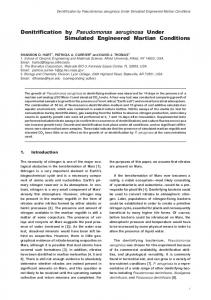
Fig. 1. Detailed physical examination revealed acutely swollen eyes with purulent discharge; examination by an ophthalmologist resulted in the diagnosis of endogenous endophthalmitis.
phy (MDCT) scan showed mechanical bowl obstruction caused by a left-sided inguinal incarcerated hernia as well as an abdominal infra-renal aortic aneurysm (4.5 cm diameter) with an intramural thrombus that lacked the signs of imminent rupture. Electrocardiography (ECG) revealed a concomitant acute anterior myocardial infarction. Laboratory tests demonstrated acute renal insufficiency, predominantly of pre-renal origin. Explorative laparotomy confirmed both an incarcerated left inguinal hernia and a gangrenous sigmoid colon that required Bassini’s hernioplastica and Hartmann’s sigmoid resection. The acute anterior myocardial infarction was treated conservatively at surgical intensive care unit (ICU) for five days. The patient had an uncomplicated postoperative recovery
on the ward, with urinary catheter in place for nine days; the patient was discharged from the hospital 14 days later. In March 2008, he presented again to the University Department of Medicine ER, this time with chills and high fever, which had persisted for four days, together with blurred vision bilaterally. The patient was in severe sepsis, febrile (38.6 °C), with arterial pressure 90/60 mm Hg, acute encephalopathy and sinus rhythm 130 bpm. Detailed physical examination revealed acutely swollen eyes with purulent discharge (Fig. 1), as well as a decreased auscultatory sound of the right basal lung. Laboratory data showed pronounced urinary tract infection (>100 leukocytes in urine sediment), serum white blood cell count of
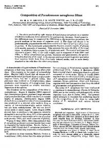
Fig. 2. The diagnosis of endogenous endophthalmitis was confirmed by orbital ultrasound. 262
Acta Clin Croat, Vol. 50, No. 2, 2011
Vesna Degoricija et al.
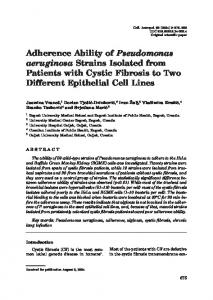
33x109/L, potassium 5.7 mmol/L, creatinine 893 µmol/L, and spontaneous prothrombin time of 68% with elevated aminotransferases, hypoproteinemia and metabolic acidosis. He was admitted to the ICU, examination by an ophthalmologist resulted in the diagnosis of endogenous endophthalmitis, which was confirmed by orbital ultrasound and MDCT scans of the orbits and brain showing bilateral endophthalmitis, however, without any trace of brain abscess (Figs. 2 and 3). Ceftriaxone treatment was started empirically for three days, one gram per day after loading dose calculated to renal function had been accomplished. Culture specimens of blood, urine and vitreous fluid were obtained and they all showed significant growth of Pseudomonas aeruginosa, with the exception of the right eye that was sterile. Tobramicin, chloramphenicol and betadine were used locally for both eyes. Ceftazidime was administered in a dosage of one to two grams two times per day, adjusted to the recovering renal function, for 16 days. The patient refused evisceration of the eyeballs. Chest x-ray revealed basal right sided pneumonia; abdominal ultrasound and MDCT scan showed no signs of postoperative infection, although an abdominal aortic aneurysm with mural thrombus was evident, albeit without any signs of possible rupture or dissection. Electrocardiography revealed sinus tachycardia of 120 bpm and a formed left ventricle (LV) aneurysm. Treatment of severe sepsis was complicated with repeated episodes of melena, the origin of which could not be established since the patient refused upper endoscopy. Consequently, anemia occurred that required correction with blood transfusions. The patient experienced full recovery from Pseudomonas aeruginosa severe sepsis again, however, now he was blind on both eyes. On discharge from the hospital, he was given advice on local application of antimicrobial therapy. Only one week later, he returned to the ER, presenting with dehydration and severe anemia. These two problems were addressed promptly and when satisfactory improvement had been achieved he was discharged from the ER. However, at the end of April 2008, he returned again to the ER and was admitted to the ICU with a combination of septic shock, concomitant bilateral septic endophthalmitis and urinary tract infection, all of which were complicated by Acta Clin Croat, Vol. 50, No. 2, 2011
Pseudomonas aeruginosa endophthalmitis following abdominal surgery
multiple organ failure syndrome and acute heart failure. He was dehydrated, somnolent and not verbally competent. Physical examination revealed decubital wound in the sacral region. On this admission, his laboratory findings were as follows: red blood cell count 2.5x10¹²/L, hemoglobin 78.3 g/L, mean cell volume 102 fL, white blood cell count 9.3x10⁹/L, platelets 89x109/L, severe metabolic acidosis and partial respiratory insufficiency with blood urea nitrogen over 60 mg/dL, creatinine 707 µmol/L and spontaneous prothrombin time 44%. Urinary culture showed Enterobacter sp. growth, whereas skin and blood cultures were sterile. Despite treatment at ICU that included targeted antimicrobial treatment, the patient’s overall condition deteriorated rapidly and he died seven days after admission to the hospital.
Discussion Pseudomonas aeruginosa is a formidable pathogen for humans because of the diversity and complexity of the factors that contribute to its virulence. Consequently, it is also a fascinating bacterial pathogen to study. Infections are most commonly nosocomial and present in a wide variety of clinical conditions including urinary tract infections (UTI), bacteremia, acute pneumonia, bone and joint infections, eye, ear and endovascular infections. Pseudomonas aeruginosa is almost a resident microbial agent in patients with cystic fibrosis, whilst patients with diabetes mellitus are also vulnerable due to the high frequency of UTI for which Pseudomonas aeruginosa is the fourth most common pathogen. The very first case report of Pseudomonas aeruginosa endogenous endophthalmitis appeared in 19683, and despite continuing investigation of this entity, only few have been reported since then. The first publication dealing with exogenous Pseudomonas aeruginosa eye infection dates back to 1964 4, and the majority of reported cases occurred after eye surgery procedures2,5,6. However, there are suggestions that it might occur as a result of trauma to the eye, or surface injury caused by extended wear contact lenses, the latter presenting most frequently as keratitis2. However, despite its affinity for the injured cornea, Pseudomonas aeruginosa infections of the eye from the bloodstream remain rare. One possible explanation for this could be 263
Vesna Degoricija et al.
Pseudomonas aeruginosa endophthalmitis following abdominal surgery
Fig. 3. Multi-detector computerized tomography scans of the orbits and brain showing bilateral endophthalmitis without any trace of brain abscess. 264
Acta Clin Croat, Vol. 50, No. 2, 2011
Vesna Degoricija et al.
its production of an enzyme that is capable of degenerating corneal protein fractions3. The high mortality associated with endogenous endophthalmitis makes it one of the most feared Pseudomonas aeruginosa infections since it is fulminant and accompanied by severe pain, chemosis, decreased visual acuity, anterior uveitis, vitreous involvement and panophthalmitis, which frequently results in blindness. There are a number of uncommon endogenous eye infections, which either evolve predominantly in or are exclusive to immune-compromised patients2. Clinical studies have detected some of the predisposing factors for these kinds of eye infection. In order of decreasing frequency, the medical conditions most commonly associated with Pseudomonas aeruginosa are diabetes mellitus, gastrointestinal disorders like gallstones, arterial hypertension, heart disease, malignancy, skin ulcer, stroke, renal failure, and drug and alcohol abuse7. The most common source of hematogenous spread is endocarditis, followed by gastrointestinal disorders (cholecystitis, abscesses), urinary tract infections8, cellulitis9, meningitis10, and pneumonia11. It is also associated with some status-post procedures like endoscopy and abdominal surgery12. Urinary tract infection where Pseudomonas is the pathogen is generally considered to be complicated UTI and it frequently serves as the nidus for Pseudomonas aeruginosa bacteremia by ascending infection 2. Since 1935, there have been approximately twenty reported cases of hematogenous spread of eye infection caused by this pathogen in immune-competent patients, most of them with unilateral endophthalmitis13. In the majority of cases, this rare condition is linked to predisposing factors and usually affects the right eye only13. The list of the potential bacteria that can give rise to endophthalmitis is diverse. One large study from 1994 identified streptococcal species as the most frequent isolates, followed by staphylococcus isolates7. Pseudomonas aeruginosa accounted for only four percent of this eye infection. An earlier study published in 1990 found staphylococcus isolates, especially Staphylococcus epidermidis, to be the most frequent pathogen8. Some recent studies revealed a growing number of fungal eye infections in apparently healthy, immune-competent patients14,15. Another study published in 2009 reported on Streptococcus pneumoniae as Acta Clin Croat, Vol. 50, No. 2, 2011
Pseudomonas aeruginosa endophthalmitis following abdominal surgery
the most frequent isolate, followed by Staphylococcus aureus and Escherichia coli13. The same study also noted few metastatic ocular infections in the immune-competent population, in which endogenous endophthalmitis of de novo onset lacked any identifiable predisposing factors. However, all of the named sources of infection and pathogens identified in these studies13-15 can cause endogenous endophthalmitis, although it is predominantly in immune-compromised hosts. Nevertheless, clinicians need to be mindful that the growing number of patients with chronic immunosuppressed medical conditions together with the ever increasing ability to prolong the life of seriously ill patients is very likely to produce an unwelcome byproduct, i.e. an increase in the incidence of Pseudomonas aeruginosa endophthalmitis16. The disease has poor visual outcomes, usually ending in the loss of vision, although the same authors have also reported success where good visual acuity has been achieved, but it requires early intervention with a combination of aggressive medical and surgical treatment12. The patient presented in this report had a stroke three years before abdominal surgery. One month after the surgery, following an uncomplicated postoperative period, he presented with severe sepsis, the origin of which was a complicated nosocomial UTI. Septic dissemination of the underlying UTI was found on MDCT and ultrasound documented bilateral endophthalmitis. Chest x-ray showed dissemination of the infection to the right basal lung. The patient had an abdominal aneurysm with intramural thrombus and ECG documented LV aneurysm, both of which are possible transmitters of bacteremia. As there was no clinical evidence of endocarditis, this cause of hematogenous spread was eliminated. Pseudomonas aeruginosa endophthalmitis is a rare condition. Nevertheless, this case of an immune-competent patient, albeit with comorbidities, should help raise awareness of this serious diagnosis and, which is at least as important, alert physicians to consider more frequently the possibility of its occurrence in the selected patient population.
265
Vesna Degoricija et al.
Pseudomonas aeruginosa endophthalmitis following abdominal surgery
References
9. Luemsamran P, Pornpanich K, Vangveeravong S, Mekanandha P. Orbital cellulitis and endophthalmitis in pseudomonas septicemia. Orbit 2008;27:455-7.
1. Reedy JS, Wood KE. Endogenous Pseudomonas aeruginosa endophthalmitis: a case report and literature review. Intensive Care Med 2000;26:1386-9.
2. Bier GP. Pseudomonas and related gram-negative bacillary infections. In: Goldman L, Ausiello D, editors. Cecil medicine. 2 3rd ed. Philadelphia: Saunders Elsevier, 2008;2236-41.
3. Ellenberger C Jr, Sturgill BC. Endogenous pseudomonas panophthalmitis. Am J Ophthalmol 1968;65: 607-11. 4. Theodore FH. Treatment of bacterial endophthalmitis following cataract extraction. Eye Ear Nose Throat Mon 1964;43:68-70.
5. Eifrig CW, Scott IU, Flynn HW Jr, Miller D. Endophthalmitis caused by Pseudomonas aeruginosa. Orbit 2008;27:455-7.
6. Pinna A, Usai D, Sechi LA, Zanetti S, Jesudasan NC, Thomas PA, Kaliamurthy J. An outbreak of postcataract surgery endophthalmitis caused by Pseudomonas aeruginosa. Ophthalmology 2009;116:2321-6.
7. Okada AA, Johnson RP, Liles WC, D’Amico DJ, Baker AS. Endogenous bacterial endophthalmitis. Report of a ten-year retrospective study. Ophthalmology 1994;101:832-8. 8. Shrader SK, Band JD, Lauter CB, Murphy P. The clinical spectrum of endophthalmitis: incidence, predisposing factors, and features influencing outcome. J Infect Dis 1990;162:115-20.
10. Kim SJ, Seo SW, Park JM, Chung IY. Bilateral endophthalmitis as the initial presentation of bacterial meningitis. Korean J Ophthalmol 2009;23:321-4. 11. Quirk JA, Beaman MH, Blake M. Communityacquired pseudomonas pneumonia in a normal host complicated by metastatic panophthalmitis and cutaneous pustules. Aust N Z J Med 1990;20:254-6.
12. Diaz-Valle D, Benitez del Castillo JM, Fernandez Acenero MJ, Santos Bueso E, Martrinez de la Casa JM, Garcia Sanchez J. Endogenous pseudomonas endophthalmitis in an immunecompetent patient. Eur J Ophthalmol 2007;17:461-3.
13. Shankar K, Gyanendra L, Hari S, Narayan SD. Culture proven endogenous bacterial endophthalmitis in apparently healthy individuals. Ocul Immunol Inflamm 2009;17:396-9.
14. Gupta P, Sachdev N, Kaur J, Dey P, Gupta V, Gupta A. Endogenous mycotic endophthalmitis in an immune-competent patient. Int Ophthamol 2009;29:315-8. 15. Rana M, Fahad B, Abid Q. Embolic aspergillus endophthalmitis in an immune-competent patient from aortic root aspergillus endocarditis. Mycoses 2008;51:352-3.
16. Chee SP, Jap A. Endogenous endophthalmitis. Curr Opin Ophthalmol 2001;12:464-70.
Sažetak OBOSTRANI ENDOGENI ENDOFTALMITIS UZROKOVAN BAKTERIJOM PSEUDOMONAS AERUGINOSA U IMUNO KOMPETENTNOG BOLESNIKA S NOZOKOMIJALNOM UROSEPSOM NAKON ABDOMINALNOG KIRURŠKOG ZAHVATA V. Degoricija, V. Škerk, Z. Vatavuk, T. Knežević, S. Šefer i Ž. Vučičević Endogeni endoftalmitis je akutna komplikacija hematogenog rasapa infekcije iz udaljenog žarišta u oči, što u najvećem broju slučajeva rezultira sljepilom. Bolest je rijetka, a najčešće se javlja u imuno kompromitiranih bolesnika. Prikazujemo imuno kompetentnog bolesnika kod kojega se nakon hitne operacije inkarcerirane ingvinalne hernije razvila nozokomijalna urosepsa komplicirana obostranim endogenim endoftalmitisom uzrokovanim bakterijom Pseudomonas aeruginosa, koji je rezultirao sljepilom. Budući da se radi o bolesniku bez prethodno opisanih predisponirajućih čimbenika za razvoj ove teške bolesti, smatramo ovaj prikaz klinički važnim za rano prepoznavanje i što ranije agresivno liječenje ove teške bolesti. Ključne riječi: Endoftalmitis; Pseudomonas aeruginosa; Kirurški postupci, operacijski – komplikacije; Bolničke infekcije; Prikaz slučaja 266
Acta Clin Croat, Vol. 50, No. 2, 2011