a Department of Obstetrics and Gynecology, AssafHarofe Medical Center, Tzrifin, ... b Department of Pathology, Sackler Faculty of Medicine at TelAviv University, ...
:" : ".~!: :,.~..
il"
BB
Biochi~i
et Biophysica fl~ta
ELSEVIER
Biochimica et Biophysica Acta 1201 (1994) 173-178
Enhancing effect of ATP on intracellular adriamycin penetration in human ovarian cancer cell lines Ron Maymon
a,*,
Batia Bar-Shira Maymon b,1, Malca Cohen-Armon c, Michael Holtzinger Judith Leibovici b
d
a Department of Obstetrics and Gynecology, AssafHarofe Medical Center, Tzrifin, Israel b Department of Pathology, Sackler Faculty of Medicine at TelAviv University, RamatAviv, Israel c Neufeld Cardiac Research Institute, Sackler Faculty of Medicine at TelAviv University, RamatAviv, Israel d Department of Obstetrics and Gynecology, Sapir Medical Center, Kfar Saba, Israel Received 1 February 1994
Abstract
Ovarian cancer has the highest mortality rate of all gynecological malignancies probably due to the evolution of clones resistant to cytotoxic drugs. Exploring possibilities to overcome such resistance constitutes a challenge in this study. We present the effect of adenosine triphosphate (ATP), serving as a chemosensitizer, in combination with adriamycin on three human ovarian cancer cell lines of epithelial origin, OC-109, OC-238 and OC-7-NU, obtained from malignant ascites of different patients, and were proven to be tumorigenic in nude mice. The three lines differ in their sensitivity to the ATP-induced increase in adriamycin accumulation. FACS analysis showed a pronounced increase in intracellular adriamycin accumulation after treatment with various concentrations of ATP. In the OC-238 line, a 50.1% increase was observed at a low ATP concentration (200/xM), whereas higher concentrations (400/.~M and 500 /zM) were needed to obtain an increase in ADR accumulation of 30% with the other two lines. Our study demonstrates that ATP improves the penetration of adriamycin at the neoplastic cellular level. Furthermore, our results may indicate that intratumoral ATP may serve as an alternative chemosensitizer which lacks the deleterious side effects of other chemosensitizing options. Keywords: Adenosine triphosphate; Adriamycin; Chemosensitizing effect; Flow cytometry; (Ovarian carcinoma)
1. Introduction
Epithelial ovarian tumors represent 25% of the female reproductive organ malignancies, accounting for a higher mortality rate than the combination of cervical and uterine cancers [1,2], and the number of cases is ever increasing [2,3]. A m o n g the explanations for this phenomenon is the clinical observation that early ovarian cancer is asymptomatic, resulting in more than 60% of the patients presenting in the late stages of the disease when the 5-year survival rate is estimated at 2 0 - 3 0 % [4]. Effective treatment revolves around ablative surgery and chemotherapy. However, a serious drawback of chemotherapy is the evolution of drug resistant clones during tumor progression
* Corresponding author. Fax: + 972 3 6409043. 1 In partial fulfillment towards a Postdoctoral Fellowship, supported by the Wolf Foundation to Promote Science and Art for the Benefit of Mankind. 0304-4165/94//$07.00 © 1994 Elsevier Science B.V. All rights reserved
SSDI 0 3 0 4 - 4 1 6 5 ( 9 4 ) 0 0 0 6 8 - 9
which may evolve towards single or multiple drugs [5,6]. This is possibly one of the major reasons for its response failure, which may be due to a decrease in intracellular drug accumulation, due either to a decrease in cell permeability [7-9] or to an increased efflux of the drug [10-12]. Thus, the use of cytotoxic drugs in conjunction with permeabilization agents may have the potential for counteracting this resistance and increasing cell permeability to the drugs. When using several tumor progression murine models, previous studies have disclosed increased cytotoxic agent activity with hyperthermia [13,14], or other chemosensitizers, such as verapamil [15] and Tween 80 [16]. Adriamycin (ADR) was used as a cytotoxic agent, because its fluorescence permits a follow-up of its intracellular accumulation by various methods, including fluorescence microscopy, FACS analysis and spectrofluorometry. The degree of combined treatment effect on the tumorigenicity of the treated cells was in correlation with the intracellular accu-
174
R. Maymon et al. /Biochimica et Biophysica Acta 1201 (1994) 173-178
mulation of ADR. Other studies [6,8,17,18] support our findings of an increased permeability to cytotoxic agents when induced by membrane-active agents [13-16]. Modulation of the membrane permeability might be a useful method for achieving a selective chemotherapeutic effect against tumor cells. Some authors indicate that brief exposure of several types of transformed 3T3, B16 melanoma, HeLa, and CHO-kl cells to external adenosine triphosphate (ATP) has markedly increased the passive membrane permeability, and allows passage through the plasma membrane of phosphorylated metabolites and ions [19-21]. Nontransformed cells, including Swiss and B A L B / c 3T3 cells and mouse embryonic fibroblasts, have not responded to ATP under the same experimental conditions [20], indicating that the difference in the cytotoxcity of exogenous ATP between normal and transformed cells may be attributed to selective permeability changes [21]. These data led some authors, such as Mure et al. [21], to consider the possible application of this procedure to chemotherapy using ATP and various anti-tumor agents like vincristine (VCR). They have demonstrated that VCR and ATP show a synergistic cytotoxic effect on clone-M3 cells, especially with low doses of VCR. This may be partially linked to the fact that ATP-dependent permeability change is enhanced by VCR or vinblastine [19]. Although the molecular mechanism for ATP control of the permeability change remains undefined [21], modulation of membrane permeability to anti-tumor agents cound be applied. The present study explored the effects of external ATP on human ovarian carcinoma cell lines' permeability to adriamycin (ADR), and compared the results with a human fibroblast cell line.
2. Materials and methods
Three ovarian carcinoma cell lines of epithelial origin were obtained from malignant ascites of three different newly diagnosed patients (OC-109, OC-238 and OC-7NU). The OC-109 line was derived from a mucinous cystadenocarcinoma, and the OC-238 and the OC-7-NU lines were derived from serous cystadenocarcinomas. Free floating cells of the ascitic fluids were centrifuged once and resuspended in DMEM supplemented with 20% fetal calf serum (FCS), 2 mM glutamine, 1 mM sodium pyruvate, non-essential amino acids, 100 U / m l penicillin, 100 /.Lg/ml streptomycin, 0.25 /~g/ml fungizone (Biolab, Jerusalem, Israel), 10 mM Hepes buffer (Sigma) and 10% autologous ascitic fluid. Cells were plated at high concentration (10 • 106/ml) in either 24 well plates (Costar) or 25 cm 2 flasks (Nunc). Cultures were incubated in a humidified, 7.5% CO 2, 37°C incubator for 4 - 7 days. At that time, non-adherent cells were discarded and medium was replaced by fresh supplemented DMEM, as described above, but without ascitic
fluid. When confluent, adherent cells were detached by a short (3-5 min) treatment with 1.25 m g / m l EDTA in phosphate-buffered saline, split and transferred to new flasks. To ascertain the tumorigenic nature of the cell lines obtained, established in vitro cultures were injected (5. 106-10 • 106 cells) subcutaneously into nude CD-1 mice. Growing tumors were observed within 2-3 weeks post-inoculation. The cell line OC-7-NU was obtained from a growing tumor in nude mice and cultured again as previously described. The presence of CA-125 marker was detected by immunohistologic staining. Fibroblast cell lines of human dermal origin were obtained from a normal skin biopsy. The samples were cut into 0.5 mm 2 pieces and were then cultured in 25 cm 2 Nunc flasks with F-10 medium (Biological Industries, Kibbutz Belt Haemek, Israel). After 10 days, the medium was changed to DMEM, and supplemented as described above. Fresh medium was replaced as needed, and the culture was continued until achieving confluency. Cells of the ovarian carcinoma lines and the human fibroblast line were then resuspended in RPMI medium supplemented with 10% FCS, 1% glutamine and 0.01% antibiotics and antimycotics (Kibbutz Beit Haemek, Israel) and plated in 75 cm 2 flasks (Nunc). Cultures were incubated at 37°C for 3 days. On the third day, cells were detached by treatment with Trypsin-EDTA solution (Kibbutz Belt Haemek, Israel), washed once with RPMI medium supplemented with 10% FCS and resuspended in fresh non-supplemented RPMI medium. Cell membranes were permeabilized in the presence of ATP by a modification of a procedure used previously in transformed cells and mast cells [22]. The efficiency of permeabilization by ATP, depended mainly on the ATP concentration, the length of exposure to ATP, the pH and the ionic strength of the incubation buffer, the temperature, and the presence of Ca and Mg ions during permeabilization. We selected conditions which allow both permeabilization and resealing of the membranes. Cells were washed twice, and resuspended in an isotonic buffer containing 169 mM glucose, 40 mM NaC1, 4.7 mM KC1, 1.18 mM KH2PO4, and 24.9 mM NaHCO 3. The pH was adjusted to 8.3 with NaOH. The cells ( 2 . 1 0 6) were incubated for 45 min at 25°C, with gentle shaking, in the presence of either 200, 400 or 500 /zm ATP (grade I, Sigma) combined with 10 or 20 /zg/ml of ADR (Farmitalia, Italy). For resealing, the permeabilization buffer was discarded and a solution of Krebs-Henseleit buffer [22] containing 1.18 mM MgC12 and 2.5 mM CaCI 2 (pH 7.4), was added. The tests were performed 10 min after resealing. Cytofluorometry was performed using a Becton Dickinson fluorescence activated cell sorter IV (FACS) with Consort 40 data processing, as previously described [13]. Adriamycin fluorescence was examined with an LP 570 filter. FACS analysis, as contrasted with biochemical de-
R. Maymon et al. / Biochimica et Biophysica Acta 1201 (1994) 173-178
175
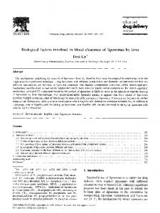
CELL LINE OC- 109
termination, can distinguish between cell populations possessing different quantities of the particular fluorescent marker. Cell viability was determined for all the concentrations of ATP and for 20 /~g/ml of ADR by trypan blue exclusion. Statistical analysis was performed using Student's t-test.
ADR !0
ADR20 NT
NT
3. Results 3.1. Effect of ATP on ADR uptake (at various concentrations) on the three human ovarian carcinoma cell lines
ATP 200 The influence of ATP on the intracellular ADR uptake of OC-109 is presented in Fig. 1. Similar results were obtained for OC-238 and OC-7-NU. The FACS analysis of ADR uptake, as a single agent, showed the same patterns of ADR intracellular accumulation in the three cell lines. Two cell populations were observed; a majority 'low permeability' population (LPP) consisting of 80%, and a 'high permeability' (HPP) minority subpopulation. Treatment in the presence of ATP showed a clear augmentation of ADR uptake by the OC-109 cells (as for the other two cell lines). Marked shifts to the right of the curves could be seen. The shift of the cell population towards a higher ADR uptake was most pronounced in the OC-238 line (Tables 1 and 2). The shift of the population towards a higher capacity to accumulate ADR (Table 1) depended upon the dose of ATP in the OC-109 and OC-7-NU lines. In the OC-238 line, maximal increase is already observed at the low ATP concentration (200 /zM). Lower concentrations did not result in any augmentative effect on permeability with any of the lines. Table 1 shows that, at different concentrations of ATP, OC-238 accumulates 2-3-times more ADR than the other two ovarian cancer lines. Similar data were repeatedly observed in four different experiments. 3.2. Effect of ATP on ADR uptake on the human fibroblast line
ATP 200
J'b
--J -J U
l
. . . .
'.
''''
ATP 400
,,,,,,llill
I
ATP 400
i
ATP 500
255
....
(I
121
II|
241
611
120
186
~1
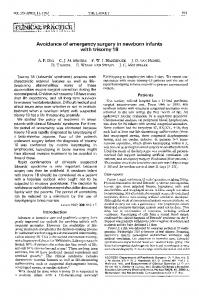
FLUORESCENCE INTENSITY We used this line as a control for the effect of ATP on the uptake of the cytotoxic agent. Fig. 2 shows the cytofluorimetric analysis of ADR accumulation alone or in combination with the different concentration of ATP. Practically no effect was exerted by ATP on the ADR uptake in this cell lines.
Fig. 1. Effect of ATP on ADR intracellular accumulation in ovarian carcinoma cell line-FACS examination. ADR concentrations 10 or 20 /~g/ml. ATP concentrations 200, 400 and 500 /zM. Fluorescence intensity is presented as log signals.
3.3. Influence of A T P and ADR on the viability of human ovarian carcinoma and fibroblast cell lines
higher rate of cell mortality was observed when the cells were treated with both agents. This effect ranged from 12.6-19% of the cells at the high concentration of ATP, with a somewhat higher effect displayed in the OC-7-NU line than in either of the others. The cell viability of the fibroblast cell line was not affected by ATP alone or when in combination with ADR. ATP alone had no effect on cell
Table 3 presents the effect of ADR alone or in conjunction with the various concentrations of ATP on the viability of OC-109, OC-238, and OC-7-NU cells. While ADR alone caused cell mortality of approximately 9%, a slightly
R. Maymon et al./Biochimica et Biophysica Acta 1201 (1994) 173-178
176
viability after resealing, either on the fibroblast cell line or on the human carcinoma cell lines.
4. Discussion
In the present study, we examined the efficacy of ADR with ATP functioning as a chemosensitizer probably by introducing changes in the plasma membrane permeability. However, ATP produced less pronounced permeability changes in the cells from a normal human fibroblast. Ozols et al. [23] recently reported on a clinical trial, using verapamil as a chemosensitizer, in combination with ADR on refractory ovarian cancer patients. The high doses of verapamil, needed to obtain an anti-tumoral effect, were found to be cardiotoxic. Another vasodilator, dipyridamole, was found to act synergistically with etoposide in a phase I trial [24]. Doselimiting toxic effects of this treatment were leukopenia and thrombocytopenia. The use of intratumoral low doses of ATP, acting as a local chemosensitizer without deleterious side effects in other critical tissue, may be a potentially promising alternative to the above options. This would not necessitate
accomodation for conditions, such as physiological saline medium, which would be obligatory in systemic treatment. Use of ATP for membrane permeability modulation, cell proliferation inhibition and cytotoxicity of anti-tumor agents has been successfully used in Chinese hamster overy cells [25,26]. Moreover, Wiley and Dubyok [27] have reported that treatment of human chronic lymphocytic leukemia lymphocytes with extracellular ATP produces large increases in cation permeability. In contrast, they have found less or no ATP-induced permeability with normal peripheral blood lymphocytes. External ATP has also been reported in modulating various cell surface-dependent properties, such as cell volume [28], ionic fluxes [29], morphology [30], histamine release [30], as well as cell aggregation [31] and virus infection [32] in various types of cells. Weisman et al. [33] have raised the possibility of external ATP activating channel formation, which allows for the free movement of Na + and K ÷, followed by an increase in the permeability of nucleotides, which then pass through the same channels. ATP can induce calcium mobilization following activation of P2-receptors [34]. Recently, it has been suggested that human ovarian cancer cells possess P2Y-receptors, al-
Table 1 Effect of ATP on adriamycin intracellular accumulation by FACS examination: increase in cell population of higher fluorescence intensity Cell line
Channels OC-109
Channels OC-238
Channels OC-7-NU
Channels OC-109
Channels OC-238
Channels OC-7-NU
ADR concentration (~g/ml)
ATP (/zM)
10 10 10 10
200 400 500
10 10 10 10
200 400 500
10 10 10 10
200 400 500
20 20 20 20
200 400 500
20 20 20 20
200 400 500
20 20 20 20
200 400 500
LPP, low permeability population; FI, fluorescence intensity.
% Cells in LPP
% Cells in range of higher FI
0-80 88.4 66.7 77.2 53.5 0-90 78.4 28.3 25.6 27.9 0-85 85.7 63.0 33,8 44.1 0-90 87.0 66.3 70.6 55.5 0-100 81.0 17,7 17,2 29.6 0-100 90.6 82.3 68.3 65.6
81-255 11.6 33.3 22.8 46.5 91-255 21.6 71.7 74.4 72.1 86-255 14.3 37.0 66.2 55.9 91-255 13.0 33.7 29.4 44.5 101-255 19.0 82.3 82.8 70.4 101-255 9.4 17.7 31.7 34.4
Increase in cells of higher FI
21.7 11.2 34.9
50.1 52.8 50.5
22.7 51.9 41.6
20.7 16.4 31.5
63.3 63.8 51.4
8.3 22.3 25.0
R. Maymon et al. /Biochimica et Biophysica Acta 1201 (1994) 173-178 CELL LINE
Table 2 Effect of ATP on adriamycin intracellular accumulation by FACS examination: shift of peak Cell line
OC-109
OC-238
OC-7-NU
OC-109
OC-238
OC-109
ADR concentr~ion (gg/ml)
ATP (~M)
Channel of LPP a
10 10 10 10 10 10 10 10 10 10 10 10 20 20 20 20 20 20 20 20 20 20 20 20
200 400 500 200 400 500 200 400 500 200 400 500 200 400 500 200 400 500
61 76 72 79 66 95 95 95 66 84 90 86 68 86 85 88 77 107 107 104 76 93 97 96
177 FB
ADR I0
Channel difference b
NI
15 11 18
255
29 29 29
J "~ ........ '''''~F-
18 24 20
AlP 200
18 17 20 30 30 27 17 21 20
tL
o
^IP 400 z
a Channel represents the fluorescence intensity peak of low permeability ~pulation (LPP). Channel difference represents the difference between the fluorescence intensity peak observed with combined ADR + ATP treatment minus the ADR treated cells.
though other receptor subtypes have not been excluded
[35]. However, despite the evidence of these studies concernAlP 500
Table 3 Effect of adriamycin alone or in conjunction with ATP on cell viability of the human ovarian carcinoma cell lines Cell line
ADR ATP concentration (/,~M) (/~g/ml)
No. of viable cells (mean 5: S.D.) 105/ml
OC-109
20 20 20 20 20 20 20 20 20 20 20 20
55.25 + 2.78 48.75 + 2.29 45.50 -t- 1.86 42.50 + 2.80 42.60+2.40 56.25 + 2.29 51.25 -t- 1.43 45.75 + 2.29 43.75 + 2.29 42.25 + 2.78 54.75 + 2.27 51.25-1-1.29 43.50 + 1.86 43.25+2.02 41.50 + 1.86
OC-238
OC-7-NU
* P < 0.025; ** P < 0.005.
200 400 500 200 400 500 200 400 500
* * *
255
Mortality (%) compared to ADR alone
6.6 12.8 12.6
** ** **
10.7 14.6 17.5
** ** **
15.1 15.6 19.0
FLUOURESCENCE IN I'[NSI TY
Fig. 2. Effect of ATP an ADR intracellular accumulation in fibroblast cell line-FACS examination. ADR concentration 1 0 / ~ g / m l . ATP concentrations 200, 400 and 500 /xM. Fluorescence intensity is presented as log signals.
ing the influence of external ATP on the cellular surface of mammalian cells, the exact mechanism and the physiological role of the ATP remains unclear [26]. Since the permeability changes have only been found in several
178
R. Maymon et a l . / Biochimica et Biophysica Acta 1201 (1994) 173-178
cultured transformed cells [19-21,26,27], an interesting question arises whether the ATP-induced change is dependent upon a malignant transformation. The ATP treatment could therefore selectively affect the transformed cells. Moreover, in combination with cytotoxic agents, ATP might permit the use of less toxic doses with more potent antineoplastic effects. An increase in ADR uptake by external ATP has been demonstrated in the present study by cytofluorometry in ovarian cancer lines. By employing this combination, it might be possible to overcome multidrug resistance to structurally dissimilar drugs. Our in vitro preliminary resuits may justify further studies concerning the possible use of this option for ovarian cancer therapy.
Acknowledgments The authors wish to thank Mrs. Sima Portnoy from the Department of Genetics, Tel Aviv University for culturing the human fibroblast cell-line, Dr. Boris Tartakovsky from the Department of Immunology, Weizmann Institute of Science, Rehovot for the preparation of the OC-109 line, Prof. I. Zanbar and Dr. I. Oschri from the Interdepartmental Core Facility of the Sackler Faculty of Medicine, Tel Aviv for their helpful advice on the use of FACS, and Sally Esakov for editing the material.
References [1] Berek, J.S., Hackner, N.F. and Lagasse, L.D. (1985) In Cancer treatment, 2nd Edn. (Haskell, C.M., ed.), pp. 409-428, Saunders, Philadelphia. [2] Lin, R.S. and Kessler, I.I. (1990) In Gynecology and obstetrics, Vol. 4, Chapt. 27 (Sciarra, J.J., ed.), pp. 1-17, Lippincot, Philadelphia. [3] Disaia, P.J. (1990) In Danforth's obstetrics and gynecology, 6th Edn. (Scott, J.R., Disaia, P.J., Hammond, C.B. and Spellacy, W.N., eds.), pp. 1067-1120, Lippincot, Philadelphia. [4] Jones, H.W. (1988) In Novak's textbook of gynecology, llth Edn, (Jones, H.W. III, Wentz, A.C. and Burnett, L.S., eds.), pp. 792-830, Williams and Wilkins, Baltimore. [5] Hoochhauser, D. and Harris A.L. (1991) Br. Med. Bull. 47, 178-196. [6] Beck, W.T. (1990). Eur. J. Cancer 26, 513-515.
[7] Ling, V. and Thompson, L.H. (1974) J. Cell Physiol. 83, 103-116. [8] Ramu, A., Shan, T.C. and Glaubiger, D. (1983) Cancer Treat. Rep. 67, 895-899. [9] Vrignaud, P., Montaudon, D., Londos-Gagliardi, D. and Robert J. (1986) Cancer Res. 46, 3258-3261. [10] Dano, K. (1973) Biochim. Biophys. Acta 323, 460-483. [11] Inaba, M., Kobayaski, H., Sakurai, Y. and Johnson, R.K. (1979) Cancer Res. 39, 2200-2203. [12] Harker, W.G. and Sikic, B.I. (1985) Cancer Res. 45, 4091-4096. [13] Leibovici, J., Klorin, G., Huszar, M., Hoenig, S., Klein, O., Michowitz, M. and Pinchassov, A. (1990) Clin. Expl. Metastasis 8, 33-46. [14] Klorin, G., Siegal, A., Bar-Shira-Maymon, B., Klein, O. and Leibovici, J. (1990) Int. J. Exp. Path. 71, 469-477. [15] Siegal, A., Hoenig, S. and Leibovici, J. (1990) Chemotherapy 36, 230-239. [16] Bar-Shira Maymon, B., Michowitz, M., Gibli, O., Klein, D. and Leibovici, J. (1992) Chemotherapy 38, 66-73. [17] Mizuno, S. and Ishida, A. (1982) Cancer Res. 42, 4726-4729. [18] Englehart, R. (1987) Cancer Res. 104, 1316-1203. [19] Kitagawa, T. and Akamatsu, Y. (1983) Biochim. Biophys. Acta 734, 25-32. [20] Kitagawa, T., Amano, F. and Akamatsu, Y. (1988) Biochem. Biophys. Acta 941, 257-263. [21] Mure, T., Sano, K. and Kltagawa, T. (1992) Jpn. J. Cancer Res. 83, 121-126. [22] Cohen-Armon, M. and Sokolovsky, M. (1991) J. Biol. Chem. 266, 2595-2605. [23] Ozols, R.F., Cunnion, R.E. and Klecker, R.W. (1987) J. Clin. Oncol. 5, 641-647. [24] Isonishi, S., Kirmani, S., Kim, S., Plaxe, S.C., Braly, P.S., McClay, E.F. and Howell, S.B. (1991) J. Natl. Cancer Inst. 83, 621-626. [25] Kitagawa, T. and Akamatsu, Y. (1981) Biochim. Biophys. Acta 649, 76-82. [26] Kitagawa, T. and Akamatsu, Y. (1986) Biochim. Biophys. Acta 360, 185-193. [27] Wiley, J.S. and Dubyak, GR. (1989) Blood 73, 1316-1323. [28] Gasic, G. and Stewart, C.C. (1966) J. Cell Physiol. 71, 239-241. [29] Weisman, G.A., Lustig, K.D., Freidberg, I. and Heppel, L.A. (1989) Enzymology 171, 857-869. [30] Kruger, P.G., Diamant, B. and Dahlquist, R. (1974) Int. Arch. Allergy Appl. Immunol. 46, 676-688. [31] Knight, V.A., Jones, B.M. and Jones, P.C. (1966) Nature 210, 1008-1010. [32] Fuchs, P., Gruber, E., Gitelman, J. and Kohn, A. (1980) J. Cell Physiol. 103, 271-278. [33] Weisman, G.A., De, B.K., Friedberg, I., Pritchard, R.S. and Heppel, L.A. (1984) J. Cell Physiol. 119, 211-219. [34] Dubyak, G.R. (1986) Arch. Biochem. Biophys. 245, 84-95. [35] Popper, L.D. and Batra, S. (1993) Cell Calcium 14, 209-218.