www.nature.com/scientificreports
OPEN
Received: 22 March 2018 Accepted: 24 July 2018 Published: xx xx xxxx
Blocking ion channels induced by antifungal lipopeptide syringomycin E with amide-linked local anesthetics Anastasiia A. Zakharova1, Svetlana S. Efimova1, Ludmila V. Schagina1, Valery V. Malev1,2 & Olga S. Ostroumova 1 The effects of the amide-linked (lidocaine (LDC), mepivacaine (MPV), prilocaine (PLC)) and ester-bound local anesthetics (benzocaine (BZC), procaine (PRC), and tetracaine (TTC)) on the pore-forming activity of the antifungal lipopeptide syringomycin E (SRE) in lipid bilayers were studied. Independently on electrolyte concentration in the membrane bathing solution the observed changes in conductance of SRE channels agreed with the altered membrane dipole potential under the action of ester-bound local anesthetics. Effects of aminoamides in diluted and concentrated solutions were completely different. At 0.1 M KCl (pH 7.4) the effects of amide-linked anesthetics were in accordance with changes in the membrane surface potential, while at 2 M KCl aminoamides blocked ion passage through the SRE channels, leading to sharp reductions in pore conductance at negative voltages and 100-fold decreases in the channel lifetimes. The effects were not practically influenced by the membrane lipid composition. The interaction cooperativity implied the existence of specific binding sites for amidebound anesthetics in SRE channels. Local anesthetics are compounds causing the suspension of impulse transmission along nerve fibers, and their molecular structure comprises hydrophobic and lipophilic parts connected by amide or ether bonds. Correspondingly, they are divided into amide-linked (e.g., LDC, MPV, and PLC) and ester-bound (e.g., BZC, PRC, and TTC) groups. The chemical structures of local anesthetics are presented on the Fig. 1a. As a rule, at physiological pH, these drugs exist in both neutral and cationic forms. Among the listed anesthetics, only BZC is excluded, which is almost completely uncharged under physiological conditions. Local anesthetics interfere with impulse conductions in nerves and muscles by binding to voltage-gated sodium channels and blocking the transient Na+ inward current1. While the major drug mechanism of action is not resolved, experimental evidence favors steric blocking, closed state stabilization, or some combination of the two. Extensive site-directed mutagenesis experiments have provided proof that LDC-like drugs bind in the inner pore2–4. However, substantial evidence supports that anesthetics generally perturb bulk membrane structure5–7 and, consequently, affect membrane transport, especially ion channels8–10. Specifically, a good correlation between the partition coefficients in lipid/water systems (and, consequently, their ability to rearrange membrane lipids) and the clinical potency of the drugs support the hypothesis that membrane lipids are the primary sites of anesthetic action11,12. Lee et al.8 proposed that local anesthetics trigger the transition of surrounding lipids to the more fluid, liquid crystalline phase, allowing the sodium channel to close, resulting in local anesthesia. Since anesthetics are present in both cationic and non-ionic forms under physiological conditions, the question of whether uncharged or charged species affect the physical properties of the membrane has been raised. Fraceto et al.13 showed that the uncharged versions of LDC and MPV increase the mobility of choline nuclei and decrease the mobility of glycerol-region hydrogens, thus modulating lipid packing. Hata et al.14,15 studied the effects of local anesthetics on the phase transition temperatures of dipalmitoylphosphatidylcholine bilayer membranes by optical and calorimetrical methods. TTC, LDC and PRC depressed the main and pre-transition 1
Institute of Cytology of the Russian Academy of Sciences, 4 Tikhoretsky prospect, St. Petersburg, 194064, Russia. Saint Petersburg State University, Institute of Chemistry, 26 Universitetskii prospect, St. Petersburg, Petergof, 198504, Russia. Anastasiia A. Zakharova and Svetlana S. Efimova contributed equally to this work. Correspondence and requests for materials should be addressed to O.S.O. (email:
[email protected]) 2
Scientific REpOrtS | (2018) 8:11543 | DOI:10.1038/s41598-018-30077-6
1
www.nature.com/scientificreports/
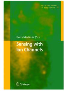
Figure 1. (a) Chemical structures of local anesthetics (LDC, PLC, MPV, PRC, BZC, and TTC). (b) Structures of tested lipopeptides (SRE, syringostatin A, syringotoxin B, and syringopeptin 22 A). The first four lipopeptides differ in the amino acid sequence between positions 2 and 6. The 3-hydroxy fatty acyl group is a derivative of decane, dodecanoic, tetra- or hexadecanoic acid. Abbreviations for nonproteinogenic amino acids: Asp(3-OH) – 3-hydroxyaspartic acid; Dab – 2,4-diaminobutyric acid; Dhb – 2,3-dehydroaminobutyric acid; Hse – homoserine; Orn – ornithine; Thr(4-Chl) – 4-chlorothreonine; aThr – allothreonine.
temperatures of the DPPC bilayers. Moreover, TTC induced complex phase behaviors of dipalmitoylphosphatidylcholine, including the formation of mixed lipid/anesthetic micelles and transition from the interdigated gel phase (rather than the ripple phase) to the liquid crystalline phase at relatively high TTC concentrations. Using high-pressure Fourier transform infrared spectroscopy, Auger et al.16 demonstrated smaller interchain interactions for dimyristoylphosphatidylcholine due to increases in both orientational and conformational disorder caused by uncharged TTC intercalation between lipid acyl chains. The authors concluded that uncharged TTC disorders myristoyl chains, while the charged form induces the formation of an interdigitated gel phase. The authors also showed that cholesterol (CHOL) prevents the formation of the interdigitated phase. In addition to modifying the elastic properties of membranes, anesthetics electrostatically interact with the lipid bilayer, i.e., the drugs alter the electrical potential at the water/lipid boundary17, termed the membrane boundary potential. The boundary potential consists of two components18–23: a surface component (ϕs), related to the surface charge of the membrane, and the dipole component (ϕd), due to specifically orientated lipid and water dipoles at the interface, which imparts a highly positive electrostatic potential to the membrane interior membrane in respect to the adjacent aqueous phase and, consequently, regulation of reconstituted ion channels24–35. Furthermore, TTC was found to neutralize the negative surface charges of cardiolipin-containing liposomes36. The authors also concluded that TTC increases surface potential more effectively than LDC37. Furthermore, both the charged and uncharged forms of TTC and LDC induce substantial changes in the membrane dipole potential38,39.
Scientific REpOrtS | (2018) 8:11543 | DOI:10.1038/s41598-018-30077-6
2
www.nature.com/scientificreports/
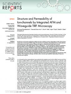
Figure 2. Current fluctuations corresponding to the openings and closings of single SRE channels in lipid bilayers in the absence (control) and presence of various local anesthetics: 10 mM LDC, PLC, MPV and PRC; 3 mM BZC; and 1 mM TTC. The membranes were composed of DOPC (a,b) or DOPC:CHOL (67:33 mol%) (c,d) and bathed in 0.1 M (a,c) or 2.0 M KCl (pH 7.4) (b,d). The transmembrane voltage was equal to −150 mV. C – closed state of the channel, O – open state of the pore.
This study aimed to establish the mechanism underlying the influence of local anesthetics on SYRingomycin E channels. The antifungal lipopeptide SRE was previously shown to form voltage-gated, predominantly anion-selective asymmetric cone-shaped channels with a narrow peptide and a wide lipid mouth40. The topology of SRE channel water pores is like that of sodium channels in open state, with a relatively wide vestibule and a narrower region, including the selectivity filter41. Moreover, SRE channels are characterized by potential and mechanical sensitivity. Altogether, these facts make SRE channels extremely convenient for studying the binding and lipid-mediated effects of local anesthetics. To test the possibility of binding, we used the drugs of two types, amide-linked (LDC, PLC, and MPV) and ester-bound anesthetics (BZC, PRC, and TTC). Binding cooperativities were evaluated by Hill’s coefficients42. Changes in the cation/anion selectivities of the channels are thought to be determined by the binding anesthetics in a narrow part of the pore. To establish whether the lipid-mediated effects of the drugs are caused by alterations in the surface or dipole components of the membrane boundary potential, two different concentrations of an electrolyte in bilayer bathing solutions were tested (0.1 and 2 M KCl). To identify the possible actions of local anesthetics on lipid packing, pure bilayers from dioleoylphosphocholine (DOPC) were compared with CHOL enriched membranes. Lipid bilayers comprising anionic phospholipids, dioleoylphosphoserine (DOPS), and raft-mimicking mixture of DOPC, sphingomyelin (SM), and CHOL were also tested.
Results and Discussion
Figure 2a demonstrates the current fluctuations corresponding to the openings and closings of single SRE channels in DOPC membranes bathed in 0.1 M KCl (pH 7.4) in the absence (control) and presence of 10 mM LDC, PLC, MPV, and PRC; 3 mM BZC; and 1 mM TTC. LDC, PLC, MPV, and TTC enhanced the amplitude of the SRE channels, while PRC did not influence this value. BZC significantly reduced the channel conductance. Figure 3a shows the corresponding G(V) curves in the absence and presence of the local anesthetics. LDC and MPV increased G by approximately 15%, while PLC and TTC enhanced G by approximately 30%. Simultaneously, PRC did not change G, while BZC reduces the channel amplitude by approximately 20%. We previously showed that local anesthetics affect the boundary potential of model lipid membranes. Changes in the boundary potential by Scientific REpOrtS | (2018) 8:11543 | DOI:10.1038/s41598-018-30077-6
3
www.nature.com/scientificreports/
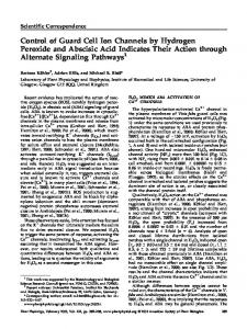
Figure 3. G(V) curves of single SRE channels in the absence (■) and presence of 10 mM LDC (◊), PLC (▼), MPV (Δ) or PRC (▲); 3 mM BZC (*); and 1 mM TTC (●). The membranes were composed of DOPC (a,b), DOPC:CHOL (67:33 mol%) (c,d), DOPS (e) or DOPC:CHOL:SM (47:33:20 mol %) (f) and bathed in 0.1 M (a,c) or 2.0 M KCl (pH 7.4) (b,d,e,f).
the positively charged amide-linked anesthetics LDC, PLC, and MPV are due to increases in the surface potential of the bilayer, while TTC increases its dipole potential17. Indeed, the surface potential increment of the DOPC membrane at 10 mM LDC, PLC and MPV was approximately 24 ± 5 mV. The increase in the dipole potential of DOPC bilayers induced by 1 mM TTC equaled 48 ± 14 mV. PRC did not obviously affect the magnitudes of the surface and dipole components of the DOPC membrane boundary potential at concentrations up to 10 mM. Here, we evaluated the changes in dipole potential induced by the addition of 3 mM BZC to the bathing solution, which reduced the dipole potential by 55 ± 8 mV. Comparing the effects of the drugs on the SRE channel current amplitude and electrical potential at the membrane/water interface, the changes in G upon the introduction of anesthetics presumably correspond to the changes in the boundary potential, its dipole or surface components. Scientific REpOrtS | (2018) 8:11543 | DOI:10.1038/s41598-018-30077-6
4
www.nature.com/scientificreports/ τ, ms* DOPC Anesthetic C, mM ρ, %# at pH 7.4 0.1 M KCl
DOPC:CHOL 2.0 M KCl
DOPC:CHOL:SM
DOPS
0.1 M KCl
2.0 M KCl 2.0 M KCl
2.0 M KCl
t−&
control
—
—
187 ± 3
74 ± 1
280 ± 6
253 ± 4
123 ± 3
198 ± 5
0.86 ± 0.07
LDC
10
75
152 ± 15
1 ± 1
161 ± 9
1 ± 1
1 ± 1
3 ± 1
0.53 ± 0.21
PLC
76
128 ± 12
16 ± 1
141 ± 3
13 ± 1
9 ± 1
15 ± 1
0.61 ± 0.09
MPV
64
152 ± 4
11 ± 1
207 ± 21
8 ± 1
2 ± 1
14 ± 1
0.75 ± 0.04