BMC Neuroscience
BioMed Central
Open Access
Research article
Enhanced expressions of microvascular smooth muscle receptors after focal cerebral ischemia occur via the MAPK MEK/ERK pathway Aida Maddahi1 and Lars Edvinsson*2 Address: 1Division of Experimental Vascular Research, BMC A13, Lund University, 221 84 Lund, Sweden and 2Department of Internal Medicine, Institute of Clinical Sciences, Lund University, Lund, Sweden Email: Aida Maddahi -
[email protected]; Lars Edvinsson* -
[email protected] * Corresponding author
Published: 15 September 2008 BMC Neuroscience 2008, 9:85
doi:10.1186/1471-2202-9-85
Received: 16 June 2008 Accepted: 15 September 2008
This article is available from: http://www.biomedcentral.com/1471-2202/9/85 © 2008 Maddahi and Edvinsson; licensee BioMed Central Ltd. This is an Open Access article distributed under the terms of the Creative Commons Attribution License (http://creativecommons.org/licenses/by/2.0), which permits unrestricted use, distribution, and reproduction in any medium, provided the original work is properly cited.
Abstract Background: MEK1/2 is a serine/threonine protein that phosphorylates extracellular signalregulated kinase (ERK1/2). Cerebral ischemia results in enhanced expression of cerebrovascular contractile receptors in the middle cerebral artery (MCA) leading to the ischemic region. Here we explored the role of the MEK/ERK pathway in receptor expression following ischemic brain injury using the specific MEK1 inhibitor U0126. Methods and result: Rats were subjected to a 2-h middle cerebral artery occlusion (MCAO) followed by reperfusion for 48-h and the ischemic area was calculated. The expression of phosphorylated ERK1/2 and Elk-1, and of endothelin ETA and ETB, angiotensin AT1, and 5hydroxytryptamine 5-HT1B receptors were analyzed with immunohistochemistry using confocal microscopy in cerebral arteries, microvessels and in brain tissue. The expression of endothelin ETB receptor was analyzed by quantitative Western blot. We demonstrate that there is an increase in the number of contractile smooth muscle receptors in the MCA and in micro- vessels within the ischemic region. The enhanced expression occurs in the smooth muscle cells as verified by colocalization studies. This receptor upregulation is furthermore associated with enhanced expression of pERK1/2 and of transcription factor pElk-1 in the vascular smooth muscle cells. Blockade of transcription with the MEK1 inhibitor U0126, given at the onset of reperfusion or as late as 6 hours after the insult, reduced transcription (pERK1/2 and pElk-1), the enhanced vascular receptor expression, and attenuated the cerebral infarct and improved neurology score. Conclusion: Our results show that MCAO results in upregulation of cerebrovascular ETB, AT1 and 5-HT1B receptors. Blockade of this event with a MEK1 inhibitor as late as 6 h after the insult reduced the enhanced vascular receptor expression and the associated cerebral infarction.
Background Acute focal cerebral ischemia results in a severely ischemic core with low residual cerebral blood flow (CBF) whereas the ischemic penumbra synaptic activity is reduced while
the residual CBF is enough to maintain membrane ionic gradients. The expansion of depolarized core coincides with the occurrence of spontaneous peri-infarct spreading depolarization [1]. The tissue viability threshold and its
Page 1 of 13 (page number not for citation purposes)
BMC Neuroscience 2008, 9:85
http://www.biomedcentral.com/1471-2202/9/85
relationship to the penumbra has focused on electrical and membrane failure in brain tissue [2,3], and therefore, it has been suggested that the ischemic depolarization increases the metabolic burden, thereby exacerbates the energy deficit, and enlarges the infarct [4]. This view has by and large neglected the fact that stroke primarily is a cerebrovascular disorder. Recently, Shin and colleagues presented data that there is neurovascular vasoconstrictor coupling during the ischemic depolarization which contributes to the hemodynamic progression of damage in focal cerebral ischemia [5]. They suggest that by reducing the adverse vascular effects of tissue depolarization is a possible way the neuroprotective drugs act to reduce the tissue injury. We have observed a rapid transcriptional upregulation of contractile endothelin-1 (ETB receptors), and angiotensin II AT1 receptors in vascular smooth muscle cells in the middle cerebral artery (MCA) leading to the ischemic region starting immediately after induction of the cerebral ischemia [6,7]. These changes result in enhanced contraction of the vasculature leading to the ischemic region, particularly because agonists for these receptor are produced in the cerebrovascular endothelium [8]. In agreement, single receptor inhibition has in the past only been found to have limited effect in reducing cerebral infarct size after focal ischemia [3]. Therefore, we hypothesize that blocking the transcriptional upregulation of endothelin, serotonin and angiotensin receptors would reduce the cerebral infarct that occurs after focal cerebral ischemia. To test this hypothesis, an animal model of consistently inducible cerebral ischemia was used: 2 hours reversible middle cerebral artery occlusion (MCAO) followed by reperfusion for 48 hours [9]. We present the novel observations that there is upregulation of the mitogen-activated protein kinase (MAPK) extracellular signal-regulated kinase (ERK1/2), the transcription factor Elk-1, and the contractile receptors for endothelin (ETA and ETB), angiotensin II AT1, and 5-hydroxytryptamine 5-HT1B receptors in both the MCA leading to the ischemic region and in microvessels within the infarct area but not in adjacent brain tissue. Systemic treatment with the MEK1 inhibitor U0126, given at the start of the reperfusion or at 6 hours afterwards abolished the enhanced receptor protein expression and reduced the infarct volume.
Results Signal transduction after MCA occlusion Distal MCAO resulted in an abrupt decrease in CBF over the dorsolateral cortex; flow was reduced to 15 ± 3% of the baseline flow in the ischemic region. After 2 hours the block was removed. The flow subsequently returned to baseline. The animal were carefully monitored during the following 48 hours and then sacrificed. We calculated the neurology score (MCAO: 4.0 ± 0.5, P < 0.05 versus sham operated animals) (Table 1), collected tissue for immunostaining and Western blot, and determination of the infarct volume (24.8 ± 1.4% of total cerebrum) and the degree of edema (Fig. 1 and 2). The physiological parameters did not differ between the groups (Table 2). There was an increase in body temperature after MCAO in all groups which are in agreement with previous studies [10]; this did not differ between the different groups in our study.
We subsequently assessed if MCAO leads to activation of pERK1/2 and pElk-1 in the smooth muscle cells of the MCA, the associated microvessels, and in brain tissue. There was weak staining of both in vehicle control (Fig. 3). The results showed that pERK1/2 and pElk-1 were markedly activated at 48 hours after the MCAO + vehicle (Fig. 3). The pERK1/2 and pElk-1 immunoreactivity were localized in the cytoplasm of the smooth muscle cells as verified by co-localization experiments with smooth muscle specific actin (Fig. 5). As can be seen in the illustration there was a significant enhanced expression of pERK1/2 (185 ± 11 %) and pElk-1 (174 ± 6 %) in the MCA leading to the infarct and in associated microvessels (130 ± 10 % and 130 ± 6 %, respectively) (Fig. 4). However, there was no significant change in their expression in associated brain tissue (106 ± 3 % and 117 ± 13 %, respectively) or in other regions of the brain. There was a weak expression of pElk-1 in cell bodies within the brain tissue around the MCA. Inhibition of signal-transduction Administration of the MEK1-specific inhibitor U0126 (30 mg/kg, intraperitoneal), which blocks the enzymatic activity of MEK1 [11] reduced both the infarct volume and the neurology score when given in conjunction with the start of the reperfusion (0 hour) or at 6 hours after the MCAO; the reductions were significant for infarct volume
Table 1: Neurological Scores measured at 0, 24 and 48 hours after MCA occlusion using an established scoring scale [26].
Neurology score
Control (n = 7)
U0126 – 0 h (n = 7)
U0126 – 6 h (n = 6)
U0126 – 12 h (n = 6)
0h 24 h 48 h
4 ± 0.0 3.8 ± 3.4 4 ± 0.5
4± 0.0 3 ± 0.6 2 ± 0.7
4 ± 0.0 3.5 ± 0.5 3.3 ± 0.7
4 ± 0.0 3.8 ± 0.3 3.8 ± 0.3
Comparison between the U0126 treated groups and vehicle treated rats (control). Data are expressed as mean ± S.D and n = number of rats.
Page 2 of 13 (page number not for citation purposes)
BMC Neuroscience 2008, 9:85
http://www.biomedcentral.com/1471-2202/9/85
Table 2: Physiological parameters
Physiological Parameters
pO2 (mmHg) pCO2(mmHg) pH Plasma glucose (mmol/L) MAP (mmHg) Temperature during operation (°C) Temperature before reperfusion (°C) Temperature after reperfusion (°C) Weight loss 48 h after occlusion (%)
Control (n = 7)
U0126, 0 h (n = 7)
U0126, 6 h (n = 6)
U0126,12 h (n = 6)
97.2 ± 2 52.6 ± 3 7.3 ± 0.01 12.1 ± 0.4 91.1 ± 2.2 37.1 ± 0.2 39.3 ± 0.1 38.1 ± 0.1 13.4 ± 1.3
108.3 ± 3.2 50.6 ± 1.9 7.3 ± 0.01 10.2 ± 0.6 92.6 ± 1.8 37.2 ± 0.1 39.2 ± 0.1 38.6 ± 0.3 8.0 ± 1.8
107.6 ± 7 49.6 ± 4.5 7.3 ± 0.02 11.1 ± 0.3 91.8 ± 2.5 37.2 ± 0.1 39.3 ± 0.2 38.1 ± 0.1 16.1 ± 3.2
120.4 ± 10 48.5 ± 0.6 7.5 ± 0.01 10.7 ± 0.4 101.4 ± 4.7 37.3 ± 0.2 38.9 ± 0.2 38.6 ± 0.2 10.3 ± 2.4
Values are the mean % of control ± s.e.m., P > 0.05 between the groups, n = number of rats.
(11.8 ± 2 % and 14.6 ± 3 %; respectively of cerebrum, P < 0.05; Fig. 1 and 2A), and neurology score (2 ± 0.7 and 3.3 ± 0.7; respectively, P < 0.05) but not for the edema (Table 1) (Fig. 2B). The administration of U0126 with start at 12 hours after the initiation of reperfusion did not result in a significantly diminished infarct volume (20.3 ± 1 % of cerebrum; Fig. 2A) or the neurology score (3.8 ± 0.3; Table 1).
muscle actin, expressed in the smooth muscle cells, revealed clear co-localization. All three receptors co-localized with the smooth muscle cells, in addition, endothelin ETB receptor protein was located in the endothelial cells of the cerebral vessels (Fig. 8); previous studies with the endothelial marker CD31 has verified this in cerebral arteries [12]. MCAO did however not show enhanced (or altered) expression of the endothelial ETB receptors.
We subsequently assessed if MEK1 inhibition altered the cerebrovascular activation of pERK1/2 and pElk-1 in the vascular smooth muscle cells after MCAO. The results showed that systemic treatment with U0126 abolished the increase in pERK1/2 and pElk-1 activation after MCAO when the treatment was given in conjunction with reperfusion (0 hour) or with start 6 hours, and for pERK1/ 2 12 hours after the start of the reperfusion (Fig. 3 and 4). In the contralateral hemisphere there was only weak pERK1/2 and pElk-1 activity at baseline in the vasculature, and this was not affected by MCAO (Fig. 3); the levels of activity were comparable to those of the vehicle control. There was no significant change in brain tissue for pERK1/ 2 and pELK-1 activity (Fig. 3).
Systemic treatment with the MEK1 inhibitor abolished the increase in receptor expression in both vascular regions when treatment was initiated either at reperfusion (0 hour) or 6 hours afterwards but not when starting 12 hours after reperfusion (Fig. 6); this correlates well with the reduction in infarct volume at the same time points (Fig. 1 and 2A).
Cerebrovascular receptor expression To investigate whether the MAPK activity leads to changes in receptor protein transcription, we analyzed the expression of vascular receptors using immunostaining and image analysis with confocal microscopy. We observed for the first time a significant increase in the expression of endothelin ETB, angiotensin AT1, and 5-hydroxytryptamine 5-HT1B receptors in smooth muscle cells not only in the ischemic MCA but also in smooth muscle cells of cerebral microvessels associated with the ischemic region (Fig. 6 and 7). There were no changes in receptor expression in contralateral vessels. Importantly, the endothelin ETA receptor expression was unchanged after MCAO and following treatment at all locations (Fig. 6 and 7). Double immunostaining for endothelin ETB, angiotensin AT1, and 5-hydroxytryptamine 5-HT1B receptors versus smooth
Western blot The Western blot experiments showed a significant increase in ETB protein level after MCAO as compare to the vehicle group (122 ± 9 %; p < 0.05). Treatment with the MEK inhibitor (U0126) given in conjunction with reperfusion (0 hour) prevented the increase in the ETB receptor protein (101 ± 5%) (Fig. 9).
Discussion and conclusion We have observed that acute cerebral infarction followed by reperfusion in the rat is accompanied by upregulation of the functional contractile phenotype and the mRNA expression of endothelin ETB and angiotensin AT1 receptors [6,7]. In the present study we show for the first time that the proteins of the contractile receptors ETB, AT1 and 5-HT1B receptors are upregulated in the smooth muscle cells of the MCA leading to the ischemic region and in microvessels associated with the focal ischemia. The colocalization studies verified that the enhanced expression is located in the smooth muscle cells. We have previously shown with confocal microscopy and double immunostaining that the enhanced receptor expression is localized to the smooth muscle cells (co-localize with smooth mus-
Page 3 of 13 (page number not for citation purposes)
BMC Neuroscience 2008, 9:85
http://www.biomedcentral.com/1471-2202/9/85
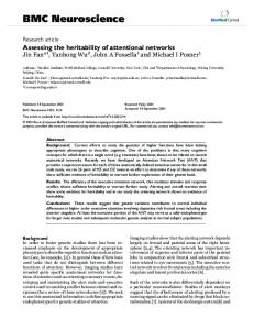
Figure cle Typical reperfusion (A) examples 1 after MCAO of coronal (0 h), brain or sections given at 6that andshow 12 hours smaller after ischemic MCAOareas and 48-h in animals reperfusion treatedversus with U0126 animalsimmediately treated withafter vehiTypical examples of coronal brain sections that show smaller ischemic areas in animals treated with U0126 immediately after reperfusion after MCAO (0 h), or given at 6 and 12 hours after MCAO and 48-h reperfusion versus animals treated with vehicle (A).
cle actin) and not to the adventitia or the endothelial cells (with CD31 staining) in conjunction with experimental subarachnoid hemorrhage [12]. The enhance receptor expression was verified by Western blot of the ETB receptor protein. This upregulation was associated with activation of the signal transduction proteins pERK1/2 and the transcription factor pElk-1. Administration of the MEK1 inhibitor U0126, acting upstream of ERK1/2, abolished their activation as well as the enhanced receptor expression, and reduced the infarct volume. Importantly this reversal
worked if U0126 was given both immediately following the reperfusion (0 hour) and at 6 hours after the insult. If the MEK1 inhibitor was given 12 hours after the start of the reperfusion there were no significant changes in infarct volume, neurology score or receptor expression but still the MAPK pERK1/2 and the transcription factor pElk1 levels were depressed. The reduction in infarct volume occurred in concert with a reduction in receptor expression and activation of ERK1/2 and Elk-1 in the middle cerebral artery and in microvessels on the ischemic side of the brain. This agrees with a previous study that showed a transient (within the first hours) increase in the MEK/ERK
Page 4 of 13 (page number not for citation purposes)
BMC Neuroscience 2008, 9:85
http://www.biomedcentral.com/1471-2202/9/85
animals A: Measurements (24.8 Figure ± 2treated 2 %) andwith treated of U0126 the brain after starting 12 damage h (20.3 at 0(%h±of (11.8 1total %)(*P ± volume) 2