REPORT
Both Sm-domain and C-terminal extension of Lsm1 are important for the RNA-binding activity of the Lsm1–7–Pat1 complex ASHIS CHOWDHURY, KALIDINDI K. RAJU,1 SWATHI KALURUPALLE, and SUNDARESAN THARUN2 Department of Biochemistry, Uniformed Services University of the Health Sciences (USUHS), Bethesda, Maryland 20814-4799, USA
ABSTRACT Lsm proteins are a ubiquitous family of proteins characterized by the Sm-domain. They exist as hexa- or heptameric RNAbinding complexes and carry out RNA-related functions. The Sm-domain is thought to be sufficient for the RNA-binding activity of these proteins. The highly conserved eukaryotic Lsm1 through Lsm7 proteins are part of the cytoplasmic Lsm1–7–Pat1 complex, which is an activator of decapping in the conserved 59–39 mRNA decay pathway. This complex also protects mRNA 39-ends from trimming in vivo. Purified Lsm1–7–Pat1 complex is able to bind RNA in vitro and exhibits a unique binding preference for oligoadenylated RNA (over polyadenylated and unadenylated RNA). Lsm1 is a key subunit that determines the RNA-binding properties of this complex. The normal RNA-binding activity of this complex is crucial for mRNA decay and 39-end protection in vivo and requires the intact Sm-domain of Lsm1. Here, we show that though necessary, the Sm-domain of Lsm1 is not sufficient for the normal RNA-binding ability of the Lsm1–7–Pat1 complex. Deletion of the C-terminal domain (CTD) of Lsm1 (while keeping the Sm-domain intact) impairs mRNA decay in vivo and results in Lsm1–7–Pat1 complexes that are severely impaired in RNA binding in vitro. Interestingly, the mRNA decay and 39-end protection defects of such CTD-truncated lsm1 mutants could be suppressed in trans by overexpression of the CTD polypeptide. Thus, unlike most Sm-like proteins, Lsm1 uniquely requires both its Sm-domain and CTD for its normal RNA-binding function. Keywords: Lsm1–7–Pat1 complex; mRNA decay; Sm-like; decapping; Lsm1
INTRODUCTION The Lsm proteins (also known as Sm-like proteins) are a ubiquitous family of proteins characterized by the presence of the Sm-domain. They typically exist as ring-shaped, six or seven-membered homo- or heteromeric complexes that bind RNA and carry out RNA-related functions (Beggs 2005; Wilusz and Wilusz 2005; Tharun 2009). The Smdomain is made up of two conserved blocks of residues (Sm-motifs I and II) separated by a nonconserved linker region of variable length (Cooper et al. 1995; Hermann et al. 1995; Seraphin 1995; Salgado-Garrido et al. 1999). The Sm-domain adopts a unique 3D structure (‘‘Sm-fold’’) consisting of an N-terminal a-helix and a strongly bent
1
Present address: Department of Biotechnology, Gokaraju Rangaraju Institute of Engineering and Technology (GRIET), Bachupally, Kukatpally, Hyderabad 500090, India. 2 Corresponding author. E-mail
[email protected]. Article published online ahead of print. Article and publication date are at http://www.rnajournal.org/cgi/doi/10.1261/rna.029876.111.
936
5-stranded b-sheet (Kambach et al. 1999; Collins et al. 2001; Mura et al. 2001; Toro et al. 2001; Schumacher et al. 2002). Here, the RNA contacting residues are present in the loop regions 3 (between b-strands b2 and b3) and 5 (between b-strands b4 and b5) (Kambach et al. 1999; Mura et al. 2001; Toro et al. 2001; Mura et al. 2003a; Mikulecky et al. 2004). In general, the Sm-domain seems to be sufficient for the RNA binding and oligomerization activities of the Lsm proteins, since most Lsm proteins are small, such that the Sm-domain (z80 residues) occupies most of the polypeptide. The spliceosomal Sm proteins and the Lsm1 through Lsm8 proteins are highly conserved eukaryotic members of the Lsm protein family. The Lsm1 through Lsm7 proteins bind to Pat1 and form the cytoplasmic Lsm1–7–Pat1 complex (Bouveret et al. 2000; Tharun et al. 2000; Tharun 2009; Totaro et al. 2011). This complex is required for normal rates of decapping in the conserved 59–39 mRNA decay pathway, wherein decapping is a key step that is dependent on prior deadenylation and permits 59–39 exonucleolytic degradation of the message body (Bouveret et al. 2000;
RNA (2012), 18:936–944. Published by Cold Spring Harbor Laboratory Press. Copyright Ó 2012 RNA Society.
CTD of Lsm1 is functionally critical
Tharun et al. 2000; Coller and Parker 2004; Garneau et al. 2007; Houseley and Tollervey 2009; Tharun 2009). Lsm1 is a key subunit that distinguishes this complex from the nuclear localized Lsm2–8 complex (made of Lsm2 through Lsm8 proteins), which is not involved in cytoplasmic mRNA decay (Bouveret et al. 2000; Tharun et al. 2000; Ingelfinger et al. 2002; Tharun 2009). In addition to being an activator of decapping, Lsm1–7–Pat1 complex also protects the 39-ends of mRNAs from trimming in vivo (Boeck et al. 1998; He and Parker 2001; Tharun et al. 2005). We showed earlier that the purified Lsm1–7–Pat1 complex is able to bind RNA in vitro and exhibits a strong binding preference for RNA substrates with oligo(A) tail (Chowdhury et al. 2007), and also showed that such RNA-binding properties of this complex are crucial for mRNA decay in vivo (Chowdhury and Tharun 2008). Mutagenic analyses revealed that the residues in loops 3 and 5 of the Smdomain of Lsm1 are critical determinants of the RNAbinding properties of the Lsm1–7–Pat1 complex (Tharun et al. 2005; Chowdhury and Tharun 2008). Intriguingly, yeast Lsm1 has extended N- and C-terminal domains (z40 and z55 residues, respectively) flanking the Sm-domain. While the N-terminal domain is absent in human Lsm1 and functionally dispensable in yeast, the C-terminal domain (CTD) is necessary for both mRNA decay and 39-end protection in yeast (Tharun et al. 2005). Consistently, human Lsm1 also carries a long CTD with moderate similarity to yeast Lsm1’s CTD (Tharun et al. 2005). Nevertheless, the function of the CTD of Lsm1 is not known. Surprisingly, our results presented here reveal that the normal RNA-binding activity of the Lsm1–7–Pat1 complex requires not only the Sm-domain, but also the CTD of Lsm1, and show that the CTD is able to support the function of this complex even when it is not contiguous with the Sm-domain of Lsm1 (in trans). Thus, the Sm-domain is not sufficient for the RNA-binding activity of Lsm1, wherein the CTD also seems to be very important. These results imply that other Sm-like proteins with extended N- or C-terminal segments could also bear functionally critical regions capable of modulating the RNA-binding activity of the Sm-domain in those segments.
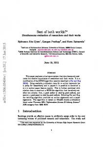
FIGURE 1. Truncation of the CTD of Lsm1 affects the decay of the endogenous GAL7 mRNA without abolishing the formation of the Lsm1–7–Pat1 complex. (A) Schematic diagram showing the extent of deletions in lsm1–27, lsm1–28, and lsm1–29. (B) Purification of the mutant Lsm1–7–Pat1 complexes. Lsm1–7–Pat1 complexes were purified from the strains indicated on top, separated by SDS-PAGE, and silver stained (upper panels). The lower band resolves into three bands in a 16% gel, part of which is shown at the bottom. (C) The endogenous GAL7 mRNA is stabilized in lsm1–27 cells. Cells were grown to log phase in galactose medium to express the GAL7 mRNA, whose transcription is then shut off by shifting the cells to glucose medium. Following this, RNA was made from the cells at different time points and subjected to Northern analysis, followed by PhosphorImager quantitation of the bands to determine the rate of disappearance of the GAL7 mRNA. The level of this mRNA in each sample was normalized for that of 7S RNA of SRP (to serve as loading control), which was determined by reprobing the blot for that RNA. A phosphorimage of the blot and a plot of the amount of RNA remaining versus time after shifting cells to the glucose medium are shown on the left and right panels, respectively.
RESULTS CTD of Lsm1 is necessary for the normal RNA-binding activity of the Lsm1–7–Pat1 complex To understand the function of the CTD of yeast (Saccharomyces cerevisiae) Lsm1, we had created the lsm1–27, lsm1–28, and lsm1–29 alleles that expressed truncated versions of Lsm1 lacking 55, 43, and 28 residues, respectively, from the C terminus, but carrying intact Sm-domain and N-terminal extension (i.e., residues 1–117) (Fig. 1A; Tharun et al. 2005). Analysis of the MFA2pG reporter mRNA revealed that
mRNA decay and 39-end protection are impaired in lsm1D cells expressing any of these alleles from a CEN vector using native promoter and UTR sequences (Tharun et al. 2005), and that such impairment is not affected by N-terminal ‘‘FLAG’’-tagging of these lsm1 alleles (Figs. 1C, 4, below; Supplemental Figs. S1E, S2B). In order to confirm that the decay of endogenous mRNA is also impaired upon truncating the CTD of Lsm1, we measured the half-life of GAL7 and GAL10 mRNAs (whose transcription can be shut off by shifting the cells to glucose medium) in lsm1–27 and www.rnajournal.org
937
Chowdhury et al.
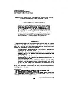
lsm1–28 mutants. We observed that both of these mRNAs are stabilized in these mutants compared with the wild-type cells (Fig. 1C; Supplemental Fig. S1E; data not shown). Western analysis revealed that the CTD-truncated mutant Lsm1 proteins are expressed in the mutant cells at levels similar to that of wild-type Lsm1 in wild-type cells (Supplemental Fig. S1B). Purification of the truncated Lsm1 containing complexes from these mutants (using the strategy we described earlier) (Chowdhury et al. 2007; Tharun 2008), followed by comparison of the SDS-PAGE band patterns of the mutant and wild-type complexes and determination of the tryptic peptide sequences (by mass spectrometry analysis) of the proteins present in the purified mutant complexes revealed that the mutant complexes contain all of the expected subunits of the Lsm1–7–Pat1 complex (Fig. 1B). Thus, inability to assemble the complex is not the likely cause of the mRNA decay and 39-end protection defects in these mutants. The purified wild-type Lsm1–7–Pat1 complex binds RNA and exhibits an intrinsic preference for oligoadenylated RNA over unadenylated RNA in vitro (Chowdhury et al. 2007). Therefore, we studied the RNA-binding activity of the mutant complexes purified from lsm1–27, lsm1–28, and lsm1–29 cells in gel-shift assays using uniformly radiolabeled PGK1 and MFA2 RNAs (42-mer RNAs derived from the 39 UTRs of the yeast MFA2 and PGK1 genes, respectively) (Chowdhury et al. 2007; Chowdhury and Tharun 2008) that lack or carry a 39-A5 tail. As seen in Figure 2A, in gelshift assays using MFA2 RNA, the mutant complexes exhibited lower RNA-binding ability than the wild-type complex with the Lsm1–27 and Lsm1–28 containing complexes exhibiting especially poor binding ability. Nevertheless, addition of an A5 tail enhanced binding of the MFA2 RNA to not only the wild-type, but also the mutant complexes. Similar results were obtained in analogous experiments using PGK1 and PGK1-A5 RNAs (Fig. 2B,C). Thus, the truncation of the CTD of Lsm1 impairs the overall RNA-binding ability of the Lsm1–7– Pat1 complex, but does not abolish the binding preference of the Lsm1–7–Pat1 complex for oligoadenylated RNA. It is known that among unadenylated RNA substrates, the wild-type Lsm1–7–Pat1 complex has a strong binding preference for those that carry a U-tract at or near the 39end over those that do not (Chowdhury et al. 2007). Since the PGK1 RNA carries a 39-U8 tract, we tested the effect of replacing this U8-tract with non-U residues (GACACCAG; PGK1-U8mut RNA). Gel-shift assays using PGK1 and PGK1-U8mut RNAs revealed that the PGK1-U8mut RNA bound more weakly than the PGK1 RNA not only to the wild-type complex, but also to the Lsm1–27 and Lsm1–28 complexes (data not shown; Supplemental Fig. S1A). This suggests that truncation of the CTD of Lsm1 does not abolish the ability of the Lsm1–7–Pat1 complex to distinguish between RNAs with and without 39-U-tracts. 938
RNA, Vol. 18, No. 5
Primary sequence of the CTD of Lsm1 is critical for the function of the Lsm1–7–Pat1 complex Past studies on other Lsm proteins support the notion that the Sm-domain is sufficient for the RNA-binding activity of the Lsm proteins. Therefore, the above results are surprising since the Sm-domain is intact in the lsm1–27, lsm1–28, and lsm1–29 alleles. Homology-based structure prediction using the Swiss-Model program (Kiefer et al. 2009) suggested that the folding of the Sm-domain of Lsm1 is not affected by the truncation of the CTD of Lsm1. To ensure that the observed functional consequences of CTD deletion are not due to any indirect effects of truncation of the Lsm1 polypeptide, we replaced the C-terminal 43 residues of S. cerevisiae Lsm1 with the corresponding region of human, Pichia stipitis or Candida glabrata Lsm1 ortholog generating the chimeric genes yyhLSM1, yyPsLSM1, and yyCgLSM1, respectively. We expressed each of these alleles using the native yeast LSM1 promoter from a CEN vector in lsm1D cells and tested its ability to support mRNA decay and 39-end protection in vivo using the MFA2pG reporter mRNA. The 39 UTR of this reporter mRNA carries a poly(G) insertion that blocks Xrn1 action in cis, such that the 59–39 decay of this mRNA results in the accumulation of a decay intermediate called poly(G) fragment (Decker and Parker 1993). The level of this fragment at steady-state is a good indicator of the efficiency of 59–39 decay in vivo, because defects in decapping and/or 59–39 exonucleolysis lead to a reduction of such levels (Hatfield et al. 1996; Tharun et al. 2005). Defects in 39-end protection cause 39-end trimming of the MFA2pG mRNA, and, therefore, also lead to the accumulation of a significant fraction of the poly(G) fragment in trimmed form, so that the size of such fraction is a measure of the status of 39-end protection in vivo (Tharun et al. 2005). Northern analysis of the RNA made from lsm1D cells expressing the chimeric LSM1 genes revealed that the accumulation of the poly(G) fragment is increased and the fraction of the poly(G) fragment in trimmed form is decreased in cells expressing yyCgLSM1 or yyPsLSM1, but not yyhLSM1, compared with the untransformed lsm1D cells (Fig. 3A). Also, the accumulation of MFA2pG mRNA in deadenylated form (which is increased by defects in decapping or 59–39 exonucleolysis) was decreased in cells expressing yyCgLSM1 or yyPsLSM1 compared with the untransformed lsm1D cells. Similar results were obtained using strains of a different genetic background (Supplemental Fig. S2A). Further, accumulation of the deadenylated form of the endogenous RPL41A mRNA was also decreased in lsm1D cells expressing yyCgLSM1 or yyPsLSM1 (but not yyhLSM1) compared with the untransformed lsm1D cells (Fig. 3B). Thus, the expression of yyCgLSM1 or yyPsLSM1, but not yyhLSM1, causes a significant suppression of the mRNA decay and 39-end protection defects in lsm1D cells. This observation is consistent with the CTD of human Lsm1 being less similar
CTD of Lsm1 is functionally critical
FIGURE 2. CTD of Lsm1 is necessary for the normal RNA-binding activity of the Lsm1–7–Pat1 complex. Increasing concentrations of Lsm1–7– Pat1 complexes purified from the strains indicated on top (or BSA in lanes marked ‘‘B’’) were subjected to gel-shift assays using uniformly radiolabeled MFA2 and MFA2-A5 RNAs (A) or PGK1 and PGK1-A5 RNAs (B,C). Plots of % RNA bound versus the concentration of the complex used are shown on the right of the phosphorimages of the gels. Bound and unbound RNA bands are indicated by brackets and asterisks, respectively, on the right. The faster moving bound RNA bands are probably due to the disassembly of the RNP complexes during the gel run. In C, the right panel shows SDS-PAGE separation of yyhLsm1 containing complex (silver stained).
than that of P. stipitis (or C. glabrata) Lsm1 to the CTD of S. cerevisiae Lsm1 (Supplemental Fig. S1C). This indicates that the primary sequence of the CTD of Lsm1 is crucial for
the functioning of the Lsm1–7–Pat1 complex. Therefore, the CTD is a functionally critical segment of the Lsm1 polypeptide and the in vivo and in vitro functional defects www.rnajournal.org
939
Chowdhury et al.
FIGURE 3. Primary sequence of the CTD is critical for Lsm1 function in vivo. (A) RNA isolated from lsm1D strains expressing different LSM1 chimeras as indicated on top were subjected to Northern analysis to reveal the MFA2pG mRNA and the poly(G) fragments. A phosphorimage of the blot is shown in the top left panel. A longer exposure of the section of the blot containing the poly(G) fragment bands is shown in the lower left panel. The fractional contribution of the poly(G) fragments to the total signal (total: full-length mRNA + trimmed and normal poly(G) fragments) was determined for each sample (following quantitation of the respective bands in the corresponding lane using the PhosPhorimager), normalized to the wild-type value and presented as a bar diagram in the top, right panel. Similarly, the fractional contribution of the trimmed poly(G) fragment to the total poly(G) fragment signal (trimmed + normal) was also determined for each sample, normalized to the value obtained for the lsm1D sample, and presented as a bar diagram in the bottom right panel. (B) RNA isolated from lsm1D strains expressing different LSM1 chimeras as indicated on top were subjected to Northern analysis to reveal the RPL41A mRNA.
pertaining to the lsm1–27, lsm1–28, and lsm1–29 alleles are not just indirect effects of polypeptide truncation. Inability of yyhLSM1 to suppress the mRNA decay defect of the lsm1D cells is not due to its insufficient expression, because the suppression was not significantly enhanced upon overexpression from high-copy 2m vectors (using the yeast LSM1 promoter or the Gal promoter) (Fig. 3; Supplemental Figs. S1D, S2A). Nevertheless, electrophoretic and mass spectrometry analyses of the yyhLsm1-containing complex purified from cells that express the yyhLSM1 allele using the yeast LSM1 promoter from a 2m vector revealed that it contains all of the subunits (Fig. 2C). Gel-shift assays revealed that similar to the Lsm1–27 or Lsm1–28-containing complex, the yyhLsm1-containing complex also has a lower RNA-binding ability than the wild-type complex, but retains the binding preference for oligoadenylated RNAs (Fig. 2C). Thus, in lsm1D cells expressing yyhLSM1, decapping may be blocked at a step after mRNA binding by the Lsm1–7–Pat1 complex. CTD of Lsm1 can function in trans The above results show that Lsm1 can fold and form the complex with other subunits, even after its CTD is deleted. 940
RNA, Vol. 18, No. 5
Therefore, we asked whether the CTD could complement the functional defects of the CTD-truncated lsm1 alleles in trans. Indeed, study of the status of the MFA2pG mRNA and the poly(G) fragments via Northern analysis revealed that expression of the C-terminal 60 residues of Lsm1 as a separate polypeptide at least partly suppresses the mRNA decay and 39-end protection defects of the CTD-truncated lsm1 mutants. This was evident from an increase in the total poly(G) fragment level and a decrease in the fraction of the poly(G) fragment in trimmed form in the mutant cells expressing the CTD peptide compared with the cells that do not (Fig. 4A). Further, in the case of both MFA2pG mRNA (Fig. 4A) and the endogenous RPL41A mRNA (Fig. 4B), accumulation of deadenylated species observed in the CTD-truncated lsm1 mutants was reduced by the expression of the CTD peptide. These results were reproducibly observed using strains of two different genetic backgrounds (Fig. 4; Supplemental S2B). Pull-down of the CTD peptide from these lsm1–27 and lsm1–28 cells coprecipitated the corresponding mutant Lsm1 proteins (Fig. 5A), suggesting that the observed phenotypic suppression indeed results from the association of the CTD peptide with the truncated Lsm1 proteins. Such association is unlikely to involve non-
CTD of Lsm1 is functionally critical
washed again. The bound RNA was then extracted and visualized by denaturing gel run and phosphorimaging. As seen in Figure 5B and Supplemental Fig. S2C, preincubation with the CTD peptide significantly enhanced RNA binding by the immobilized Lsm1–27 complex, but not the wild-type complex. Therefore, the CTD suppresses the mRNA decay and 39-end protection defects of the CTD-truncated lsm1 mutants in trans by virtue of its ability to associate with the mutant Lsm1–7–Pat1 complex, and thereby facilitate RNA binding by that complex. DISCUSSION The Lsm1–7–Pat1 complex is unique among the Sm-like protein complexes in being able to distinguish oligoadenylated RNA from unadenylated and polyadenylated RNAs. Such ability and the overall RNA-binding activity of this complex are crucial for mRNA decay and 39-end protection in vivo and are critically dependent on the residues in the Sm-domain of the Lsm1 subunit (Tharun et al. 2005; Chowdhury and Tharun 2008). The key finding presented here is that the CTD of Lsm1 is also required (in addition to the Smdomain) for the normal RNA-binding FIGURE 4. The 60-mer CTD polypeptide suppresses the mRNA decay and 39-end protection activity of this complex. This explains defects of the CTD-truncated lsm1 alleles in trans. MFA2pG mRNA and the poly(G) fragments the mRNA decay and 39-end protection (A) or RPL41A mRNA (B) present in the RNA isolated from the CTD-truncation mutants of lsm1 (expressing or not expressing 6x-His tagged CTD peptide) and control strains (indicated defects of CTD-truncated lsm1 mutants. on top) were visualized by Northern analysis. A phoshorimage of the blot and bar diagrams An important question raised by showing the accumulation of the poly(G) fragments (quantitated as described for Fig. 3) are these studies is how the CTD of Lsm1 shown in the top and bottom panels, respectively, in A. In B, the top and bottom panels show affects RNA binding by the Lsm1–7– RNA from strains of two different strain backgrounds. Pat1 complex. Our studies support the idea that the CTD acts primarily by facilitating RNA-binding activity of the Sm-domain of native interactions because expression of the CTD peptide Lsm1 (or other subunits of the complex) rather than did not significantly affect mRNA decay or 39-end trimforming an additional RNA-binding surface of its own ming in wild-type (data not shown) and lsm1D (Fig. 4) cells for the following reasons. Although the deletion of the (as revealed by the analysis of MFA2pG and RPL41A CTD of Lsm1 results in a severe impairment of the RNAmRNAs) and the pull-down of the CTD peptide from binding activity of the Lsm1–7–Pat1 complex, it abolishes such wild-type cells did not coprecipitate the wild-type neither of the two important RNA-binding properties of Lsm1 (Fig. 5A). this complex, namely, the strong binding preference for We then asked whether the CTD peptide could compleoligoadenylated RNAs over unadenylated RNAs and the ment the RNA binding defect of the purified Lsm1–27 binding preference for unadenylated RNAs carrying a complex in vitro. The Lsm1–27 complex was immobilized U-tract near the 39-end over those that do not (Chowdhury onto the anti-FLAG antibody matrix before or after incubatet al. 2007). Thus, the complexes purified from the CTDing it with a synthetic peptide containing the C-terminal truncated lsm1 mutants, lsm1–27 and lsm1–28, exhibit 55 residues of yeast Lsm1. After washing, the matrix was weaker affinity (compared with the wild-type complex) incubated with uniformly radiolabeled PGK1 RNA and www.rnajournal.org
941
Chowdhury et al.
FIGURE 5. CTD peptide associates with the mutant Lsm1–7–Pat1 complex (containing CTD-truncated Lsm1) and enhances its RNAbinding activity. (A) Lysates from strains (indicated on top) expressing untagged or His-tagged CTD polypeptide and proteins pulled down from such lysates using the Ni-NTA matrix were subjected to Western analysis using anti-FLAG antibodies. (B) Purified wild-type and Lsm1–27 containing complexes or BSA were incubated with the antiFLAG antibody matrix (after or without preincubation with synthetic CTD peptide) and the ability of the immobilized material to bind radiolabeled PGK1 RNA was studied as described in the text. RNA bound to the matrix in each experiment was visualized on a denaturing gel (top) and its amount (quantitated using a PhosPhorimager) was shown as a bar diagram below the corresponding lane.
for all types of RNA substrates studied, i.e., RNAs with and without oligo(A) tail and unadenylated RNAs with and without 39-U-tracts. However, importantly, the ability of the Lsm1–7–Pat1 complex to recognize the oligo(A) tail can be abolished by mutating the predicted RNA-binding pocket residues located in loops 3 or 5 (while keeping the CTD intact) of the Sm domain of Lsm1, such that the mutant complex from lsm1–14 cells exhibits normal affinity for unadenylated and polyadenylated RNAs, but reduced affinity for oligoadenylated RNAs, so that all of these RNA substrates are bound with comparable affinities (Chowdhury and Tharun 2008). This underscores the importance of the Sm domain of Lsm1 for such recognition. Further, some Sm-domain mutations of Lsm1 (in the presence of intact CTD) can result in almost complete loss of the RNAbinding ability of the Lsm1–7–Pat1 complex (Chowdhury and Tharun 2009). Finally, the CTD of Lsm1 does not 942
RNA, Vol. 18, No. 5
contain any recognizable motifs. In any case, we cannot completely rule out the possibility of some of the CTD residues contacting RNA directly, although our efforts to show direct binding of RNA by the CTD peptide has not been successful so far. The observation that, in addition to the Sm-domain, the CTD of Lsm1 is also required for the normal in vivo functions of the Lsm1–7–Pat1 complex in mRNA decay and 39-end protection and for the normal RNA-binding activity of the purified Lsm1–7–Pat1 complex in vitro indicates that the scenario of Lsm1 is in contrast to many other Lsm family members wherein the Sm-domain generally seems to be sufficient for function. Most Sm-like proteins are very small, such that the Sm-domain occupies almost the whole protein, although some Lsm family members do carry extended N- or C-terminal segments like Lsm1. For example, the Lsm4 subunit of the Lsm1–7–Pat1 complex has a C-terminal extension following its Sm-domain. However, deletion of this C-terminal segment does not affect mRNA decay in vivo (Decker et al. 2007), suggesting that it is unlikely to cause a significant impairment of the RNAbinding activity of the Lsm1–7–Pat1 complex. Similarly, the essential protein Lsm8, which is the key distinguishing subunit of the nuclear Lsm2–8 complex (that forms the core of the U6 snRNP), also carries a C-terminal extension following its Sm-domain. However, deletion of this C-terminal segment does not affect the viability in yeast, suggesting that it has a minimal effect on the splicing function of the U6 snRNP (Reijns et al. 2009). Nevertheless, the E. coli Hfq protein presents a different picture. While Hfq’s Sm-domain residues form two RNA-binding surfaces that bind small noncoding regulatory RNAs (ncRNAs) and poly(A), the CTD residues form a third RNA-binding surface that binds mRNAs, so that deletion of the CTD does not affect ncRNA or poly(A) binding and affects only mRNA binding (Mikulecky et al. 2004; Vecerek et al. 2008). This is in contrast to the CTD of Lsm1, which, as revealed by our studies, is necessary for the overall RNA-binding activity of the Lsm1–7–Pat1 complex, i.e., it is required for binding RNAs with and without oligo(A) tail and unadenylated RNAs with and without 39-U-tracts with wild-type affinities. Our pull-down experiments show that the CTD peptide expressed in trans associates with the CTD-truncated mutant Lsm1 proteins (Lsm1–27 and Lsm1–28), but not with the wild-type Lsm1. This implies that the CTD of Lsm1 makes specific contacts with one or more sites on the Lsm1– 7–Pat1 complex. At present we do not know which subunits carry such sites. Structural studies on the SmAP3 (archaeal Lsm protein) homoheptamer and the homooctamer formed by yeast Lsm3 in bacteria reveal that the residues in the C-terminal extension could facilitate intersubunit interactions within the Lsm ring and between two Lsm rings (Mura et al. 2003b; Naidoo et al. 2008). These observations suggest that the CTD of Lsm1 may also make contacts with the Lsm2 through Lsm7 or Pat1 subunits. In any case, the CTD of
CTD of Lsm1 is functionally critical
Lsm1 is not required for the Lsm1–7– TABLE 1. Plasmids used in this study Pat1 complex formation, since our rePlasmid Insert Vector Additional details Source sults show that both Lsm1–27 (which a lacks the entire CTD) and yyhLsm1 pST11 LSM1 pRS416 Insert driven by Tharun et al. (2005) (wherein the CTD is replaced with pST17 FLAG-LSM1 native yeast Chowdhury et al. (2007) pST80 FLAG-lsm1–27 LSM1 promoter This study human Lsm1’s CTD, which is functionpST81 FLAG-lsm1–28 ally deficient in the context of the yeast pST94 FLAG-lsm1–29 Lsm1) are able to form the Lsm1–7– pST308 FLAG-lsm1–27 pRS413a Pat1 complex. Thus, the interactions of pST309 FLAG-lsm1–28 the CTD of Lsm1 (with the Sm domain pST324 FLAG-lsm1–27 pRS317a pST325 FLAG-lsm1–28 of Lsm1 or with other subunits), though pST221 FLAG-yyCgLSM1 pRS416a important functionally, do not seem to pST271 FLAG-yyPsLSM1 be necessary for the integrity of the pST215 FLAG-yyhLSM1 Lsm1–7–Pat1 complex. These results pST248 FLAG-yyhLSM1 pRS426a are consistent with studies on other SmpST246 FLAG-yyhLSM1 pESC-URAb Insert driven by pST301 6xHis-CTD Gal promoter like proteins, which also show that the (C-terminal 60 Sm-domain is sufficient for oligomeriresidues of zation. For example, although human yeast LSM1) Sm-B and Sm-D3 proteins carry long a Sikorski and Hieter (1989). CTDs, their CTDs are dispensable for b Agilent Technologies. B-D3 subcomplex formation (Hermann et al. 1995; Kambach et al. 1999). Apart from Lsm1, another subunit Purification of Lsm1–7–Pat1 complex, preparation of radiothat is likely to be very critical for the Lsm1–7–Pat1 labeled RNAs, gel-shift assays, mRNA stability measurement and complex function is Pat1. pat1D mutant has a strong Northern, pull-down, and Western analyses were done as described mRNA decay and 39-end protection phenotypes like the (Chowdhury et al. 2007; Chowdhury and Tharun 2008; Chowdhury lsm1D mutant (Bonnerot et al. 2000; Bouveret et al. 2000; and Tharun 2009). Tharun et al. 2000). Yeast Pat1 can associate with poly(U) sepharose, suggesting that it is an RNA-binding protein (Pilkington and Parker 2008). However, the contribution SUPPLEMENTAL MATERIAL of Pat1 to the RNA binding and other functions of Lsm1– Supplemental material is available for this article. 7–Pat1 complex is not known. It is possible that Pat1 facilitates the RNA binding by the Lsm1–7–Pat1 complex by providing an additional RNA-binding surface of its own. ACKNOWLEDGMENTS Overall, these studies suggest that polypeptide segments This work was supported by funds from NIH RO1 grant that modulate the RNA-binding functions of the Sm(GM072718) and USUHS exploratory grant (RO71JX) to S.T. domain could occur in the N- and C-terminal extensions We thank Dr. Roy Parker for his comments on the manuscript. (regions that flank the Sm-domain) of Sm-like proteins, and therefore have important implications to the biology Received August 13, 2011; accepted February 24, 2012. of this large ubiquitous family of proteins. MATERIALS AND METHODS Strains needed for Lsm1–7–Pat1 complex purifications were made by introducing the plasmids expressing different lsm1 alleles into yST247 (Chowdhury et al. 2007), which is in the genetic background of yRP841 (Hatfield et al. 1996). Strains used for the experiments shown in Figures 1C, 3B, 4A,B (top) and Supplemental Figures S1 and S2A, are also in this background and were generated by transforming the lsm1D strain yRP1365 (Tharun et al. 2000) with appropriate plasmids. Strains used for experiments shown in Figures 3A and 4B (bottom) and Supplemental Figure S2B were made by transforming yST188 (trp1D1, his3-11,-15, ura3-1, leu2-3,-112, ade2-1, can1-100, lsm1D::TRP1, [pGal-MFA2pG]), which is in the genetic background of BMA64 (Mayes et al. 1999). Plasmids used in this study are described in Table 1.
REFERENCES Beggs JD. 2005. Lsm proteins and RNA processing. Biochem Soc Trans 33: 433–438. Boeck R, Lapeyre B, Brown CE, Sachs AB. 1998. Capped mRNA degradation intermediates accumulate in the yeast spb8-2 mutant. Mol Cell Biol 18: 5062–5072. Bonnerot C, Boeck R, Lapeyre B. 2000. The two proteins Pat1p (Mrt1p) and Spb8p interact in vivo, are required for mRNA decay, and are functionally linked to Pab1p. Mol Cell Biol 20: 5939–5946. Bouveret E, Rigaut G, Shevchenko A, Wilm M, Seraphin B. 2000. A Sm-like protein complex that participates in mRNA degradation. EMBO J 19: 1661–1671. Chowdhury A, Tharun S. 2008. lsm1 mutations impairing the ability of the Lsm1p-7p-Pat1p complex to preferentially bind to oligoadenylated RNA affect mRNA decay in vivo. RNA 14: 2149–2158.
www.rnajournal.org
943
Chowdhury et al.
Chowdhury A, Tharun S. 2009. Activation of decapping involves binding of the mRNA and facilitation of the post-binding steps by the Lsm1-7-Pat1 complex. RNA 15: 1837–1848. Chowdhury A, Mukhopadhyay J, Tharun S. 2007. The decapping activator Lsm1p-7p-Pat1p complex has the intrinsic ability to distinguish between oligoadenylated and polyadenylated RNAs. RNA 13: 998–1016. Coller J, Parker R. 2004. Eukaryotic mRNA decapping. Annu Rev Biochem 73: 861–890. Collins BM, Harrop SJ, Kornfeld GD, Dawes IW, Curmi PM, Mabbutt BC. 2001. Crystal structure of a heptameric Sm-like protein complex from archaea: implications for the structure and evolution of snRNPs. J Mol Biol 309: 915–923. Cooper M, Johnston LH, Beggs JD. 1995. Identification and characterization of Uss1p (Sdb23p): a novel U6 snRNA-associated protein with significant similarity to core proteins of small nuclear ribonucleoproteins. EMBO J 14: 2066–2075. Decker CJ, Parker R. 1993. A turnover pathway for both stable and unstable mRNAs in yeast: Evidence for a requirement for deadenylation. Genes Dev 7: 1632–1643. Decker CJ, Teixeira D, Parker R. 2007. Edc3p and a glutamine/ asparagine-rich domain of Lsm4p function in processing body assembly in Saccharomyces cerevisiae. J Cell Biol 179: 437–449. Garneau NL, Wilusz J, Wilusz CJ. 2007. The highways and byways of mRNA decay. Nat Rev Mol Cell Biol 8: 113–126. Hatfield L, Beelman CA, Stevens A, Parker R. 1996. Mutations in transacting factors affecting mRNA decapping in Saccharomyces cerevisiae. Mol Cell Biol 16: 5830–5838. He W, Parker R. 2001. The yeast cytoplasmic LsmI/Pat1p complex protects mRNA 39 termini from partial degradation. Genetics 158: 1445–1455. Hermann H, Fabrizio P, Raker VA, Foulaki K, Hornig H, Brahms H, Luhrmann R. 1995. snRNP Sm proteins share two evolutionarily conserved sequence motifs which are involved in Sm proteinprotein interactions. EMBO J 14: 2076–2088. Houseley J, Tollervey D. 2009. The many pathways of RNA degradation. Cell 136: 763–776. Ingelfinger D, Arndt-Jovin DJ, Luhrmann R, Achsel T. 2002. The human LSm1-7 proteins colocalize with the mRNA-degrading enzymes Dcp1/2 and Xrn1 in distinct cytoplasmic foci. RNA 8: 1489–1501. Kambach C, Walke S, Young R, Avis JM, de la Fortelle E, Raker VA, Luhrmann R, Li J, Nagai K. 1999. Crystal structures of two Sm protein complexes and their implications for the assembly of the spliceosomal snRNPs. Cell 96: 375–387. Kiefer F, Arnold K, Kunzli M, Bordoli L, Schwede T. 2009. The SWISS-MODEL Repository and associated resources. Nucleic Acids Res 37: D387–D392. Mayes AE, Verdone L, Legrain P, Beggs JD. 1999. Characterization of Sm-like proteins in yeast and their association with U6 snRNA. EMBO J 18: 4321–4331. Mikulecky PJ, Kaw MK, Brescia CC, Takach JC, Sledjeski DD, Feig AL. 2004. Escherichia coli Hfq has distinct interaction surfaces for DsrA, rpoS and poly(A) RNAs. Nat Struct Mol Biol 11: 1206–1214.
944
RNA, Vol. 18, No. 5
Mura C, Cascio D, Sawaya MR, Eisenberg DS. 2001. The crystal structure of a heptameric archaeal Sm protein: Implications for the eukaryotic snRNP core. Proc Natl Acad Sci 98: 5532–5537. Mura C, Kozhukhovsky A, Gingery M, Phillips M, Eisenberg D. 2003a. The oligomerization and ligand-binding properties of Sm-like archaeal proteins (SmAPs). Protein Sci 12: 832–847. Mura C, Phillips M, Kozhukhovsky A, Eisenberg D. 2003b. Structure and assembly of an augmented Sm-like archaeal protein 14-mer. Proc Natl Acad Sci 100: 4539–4544. Naidoo N, Harrop SJ, Sobti M, Haynes PA, Szymczyna BR, Williamson JR, Curmi PM, Mabbutt BC. 2008. Crystal structure of Lsm3 octamer from Saccharomyces cerevisiae: implications for Lsm ring organisation and recruitment. J Mol Biol 377: 1357–1371. Pilkington GR, Parker R. 2008. Pat1 contains distinct functional domains that promote P-body assembly and activation of decapping. Mol Cell Biol 28: 1298–1312. Reijns MA, Auchynnikava T, Beggs JD. 2009. Analysis of Lsm1p and Lsm8p domains in the cellular localization of Lsm complexes in budding yeast. FEBS J 276: 3602–3617. Salgado-Garrido J, Bragado-Nilsson E, Kandels-Lewis S, Seraphin B. 1999. Sm and Sm-like proteins assemble in two related complexes of deep evolutionary origin. EMBO J 18: 3451–3462. Schumacher MA, Pearson RF, Moller T, Valentin-Hansen P, Brennan RG. 2002. Structures of the pleiotropic translational regulator Hfq and an Hfq-RNA complex: a bacterial Sm-like protein. EMBO J 21: 3546–3556. Seraphin B. 1995. Sm and Sm-like proteins belong to a large family: identification of proteins of the U6 as well as the U1, U2, U4 and U5 snRNPs. EMBO J 14: 2089–2098. Sikorski RS, Hieter P. 1989. A system of shuttle vectors and yeast host strains designed for efficient manipulation of DNA in Saccharomyces cerevisiae. Genetics 122: 19–27. Tharun S. 2008. Purification and analysis of the decapping activator Lsm1p-7p-Pat1p complex from yeast. Methods Enzymol 448: 41–55. Tharun S. 2009. Roles of eukaryotic Lsm proteins in the regulation of mRNA function. Int Rev Cell Mol Biol 272: 149–189. Tharun S, He W, Mayes AE, Lennertz P, Beggs JD, Parker R. 2000. Yeast Sm-like proteins function in mRNA decapping and decay. Nature 404: 515–518. Tharun S, Muhlrad D, Chowdhury A, Parker R. 2005. Mutations in the Saccharomyces cerevisiae LSM1 gene that affect mRNA decapping and 39 end protection. Genetics 170: 33–46. Toro I, Thore S, Mayer C, Basquin J, Seraphin B, Suck D. 2001. RNA binding in an Sm core domain: X-ray structure and functional analysis of an archaeal Sm protein complex. EMBO J 20: 2293–2303. Totaro A, Renzi F, La Fata G, Mattioli C, Raabe M, Urlaub H, Achsel T. 2011. The human Pat1b protein: a novel mRNA deadenylation factor identified by a new immunoprecipitation technique. Nucleic Acids Res 39: 635–647. Vecerek B, Rajkowitsch L, Sonnleitner E, Schroeder R, Blasi U. 2008. The C-terminal domain of Escherichia coli Hfq is required for regulation. Nucleic Acids Res 36: 133–143. Wilusz CJ, Wilusz J. 2005. Eukaryotic Lsm proteins: lessons from bacteria. Nat Struct Mol Biol 12: 1031–1036.