classified by using PNN-RBF training and classification method, whether the disease is ... medical images but, the cost of dual reading process is extremely high.
Available online at www.sciencedirect.com
ScienceDirect Procedia Computer Science 50 (2015) 388 – 394
2nd International Symposium on Big Data and Cloud Computing (ISBCC’15)
Brain Image Classification using Learning Machine Approach and Brain Structure Analysis A.Veeramuthua, S.Meenakshib, V. Priya Darsinic a b
Research Scholar, Department of IT, Sathyabama University, Chennai-600119, India Professor/Head, Department of IT, SRR Engineering College, Chennai-603103, India c PG Student, Department of IT, Sathyabama University, Chennai-600119, India
Abstract Computer Aided Diagnosis (CAD) for functional brain images helps to analysis the structure of the brain images. The Multi Level Discrete Wavelet Transform method helps to decompose image and then the features are extracted. The brain image is classified by using PNN-RBF training and classification method, whether the disease is of normal, benign or malignant stages. This method helps to detect the disease in early stage itself. The Fisher Discriminant Ratio (FDR) method is used to reduce t he computational cost by identifying the relationships among discriminate brain areas. The Morphological Filter technique is used to segment the images according to the Region of Interest (ROI), which is applied to abnormal images. This method yields better accuracy and sensitivity to find the discriminate brain areas. The early diagnosis of the tumor in brain is possible. © 2015 2015 The TheAuthors. Authors.Published Publishedby byElsevier ElsevierB.V. B.V.This is an open access article under the CC BY-NC-ND license © Peer-review under responsibility of scientific committee of 2nd International Symposium on Big Data and Cloud Computing (http://creativecommons.org/licenses/by-nc-nd/4.0/). Peer-review (ISBCC’15).under responsibility of scientific committee of 2nd International Symposium on Big Data and Cloud Computing (ISBCC’15) Keywords: Computer Aided Diagnosis, Discrete Wavelet Transform, PNN-RBF Training, Classification, FDR, Morphological Filter, ROI;
1. Introduction Automated detection, classification, and segmentation of tumor cells in dissimilar medicinal brain images are aggravated by the need of high accuracy when dealing with a human life. Monitoring and diagnosing the diseases like Alzheimer’s disease (AD) depends on the detecting the presence of specific features by a human observer. Due to huge number of patients in intensive care units is need continuous observation in such conditions, a number of techniques for automated diagnostic system have been developed in recent years attempt to solve this kind of problems by the help of Computer Aided Diagnosis (CAD). The tumor detection can be better by dual reading the medical images but, the cost of dual reading process is extremely high. So, a good software is needed to assist human in medicinal institutions is required these days.
1877-0509 © 2015 The Authors. Published by Elsevier B.V. This is an open access article under the CC BY-NC-ND license (http://creativecommons.org/licenses/by-nc-nd/4.0/). Peer-review under responsibility of scientific committee of 2nd International Symposium on Big Data and Cloud Computing (ISBCC’15) doi:10.1016/j.procs.2015.04.030
A. Veeramuthu et al. / Procedia Computer Science 50 (2015) 388 – 394
389
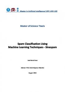
In this paper focussed on automatic classification and segmentation of brain images by extracting the particular features of the input image. The discriminant brain areas are detected, and then the performance of the system are analyzed. At present there are limited methods far and widely accepted for tumour detection. Therefore automatic, accurate, and reliable methods for tumour detection are very important and needed. In functional brain images classification and the structure analysis the Multi Level Wavelet Transform technique is used, which decompose the signal and function into different frequency sub-bands that lead to set of wavelet coefficient. The GLCM feature extraction method is used to extract the texture feature of an image which is based on the condition probability density function, discussed in M. M. Mokji et al. [1] the positions of pixels which have similar gray level values are taken in this method. The Probabilistic Neural Network-Radial Based Function (PNNRBF) technique is used for training and classification process of brain images. Then the morphological filter method is used to analysis brain structure, which helps to find the discriminant areas in functional brain images. This paper is organized as follows. Chapter 2 discussed about the related work of this paper, chapter 3 deals about proposed method, in chapter 4 experimental setup discussed in detail, in chapter 5 discussed about performance analysis, finally in chapter 6 concluded the paper. 2. Related Works Currently there are several algorithms, methods, and techniques are proposed to predict and classify the tumour regions in functional brain images. The Non-negative Matrix Factorization (NMF) techniques are used in normalized data where the features of each subject’s relevant components are extracted. Curse of dimensionality, which related to large dimensionality of the input data, gets reduced. To reduce the number of features, the NMF method is used and by using Support Vector Machine the reduced features are classified. This method gives up to 94% of accuracy in high specificity and sensitivity values, P. Padilla et al. [2]. The Association Rules (AR) and Neural Network (NN) techniques are implemented in brain images, Murat Karabatak et al. [3]. The dimension of dataset gets reduced by using AR method and for efficient classification, NN technique is used. The AR+NN System performance is compared with that of other feature selection and classification methods is done, which gives 98.61% of accuracy. To extract the relevant features from the image database, the Independent Component Analysis (ICA) is used and also it helps to reduce the features of space dimension. These methods detect the error below 9% and detect the pattern in unsupervised manner, discussed in Alvarez Illan et al. [4]. Since the local low-resolution face images are not robust and also not adequate, so GLL method is proposed. The GLL fuses on Gabor filter, Local Binary Pattern (LBP), and Local Phase Quantization (LPQ). Gabor filter, operates on Gabor wavelet functions where t wo scales and eight orientations are used to capture the face image’s salient visual properties. By using Gabor features, to get blur invariant property the LBP features and LPQ features are extracted. The information in the spatial domain the different scales and orientations which provide better accuracy in discriminative areas, discussed in Shu-Ren Zhou et al. [5]. 3. Proposed Method The proposed method for brain image classification and structure analysis consists of a) Discrete Wavelet Transform b) GLCM feature extraction c) PNN-RBF Training and Classification d) Discriminant Area Detection, which is clearly shown in architecture, see in Fig. 2. 3.1 Discrete wavelet Transform (DWT) The DWT method is a Spectral Estimation technique where it decompose signal or function into sub-bands of different types which lead to set of wavelet coefficient. The wavelet coefficients characterize the signals by decomposing the low level physical features by repeatedly filtering the coefficients of the image in row by row and column by column using basic fashion. The wavelet, ψ (t) function is satisfied by the below equation (1).
390
A. Veeramuthu et al. / Procedia Computer Science 50 (2015) 388 – 394
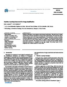
ݐെݒ ൰�������������������������������ሺͳሻ ݑ ξݑ Where u, v are the sub-bands for low level feature and high level feature. The two dimensional images in DWT consists of various sub band details like high-high level gives information about frequency diagonal band, low-low level gives information about frequency approximation band, high-low level gives the detail about frequency vertical, and low-high level gives information about frequency horizontal detail band which is represented in Fig.1. This method is used to retrieve better image with a low cost computation. This transforms discrete time signals to discrete wavelet representation. ߰௨ǡ௩ ሺݐሻ ൌ
ͳ
߰൬
Fig. 1 Discrete wavelet transform
3.2 GLCM Feature Extraction The Gray Level Co-occurrence Matrix (GLCM) is discussed in M. M. Mokji et al. [1] which are based on the method of condition probability density function. It gives the information about the position of the pixel which has the similar gray level matrix. The direction and the distances are represented in Co-occurrence matrix function. ሺݑǡ �ݒȁ�݀ǡ ߠሻ
ሺǡ �ȁǡ Ʌሻ ����������������������������ሺʹሻ σݑσݒሺǡ �ȁǡ Ʌሻ
The distance and direction are represented as u, v where gray level pixel rate are calculated. The relative frequency of two pixels separated by distance, d and matrix element, p (u, v| d, θ) and θ is represented as the direction specific particular angle which is discussed in Jeyanthi Prabhu et al. [6]. In GLCM texture feature are extracted by using statistical distribution that can be observed by the combination of intensity at specified position which is relative to each other in the image. Thus GLCM is used for extracting texture feature by converting RGB images into gray scale images. 3.3 PNN -RBF Training and Classification Probabilistic Neural Network-Radial Basis Function (PNN-RBF) method is proposed for classification process, this method consists of three layers like input, hidden, and output layers. In hidden layer the neurons of the Gaussian Transfer function outputs are inversely proportional to the midpoint of the neurons. The RBF networks are related to K-Means clustering and PNN/GRNN networks. The main advantage of RBF networks is having a variable number of neurons that is the number of training points are typically less. The parameters involved in training, the number of neural network in hidden layers is the coordinates of the midpoint, RBF function is found using equation (3),
A. Veeramuthu et al. / Procedia Computer Science 50 (2015) 388 – 394
391
ܹ݄݁݅݃ �ݐൌ �� �ሺ݀݅݁ܿ݊ܽݐݏሻ�������������������������ሺ͵ሻ Then the radius of each RBF function in terms of dimension is calculated and the weight is applied to output of RBF function in the summation layer. The optimal weight between hidden and summation layer in the neurons are calculated using ridge regression, which minimize the Generalized Cross Validation (GCV). Due to large spread neurons the distance from large spread neurons point have greater influence. The prediction value of the new point is found by summing the output values using RBF function and then the computed weights of each neuron is multiplied. In PNN-RBF one or more RBF neurons are taken in the space which is described by prediction values, since space has many dimensions. The midpoint of the each neuron is evaluated by using Euclidean distance, and to compute the weight of each neuron, a radial basis function (RBF) is applied. The RBF is so named because the radius distance is the argument to the function.
Fig. 2 Architecture of classification and structure analysis
3.4 Discriminant Area Detection The brain structural analysis is the process which focuses on Region of Interest (ROI). The Morphological filter in image process helps to remove the imperfection in the binary images and also these techniques can be extended in gray scale images. The morphological filter method is based only on pixel values where one pixel area is 0.264. This method focus on structural elements of all possible location in the image and it is compared with neighbour pixels, where it operates weather the element fits or hits the neighbourhood elements. In this method the matrix dimension specifies the size and the pattern where one or zero is specified from the shape of the structural elements. Then the origin of the pixel is calculated. The morphological filtering process consists of methods like Dilation and Erosion. Dilation process will add pixel to the images and Erosion process will remove the unwanted pixels. The combination of this method helps to segment the images.
392
A. Veeramuthu et al. / Procedia Computer Science 50 (2015) 388 – 394
4. Experimental Setup and Results In Fig. 3 the classification and decomposition of input image is shown. In Fig. 4 shows the images segmentation according to the region of interest (ROI).
Fig. 3 Classification and decomposition of input image
Fig. 4 Region based image segmentation
A. Veeramuthu et al. / Procedia Computer Science 50 (2015) 388 – 394
393
Fig. 5 Segmented area calculation
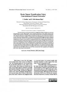
In Fig. 5 morphological filtering method is used for calculating the pixel area for each segmented images and the sensitivity, accuracy, specificity, and t-area are calculated and validated for the given input image. 5. Performance Analysis
Fig. 6 Performance analysis
394
A. Veeramuthu et al. / Procedia Computer Science 50 (2015) 388 – 394
The performance for brain functional image classification and brain structures are analyzed by using discrete wavelet transform (DWT) where the features are extracted. The Energy feature is 0.0001, the contrast feature is 4.9848, the correlation feature is 0.0004, the homogeneity feature is 0.0004, and the entropy feature is 0.0055. By using segmentation and classification method, the number of defected cells for benign stage is calculated as 2169 cells, the tumor area is calculated as 12.2951mm, the sensitivity is achieved for the given input image is 100%, and the specificity is calculated as 75%, the accuracy is calculated as 90%, and the t-area is calculated as 11% as shown in Fig. 6. 6. Conclusion The Probabilistic Neural Network-Radial Basis Function method is proposed to increase the classification accuracy in tumor functional brain images. The classification is done by extracting the features by using multi level wavelet method. Then the morphological filtering technique is used to segmentation process where the Region of Interest areas of the brain functional images are compared with the neighbourhood pixels. This method yields an accuracy of 98% sensitivity of 100% for the functional brain images. This technique is an efficient method to diagnosis the tumor region in early stage itself. References 1. Mokji M.M, Abu Bakar S.A.R. Gray Level Co-Occurrence Matrix Computation Based On Haar Wavelet, Computer Graphics, Imaging and Visualisation (CGIV ), IEEE, 2007, p.273-279. 2. Padilla P, Gorriz J.M, Ramirez J, Lang E.W., Chaves R, Segovia F, Lopez M, Salas-Gonzalez D, Alvarez I. Analysis of SPECT brain images for the diagnosis of Alzheimer’s disease based on NMF for feature extraction, Neuroscience Letters, 2010, p.192–196. 3. Murat Karabatak, Cevdet Ince M. A new feature selection method based on association rules for diagnosis of erythemato-squamous diseases, Expert system with application, vol. 36, 2009, p.12500-12505. 4. Alvarez Illan I, Gorriz J.M, Ramirez J, Salas-Gonzalez D, Lopez M, Segovia F, Padilla P, Puntonet C.G. Projecting independent components of SPECT images for computer aided diagnosis of Alzheimer’s disease, Pattern Recognition Letters, 2010, p.1342–1347. 5. Shu-Ren Zhou, Jian-Ping, Yin Jian-Ming Zhang. Local binary pattern (LBP) and local phase quantization (LBQ) based on Gabor filter for face representation, Sci Verse Science Direct Neuro Computing, 2013, p.206-264. 6. Jeyanthi Prabhu and Jawahar Senthil Kumar. Wavelet based content based image retrieval using color and texture feature extraction by gray level co ocurence matrix and color coocurence matrix, Journal of Computer Science, Science Publication 10 (1), 2014, p.15-22.