Rev. Inst. Med. trop. S. Paulo 45(5):295-297, September-October, 2003
BRIEF COMMUNICATION DIAGNOSIS OF ABDOMINAL ANGIOSTRONGYLIASIS BY PCR FROM SERA OF PATIENTS
Ana Cristina Arámburu da SILVA(1), Carlos GRAEFF-TEIXEIRA(1) & Arnaldo ZAHA(2)
SUMMARY Abdominal angiostrongyliasis is a zoonotic infection caused by an intra-vascular nematode parasitic of wild rodents, Angiostrongylus costaricensis. No parasitological diagnosis is currently available and immunodiagnosis presents several drawbacks. Primers constructed based on a congeneric species, A. cantonensis, were able to amplify a 232 bp fragment from serum samples of 3 patients with histopathological diagnosis. Extraction was better performed with DNAzol and the specificity of the primers was confirmed by Southern blot. This disease has been diagnosed with frequency in south of Brazil, thus, this method appears like the important and unpublished alternative to improve diagnostic of disease. KEYWORDS: Angiostrongylus costaricensis; Eosinophilic gastroenteritis; PCR.
INTRODUCTION The intra-arterial nematode Angiostrongylus costaricensis, is normally a rodent parasite with a molluscan secondary host. Man could be infected after ingestion of food or water contaminated with third stage larva (L3) eliminated in the mucous secretions of slugs, especially from the family Veronicellidae10. The parasite has been reported from Southern United States to Northern Argentina3,13 and in Brazil there is an endemic area in the Southern States of Paraná, Santa Catarina and Rio Grande do Sul5,11. The huge inflammatory reaction in human tissues prevents the elimination of larvae in the feces. Definitive diagnosis is only achieved with the pathological examination of tissue fragments ressected during surgical treatment in complicated clinical courses (intestinal perforation and/or obstruction)6. Immunodiagnostic tests show many difficulties with cross-reacting antibodies, diversity of the humoral reactivity and the eventual persistence of antibodies for several months after the acute phase4,7. In this setting, tests for detection of nucleic acids in serum, amplified by the polymerase chain reaction (PCR) may be of great value for the diagnosis of this zoonotic parasitosis. MATERIAL AND METHODS The Santa Rosa strain of A. costaricensis has been maintained in the laboratory since 1992, with passages through Swiss mice and Oligoryzomis nigripes as definitive hosts and veronicelid slugs, Phyllocaulis soleiformis, as intermediate hosts. Other parasites were
obtained at local butchers (Ascaris suum) or at the experimental surgery center, PUCRS Medical School (Ancylostoma caninum). Larva from Strongyloides ratti, Toxocara canis and Echinococcus granulosus were kindly provided, respectively by Dulcinéia Barbosa de Campos (Instituto de Patologia e Saúde Pública da Universidade Federal de Goiás, Goiânia, Brazil), Guita Elefant (Instituto de Medicina Tropical de São Paulo, São Paulo, Brazil) and Henrique B. Ferreira (Centro de Biotecnologia, UFRGS, RS, Brazil). In Asia and Pacific Islands another metastrongylid produces human disease: Angiostrongylus cantonensis provoking eosinophilic meningoencephalitis due to the passage of its larval stage in the central nervous system9. The sequence of the primers employed in the present experiments was chosen from a mRNA sequence from A. cantonensis adult worms, [Genbank accession number U17585] that encodes a 66 kDa native protein 2. The oligonucleotides were synthesized by MGIF (Molecular Genetics Information Facility of Georgia, USA): AC1: 5’ CTCGGCTTAATCTTTGCGAC-3’ and AC2: 5’AACGAGCGGCAGTAGAAAAA-3’. AC2. The amplification by polymerase chain reaction (PCR) was performed with 150 pmol of each primer, 1 U Taq DNA Polymerase (Cenbiot, UFRGS) and 10 µL of the DNA extracted from serum samples. The reaction was performed through 35 cycles: 2 min at 94 °C; 2 min at 58 °C and 3 min at 72 °C in a PTC-100 (MJ Research). For sensitivity determination of the test serial dilution of genomic A. costaricensis DNA was submitted to the PCR amplification. The specific fragment was detected up to the estimated concentration of 18.5 ng/µL (data not shown).
(1) Faculdade de Biociências e Instituto de Pesquisas Biomédicas, PUCRS. (2) Centro de Biotecnologia, UFRGS. Correspondence to: Ana Cristina Arámburu da Silva, Av. Ipiranga 6690, 90619-000 Porto Alegre, RS, Brasil. E-mail:
[email protected]
SILVA, A.C.A.; GRAEFF-TEIXEIRA, C. & ZAHA, A. - Diagnosis of abdominal angiostrongyliasis by PCR from sera of patients. Rev. Inst. Med. trop. S. Paulo, 45(5):295-297, 2003.
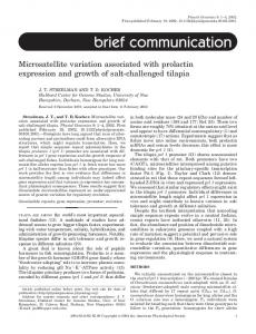
From January to April 1998, serum samples from three patients with histopathological confirmed diagnosis of abdominal angiostrongyliasis were collected 7 days after surgery (dps); from one patient it was possible to collect samples from 14 and 21 dps. Two negative controls were processed with the same procedures used to the positive samples. DNA extraction in 500 µL of the biological material was tested with the following procedures: DNAzol (Gibco, BRL), Proteinase K (AUSUBEL et al. 1989), simple boiling8 and Kit Sephaglas Band Prep (Pharmacia Biotech). The best results were obtained with DNAzol extraction (Fig. 1).
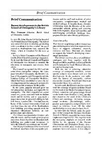
Fig. 2 - (A) 2% agarose gel electrophoresis showing the products of serum samples from 8 healthy individuals from the endemic area of Guaporé (RS, Brazil) with a positive IgGELISA for abdominal angiostrongyliasis (lanes 1-8); lane 9, negative control: only reaction buffer plus water; lane 10, positive control: 18.5 ng of A. costaricensis DNA. 10 µL of each PCR product was applied to each lane, and the result was revealed using UV transillumination after ethidium bromide staining. (B) Southern hybridization performed on samples of A.
also a not well-adapted host for A. costaricensis and morbidity is associated with the presence of the parasites. The variability of anti-A. costaricensis antibody response among infected individuals, prevents a better performance of immunodiagnostic tests. Therefore, a PCR-based molecular diagnostic test for abdominal angiostrongyliasis has a huge potential for clinical use and for epidemiological studies. Fig. 1 - (A) 2% agarose gel electrophoresis showing the PCR products of three patients with Angiostrongyliasis. Lane 1, DNA from A. costaricensis (positive control); lane 2, DNA from Echinococcus granulosus with primers 946 AG6 and 1384 AG6; lanes 3 and 4, negative controls (without template); lanes 5-10, infected sera samples: lane 5, patient C; lane 6, patient A; lane 7, patient B at 7 days post surgery (dps); lane 8, 14 dps; lane 9, 21 dps; lane 10, patient B.10 µL of each PCR product was applied to each lane, and the result was revealed using UV transillumination after ethidium bromide staining. A fragment of 232-bp DNA was observed with A. costaricensis. (B) Southern hybridization performed on samples of A.
The specificity of PCR products obtained from the serum samples was confirmed by Southern blot. An estimated amount of 100 ng of genomic DNA of A. costaricensis were electrophoresed and 10 µL of each amplified product of serum transferred to a nitrocellulose membrane (Nytran, Schleicher & Schuell) and hybridized with a total A. costaricensis DNA probe 100 ng labelled with dATP-α- 32P. The membrane was exposed to Kodak X-Omat TM film for 24 hours at room temperature for autoradiography (Fig. 1). Serum samples collected in a previous epidemiological study (Guaporé, RS, Brazil) from asymptomatic individuals: 8 positive for IgG antibodies (ELISA)4 were also negative by PCR, confirming the value of this assay (Fig. 2). RESULTS AND DISCUSSION The primers were able to amplify a 232 bp fragment of genomic DNA from A. costaricensis and no amplification was detected when the primers were used with DNA from Strongyloides ratti, Ancylostoma caninum, Ascaris suum and Toxocara canis. PCR products were detected in the three patients with confirmed diagnosis of abdominal angiostrongyliasis. The murine experimental infection is associated with high mortality surviving animals, usually those with a low parasitic burden12. Man, is 296
RESUMO Diagnóstico da angiostrongilíase abdominal utilizando PCR em soro de pacientes Angiostrongilíase abdominal é uma infecção zoonótica causada por um parasito nematódeo intravascular de roedores silvestres, Angiostrongylus costaricensis. Nenhum diagnóstico parasitológico é atualmente disponível e o imunodiagnóstico apresenta alguns obstáculos. Oligonucleotídeos foram construídos baseados em um gênero específico, Angiostrongylus cantonensis, e foi capaz de amplificar um fragmento de 232 bp de amostras de soro de 3 pacientes com diagnóstico histopatológico. O melhor método de extração foi com DNAzol e a especificidade dos oligonucleotídeos foi confirmada por Southern Blot. A doença tem sido diagnosticada com freqüência no sul do Brasil, assim, este método surge como uma importante e inédita alternativa no auxílio do diagnóstico desta doença. ACKNOWLEDGEMENTS Finantial support: FAPERGS 97.0476.0 and 00.1272.6, CNPq, CAPES (Probral 055/97) and HSL-PUCRS. REFERENCES 1. AUSUBEL, F.M.; BRENT, R.; KINGSTON, R.E. et al. - Current protocols in molecular biology. New York, John Wiley, 1989. 2. BESSARAB, I.N. & JOSHUA, G.W.P. - Stage-specific gene expression in Angiostrongylus cantonensis: characterisation and expression of an adult-specific gene. Molec. Biochem. Parasit., 88: 73-84, 1997. 3. DEMO, O.J. & PESSAT, A.O.N. - Angiostrongilosis abdominal. Primeiro caso humano encontrado en Argentina. Prensa méd. argent., 73: 732-738, 1986.
SILVA, A.C.A.; GRAEFF-TEIXEIRA, C. & ZAHA, A. - Diagnosis of abdominal angiostrongyliasis by PCR from sera of patients. Rev. Inst. Med. trop. S. Paulo, 45(5):295-297, 2003.
4. GEIGER, S.M.; LAITANO, A.C.; SIEVERS-TOSTES, C. et al. - Detection of the acute phase of abdominal angiostrongyliasis with a parasite-specific IgG Enzyme Linked Immunosorbent Assay. Mem. Inst. Oswaldo Cruz, 96: 515-518, 2001. 5. GRAEFF-TEIXEIRA, C.; CAMILLO-COURA, L. & LENZI, H.L. - Clinical and epidemiological aspects of abdominal angiostrongyliasis in southern Brazil. Rev. Inst. Med. trop. S. Paulo, 33: 373-378, 1991a. 6. GRAEFF-TEIXEIRA, C.; CAMILLO-COURA, L. & LENZI, H.L. - Histopathological criteria for the diagnosis of abdominal angiostrongyliasis. Parasit. Res., 77: 606611, 1991b. 7. GRAEFF-TEIXEIRA, C.; AGOSTINI, A.A.; CAMILLO-COURA, L. & FERREIRADA-CRUZ, M.F. - Seroepidemiology of abdominal angiostrongyliasis: the standardization of an immunoenzymatic assay and prevalence of antibodies in two localities in Southern Brazil. Trop. Med. Int. Hlth., 2: 254-260, 1997. 8. KOCAGÖZ, T.; YILMAZ, E.; OZKARA, S. et al. - Detection of Mycobacterium tuberculosis in sputum samples by polymerase chain reaction using a simplified procedure. J. clin. Microbiol., 31: 1435-1438, 1993.
9. KOO, J.; PIEN, F. & KLIKS, M.M. - Angiostrongylus (Parastrongylus) eosinophilic meningitis. Rev. infect. Dis., 10: 1155-1162, 1988. 10. MORERA, P. - Life history and redescription of Angiostrongylus costaricensis Morera and Céspedes, 1971. Amer. J. trop. Med. Hyg., 22: 613-621, 1973. 11. PENA, G.P.M.; ANDRADE FILHO, J.S. & ASSIS, S. C. - Angiostrongylus costaricensis: first record of its occurrence in the State of Espírito Santo, Brazil and review of its geographic distribution. Rev. Inst. Med. trop. S. Paulo, 37: 369-374, 1995. 12. SANTOS, F.T.; PINTO, V.M. & GRAEFF-TEIXEIRA, C. - Evidences against a significant role of Mus musculus as natural host for Angiostrongylus costaricensis. Rev. Inst. Med. trop. S. Paulo, 38: 171-175, 1996. 13. UBELAKER, J.E. & HALL, N.M. - First report of Angiostrongylus costaricensis Morera and Céspedes, 1971 in the United States. J. Parasit., 65: 307, 1979. Received: 15 September 2003 Accepted: 13 October 2003
297