brief communication Oncogenic properties of PPM1D located within a breast cancer amplification epicenter at 17q23
tion5,6. These two genes are adjacent, located within 100 kb of each other. We determined that both APPBP2 and PPM1D were co-amplified more than 2.5-fold in 27 of 164 primary breast tumors (16%) and that both genes were overexpressed in amplified tumors and cell lines (Table 1). We examined a panel of tumors and breast cancer cell lines to determine whether APPBP2 or PPM1D were within an epicenter of maximal amplification, or, conversely, whether other nearby genes were more frequently or more highly amplified. We measured DNA copy number for 16 different sequences distributed throughout a region of 2.2 Mb surrounding APPBP2 and PPM1D. This region includes the immortalizing gene TBX2, which represses CDKN2A (p19ARF) expression7, and the p70 S6 kinase gene RPS6KB1, which, owing to its role in mitogenic signaling, may have oncogenic functions8. All of the samples showed as high or higher amplification of APPBP2 and PPM1D than
We found that PPM1D, encoding a serine/threonine protein phosphatase, lies within an epicenter of the region at 17q23 that is amplified in breast cancer. We show that overexpression of this gene confers two oncogenic phenotypes on cells in culture: attenuation of apoptosis induced by serum starvation and transformation of primary cells in cooperation with RAS.
We used DNA microarray analysis1 to identify genes that are both amplified and overexpressed in the breast cancer cell line MCF7. The four genes that were the most amplified and overexpressed were NCOA3, ZNF217, PPM1D and APPBP2 (see Fig. 1a and Web Table A online). The genes NCOA3 and ZNF217 are both located on chromosomal region 20q and undergo
amplification in breast cancer; when overexpressed, these genes confer cellular phenotypes consistent with a role in tumor formation2–4. Neither PPM1D nor APPBP2 have been directly implicated in breast cancer, but both genes are located on chromosomal region 17q23, one of the regions most commonly amplified in breast cancer, and have been seen to undergo amplifica-
c
a gene expression
NCOA3 NCOA3 APPBP2 PPM1D ZNF217 RAS
RAS + PPM1D
PPM1D
d viable cells (x 10,000)
200
DNA copy number
b 25
DNA copy number
© 2002 Nature Publishing Group http://genetics.nature.com
Published online: 20 May 2002, DOI: 10.1038/ng888
150
100
50
20
0 0.1%
15
0.3%
1%
2%
5%
10%
serum concentration
10 5
e
0 0
500
1,000
CLTC RPS6KB1 USP APPBP2 PPM1D
1,500 FLJ21857
2,000 TBX2TBX4
kb
B
0 T2
B
5 1 17 36 MB MB A A MD MD
34
4
7 T4
M
7 CF
B1
AM MD
anti-PPM1D anti–β-tubulin
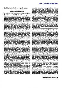
Fig. 1 APPBP2/PPM1D amplicon characterization, assays for APPBP2 and PPM1D oncogenic function and detection of PPM1D protein in breast cancer cell lines. a, Gene expression in the breast cancer cell line MCF7, as compared with normal human mammary epithelial cells (ordinate, log10 scale), is plotted against DNA copy number of MCF7 as compared with normal human DNA (abscissa, log10 scale). Genes that seemed to be amplified by more than 2.3-fold at the DNA level, and overexpressed by more than 6.5-fold, are shown in bold. Two distinct cDNA clones correspond to NCOA3. For the underlying data for all 7,316 cDNAs, see Web Table A online. b, High-resolution DNA copy-number profiles for breast cancer cell lines MCF7 (green diamonds) and ZR75-30 (red triangles) and primary invasive breast tumors 87-637 (blue squares) and 87-320 (gray squares) are shown for a 2.2-Mb region surrounding APPBP2 and PPM1D. c, Typical transformed foci of mouse embryo fibroblasts that were infected with retroviral constructs containing PPM1D and mutationally activated RAS, along with representative areas of surviving cells after infection with either the RAS or PPM1D vectors alone (APPBP2 had no effects in this assay). d, Number of viable cells after 48 h of incubation in the presence of the indicated serum concentrations. The empty pLPC (white bars), pLPC-PPM1D (gray bars) and pLPC-APPBP2 (black bars) vectors were introduced by retroviral transfection into the C8 line of mouse embryo fibroblasts transformed with E1A and RAS14. These cells undergo apoptosis when starved for serum14. e, Immunoblot detection of PPM1D levels in breast cancer cell lines. Anti-PPM1D antibody detected expression in cell lines harboring amplified PPM1D genomic DNA (BT474, MCF7 and MDAMB361), but not in cell lines that were single-copy at the PPM1D locus (BT20, MDAMB134 and MDAMB175). An immunoblot with anti-tubulin antibody was used to control for protein content.
nature genetics • volume 31 • june 2002
133
brief communication Table 1 • Amplification and expression of APPBP2 and PPM1D in human tumors and breast cancer cell lines
© 2002 Nature Publishing Group http://genetics.nature.com
Tumor sample/cell line 87-320 (breast tumor) 87-637 (breast tumor) 88-349 (breast tumor) 90-603 (breast tumor) 88-218 (breast tumor) MCF7 (breast cell line) BT474 (breast cell line) MDAMB175 (breast cell line) WA12-2 (prostate tumor) WA13-1 (prostate tumor) WA20-10 (prostate tumor)
Relative DNA copy number*
Relative APPBP2 expression*
8.1 9.8 2.8 1.1 0.8 11 3.4 1.0 1.1 2.0 1.1
8.3 4.0 2.3 1.0 0.9 29 4.8 1.5 1.1 2.0 1.4
Relative PPM1D expression* 3.2 5.2 6.0 0.4 0.6 66 19 4.1 140 41 2.1
*Relative DNA and RNA copy numbers were determined by quantitative PCR (see Web Note A online).
they did for other genes in the surrounding region, including TBX2, its homolog TBX4 and RPS6KB1 (Fig. 1b). One breast cancer cell line showed an amplicon of 500–600 kb that contains only APPBP2, PPM1D and the 5′ portion of a ubiquitin-specific protease gene (Fig. 1b). This gene was excluded from the 1-Mb amplicon of a primary tumor sample that did include APPBP2, PPM1D, a gene termed FLJ21857 and TBX2 (Fig. 1b). Another primary tumor sample contained its amplification maximum at the region containing APPBP2 and PPM1D (Fig. 1b). This analysis demonstrates that APPBP2 and PPM1D lie within an epicenter of maximal amplification and thus supports the likelihood that they are target-amplified cancer genes that provide a selective advantage to tumor cells. To determine whether APPBP2 or PPM1D have oncogenic properties that could provide a selective advantage to tumor cells, we overexpressed these genes in different cell types and examined various properties associated with tumorigenesis. Neither PPM1D nor APPBP2 overexpression malignantly transformed NIH-3T3 cells, as judged by growth in soft agar, growth in low serum or tumor formation in athymic mice. However, PPM1D, but not APPBP2, cooperated with RAS to transform primary mouse fibroblasts (Fig. 1c). PPM1D did not immortalize these cells, as the PPM1D/RAS-transformed colonies did not survive beyond seven passages. In this respect, PPM1D is a relatively weak oncogene. However, we observed an additional oncogenic phenotype upon overexpression of PPM1D (but not APPBP2) in RAS/E1Atransformed primary fibroblasts. Overexpression of PPM1D significantly attenuated apoptosis induced by serum starvation (Fig. 1d). These results indicate that PPM1D has oncogenic properties; taken together with its location within an amplification epicenter, these findings demonstrate that PPM1D provides a selective advantage upon genomic amplification. Our claim that PPM1D is a functionally 134
important amplified oncogene is further supported by our finding that PPM1D, but not APPBP2, is frequently overexpressed in late-stage prostate cancers in the absence of gene amplification (Table 1). Overexpression in the absence of gene amplification is a hallmark of amplified oncogenes such as MYC and CCND1 (ref. 9). In addition, we detected overexpression of the PPM1D protein in breast cancer cell lines harboring genomic amplification of PPM1D (Fig. 1e). The gene APPBP2 encodes a microtubule-binding protein that mediates intracellular protein targeting10. Its amplification is probably a secondary consequence of its nearness to PPM1D, as it did not induce any oncogenic phenotype when overexpressed. The gene PPM1D, which is induced by activation of wildtype p53 function, encodes a serine/threonine phosphatase that, when overexpressed, attenuates p53-mediated transcription and irradiation-induced apoptosis11. Similar to MDM2, PPM1D may be oncogenic largely as a result of its negative effect on p53 function; indeed, both of the oncogenic phenotypes we observed could be explained by loss of p53 function. A similar conclusion has recently been reached by others15. The gene TBX2 is in the center of a distinct amplification maxima in the MCF7 cancer cell line (Fig. 1a) and is frequently co-amplified in tumors in which PPM1D is amplified (6 of 11 tested). Notably, PPM1D and TBX2 have overlapping oncogenic properties, in that both genes act in combination with RAS to transform primary fibroblasts7. However, TBX2 is a potent immortalizing gene, whereas PPM1D is not, and PPM1D overexpression attenuates serum-starvation induced apoptosis, and this has not yet been tested for TBX2 (ref. 7). Amplification of TBX2 seems to occur exclusively in tumors that contain wildtype p53 protein7, and coamplification of PPM1D and TBX2 may allow cells to more effectively escape from normal p53 tumor-suppressor function than amplification of either gene alone.
The previously reported finding that breast cancers that harbor 17q23 amplifications have a poor prognosis12,13 indicates that PPM1D may provide a clinically important target for the development of anticancer therapeutics. Note: Supplementary information is available on the Nature Genetics website. Acknowledgments
We thank Jodi Harris for preparation of the figures. Competing interests statement
The authors declare that they have no competing financial interests. Jing Li1, Ying Yang1, Yue Peng1, Richard J. Austin2, Winfried G. van Eyndhoven1, Ken C.Q. Nguyen1, Tim Gabriele2, Mila E. McCurrach3, Jeffrey R. Marks4, Timothy Hoey2, Scott W. Lowe3 & Scott Powers1 1Tularik
Inc., Genomics Division, 266 Pulaski Road, Greenlawn, New York, USA. 2Tularik Inc., South San Francisco, California, USA. 3Cold Spring Harbor Laboratory, Cold Spring Harbor, New York, USA. 4Department of Surgery, Duke University Medical Center, Durham, North Carolina, USA. Correspondence should be addressed to S.P. (e-mail:
[email protected]). Received 8 November 2001; accepted 28 March 2002. 1. 2. 3. 4. 5. 6. 7. 8. 9. 10.
11. 12. 13. 14. 15.
Pollack, J.R. et al. Nature Genet. 23, 41–46 (1999). Anzick, S.L. et al. Science 277, 965–968 (1997). Collins, C. et al. Proc. Natl Acad. Sci. USA 95, 8703–8708 (1998). Nonet, G.H. et al. Cancer Res. 61, 1250–1254 (2001). Wu, G. et al. Cancer Res. 61, 4951–4955 (2001). Monni, O. et al. Proc. Natl Acad. Sci. USA 98, 5711–5716 (2001). Jacobs, J.J. et al. Nature Genet. 26, 291–299 (2000). Blume-Jensen, P. & Hunter, T. Nature 411, 355–365 (2001). Schwab, M. Semin. Cancer Biol. 9, 319–325 (1999). Zheng, P., Eastman, J., Vande Pol, S. & Pimplikar, S.W. Proc. Natl Acad. Sci. USA 95, 14745–14750 (1998). Takekawa, M. et al. EMBO J. 19, 6517–6526 (2000). Bärlund, M. et al. J. Natl Cancer Inst. 92, 1252–1259 (2000). Latham, C. et al. Cancer Genet. Cytogenet. 127, 16–23 (2001). Lanni, J.S., Lowe, S.W., Licitra, E.J., Liu, J.O. & Jacks, T. Proc. Natl Acad. Sci. USA 94, 9679–9683 (1997). Bulavin, D.V. et al. Nature Genet. 31, 210–215 (2002).
nature genetics • volume 31 • june 2002