Brief Communication A new model of self-expanding tracheal stent made in Brazil: an experimental study in rabbits* Novo modelo de endoprótese traqueal autoexpansível de fabricação nacional: estudo experimental em coelhos
Celso Murilo Nálio Matias de Faria, Olavo Ribeiro Rodrigues, Helio Minamoto, Patricia Maluf Cury, José de Mendonça Costa Neto, Domingo Marcolino Braile
Abstract We aimed to test a new model of self-expanding tracheal stent so that it might be made available for clinical use. Using direct laryngoscopy, we placed polyurethane-coated, nitinol stents into the middle third of the trachea in 25 New Zealand rabbits. After a mean observation period of 26 days, we evaluated stent migration, degree of expansion, attachment, adherence, formation of granulation tissue, presence of inflammatory infiltrate, parietal involvement, and epithelial lining. The results showed complete radial expansion, little adherence to the tracheal mucosa, and low tissue attachment, as well as high rates of granuloma formation and stent migration. This new model proved to be biocompatible and showed a behavior similar to that of other stents on the market. Keywords: Prosthesis implantation; Tracheal stenosis; Rabbits; Stents.
Resumo Objetivamos testar um novo modelo de endoprótese traqueal autoexpansível para que esse possa ser futuramente disponibilizado para o uso clínico. As endopróteses de nitinol revestidas de poliuretano foram alocadas no terço médio da traqueia de 25 coelhos da raça Nova Zelândia sob laringoscopia direta. Após um período de observação médio de 26 dias, avaliou-se a migração da prótese, grau de dilatação, incorporação, aderência, formação de tecido de granulação, presença de infiltrado inflamatório, envolvimento parietal e revestimento epitelial. Os resultados demonstraram completa expansibilidade radial, pouca aderência à mucosa traqueal e baixa incorporação tecidual, assim como alta taxas de formação de granulomas e de migração. Esse novo modelo demonstrou ser biocompatível e teve comportamento semelhante ao de outras próteses disponíveis no mercado. Descritores: Implante de prótese; Estenose traqueal; Coelhos; Stents.
The objective of the present study was to test a new model of self-expanding tracheal stent(1) made in Brazil so that it might be made available for clinical use.(2) We placed tracheal stents in 25 New Zealand rabbits (Oryctolagus cuniculus; mean weight, 3,520 g). After an observation period of 15-35 days (mean, 26 days), we evaluated the stents. The self-expanding stents(3) were made from a single nitinol (nickel-titanium) filament(4) and were coated with polyurethane. The stents were 20 mm in length by 8 mm in external diameter after release. This new model was designed,
manufactured, and provided by Braile Biomédica, located in the city of São José do Rio Preto, Brazil. Using laryngoscopy and an applicator, we placed the stents with the aid of a guide wire inserted into the lumen of the applicator. The stents were placed into the middle third of the trachea in the rabbits, stent placement having been confirmed by neck and chest X-ray examination. By means of macroscopic examination, we analyzed stent migration, degree of expansion, attachment/adherence, and formation of granulation tissue. By means of microscopic examination, we analyzed presence of inflammatory
* Study carried out at the São José do Rio Preto School of Medicine, São José do Rio Preto, Brazil. Correspondence to: Celso Murilo Nálio Matias de Faria. Rua Alcides Rossani, 853, Vilage LaMontagne, CEP 15093-520, São José do Rio Preto, SP, Brasil. Tel. 55 17 2139-8328. E-mail:
[email protected] Financial support: The stents used in the present study were provided by Braile Biomédica. Submitted: 26 July 2011. Accepted, after review: 28 February 2012.
J Bras Pneumol. 2012;38(2):214-217
A new model of self-expanding tracheal stent made in Brazil: an experimental study in rabbits
infiltrate, cellularity, parietal involvement, and mucosal epithelial lining. After the observation period, we sacrificed the animals by administering intravenous thiopental. Subsequently, we examined postmortem neck and chest X-rays in order to analyze stent migration. After the postmortem neck and chest X-ray examination, we removed the laryngotracheal complex in order to analyze the degree of expansion of the stents, the attachment/adherence of the stents to the tracheal wall, and the presence of granulation tissue. We performed histological examination in order to evaluate the inflammatory infiltrate and its cellularity, as well as the extent of the inflammatory process in the tracheal wall (parietal involvement), together with the changes in the epithelial lining.(5) Of the 25 rabbits employed, 12 (48%) failed to meet the study criteria, having been excluded from the analysis of the results because of incorrect stent placement (in 10) and death from pneumonia before 15 days of observation (in 2). Incorrect stent placement occurred at the beginning of the experiment. By modifying the applicator and mastering the placement technique, we were able to solve the problem. For the evaluation of the results, we included 13 animals that survived the observation period. Our macroscopic and microscopic findings are shown in Table 1. Stent migration was observed in 5 of 6 animals (84%). Of the 13 animals that survived the observation period, 7 had rigor mortis, which precluded hyperextension of the neck for X-ray examination and, consequently, the determination of the exact position of the stents. Therefore, stent migration was analyzed in the 6 remaining animals only. Covered and self-expanding stents have been reported to show high migration rates, our results being therefore consistent with those reported in the literature (Figure 1).(6,7) We determined the degree of expansion by measuring the external diameter of the stents. We found that the stents expanded in 100% of the cases, expansion having been complete in 54% and nearly complete in 46%. This small difference can be attributed to the size of the stent, which is purposely larger than the trachea so that fixation can occur by radial force. The stents tested showed low tissue attachment (16%). Adherence to the tracheal mucosa remained but was easily removed from the tracheal lumen.
215
Table 1 - Macroscopic and microscopic findings. Macroscopic findings % Stent migration 84 Complete expansion 54 Partial expansion 46 Tissue attachment 16 Adherence 84 Granuloma formation 77 Microscopic findingsa Cellularity Polymorphonuclear cells 67 Mononuclear cells 33 Parietal involvement Mucosa 42 Mucosa + submucosa 33 All layers 25 Epithelial lining Erosion 8 Ulceration 42 Regenerative hyperplasia 33 Squamous metaplasia 17 a
Evaluation of the tracheal segment containing the stent.
This occurs with covered stents, coating polymers preventing tissue from entering the metallic platform and proliferating among the meshes. Treatment with tracheal stents has been associated with granuloma formation.(5,6,8) In the present study, the rate of granuloma formation was 77%, granulomas having been found in the proximal and distal ends of the stents. Histological examination revealed the presence of inflammatory infiltrate in all of the cases, polymorphonuclear cells having predominated. This indicated an acute phase response to the (repetitive and persistent) injury caused by the stents. Regarding the extent of the inflammatory process in the tracheal wall, we found that the inflammatory process was limited to the mucosa and submucosa (inner third of the tracheal wall) in 75% of the animals and was extensive (involving all of the tracheal wall layers) in only 17%. The most common changes in the respiratory epithelial lining that was in contact with the stent were epithelial erosion and ulceration, which resulted from mechanical injury to the respiratory epithelium. We found a high rate of squamous metaplasia (67%). Squamous metaplasia was found in the tracheal segment that was not in contact with the stent, resulting from (and being indicative of) chronic inflammation.
J Bras Pneumol. 2012;38(2):214-217
216
Faria CM, Rodrigues OR, Minamoto H, Cury PM, Costa Neto JM, Braile DM
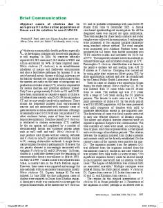
A
B
Figure 1 - Rabbit neck and chest X-rays showing the stent immediately after its release (in A) and 30 days after its placement (in B). Note that the stent moved caudally.
The polyurethane-coated, nitinol stents investigated in the present study showed a behavior similar to that of other stents on the market.(9) Further studies investigating the biocompatibility of this new model of tracheal stent and employing different stent sizes, diameters, and radial forces, as well as different animal models, can provide additional information before the stents are made available for clinical use. The self-expanding, polyurethane-coated, nitinol stents investigated in the present study proved easy to apply, release, and place in the tracheal lumen of rabbits. The stents showed good radial expansion, having adapted to the diameter of the trachea. The results showed high rates of granuloma formation and stent migration, as well as low permeability. In addition, the results showed good adherence to the tracheal mucosa and low tissue attachment. The stent proved to be biocompatible.
References 1. Terra RM, Minamoto H, Jatene FB. Intratracheal stent: prosthesis or orthesis? J Bras Pneumol. 2006;32(6):606-7. Pmid:17438639. http://dx.doi.org/10.1590/ S1806-37132006000600024
J Bras Pneumol. 2012;38(2):214-217
2. Xavier RG, Sanches PR, de Macedo Neto AV, Kuhl G, Vearick SB, Michelon MD. Development of a modified Dumon stent for tracheal applications: an experimental study in dogs. J Bras Pneumol. 2008;34(1):21-6. Pmid:18278372. http://dx.doi.org/10.1590/S1806-37132008000100005 3. Saueressig MG, Macedo Neto AV, Moreschi AH, Xavier RG, Sanches PR. Correção das estenoses traqueobrônquicas mediante o emprego de órteses. J Pneumol. 2002;28(2):84-93.http://dx.doi.org/10.1590/ S0102-35862002000200005 4. Buehler WJ, Gilfrich JW, Wiley RC. Effect of low-temperature phase changes on the mechanical properties of alloys near composition TiNi. J Appl Phys. 1963;34(5):1475-8. 5. Sara C. Histological changes in the trachea and bronchi with tracheostomy. Med J Aust. 1967;1(23):1174-7. PMid:6029499. 6. Rafanan AL, Mehta AC. Stenting of the tracheobronchial tree. Radiol Clin North Am. 2000;38(2):395-408.http:// dx.doi.org/10.1016/S0033-8389(05)70170-6 7. Saito Y, Imamura H. Airway stenting. Surg Today. 2005;35(4):265-70. PMid:15815840. http://dx.doi. org/10.1007/s00595-004-2942-y 8. Mitsuoka M, Hayashi A, Takamori S, Tayama K, Shirouzu K. Experimental study of the histocompatibility of covered expandable metallic stents in the trachea. Chest. 1998;114(1):110-4. Pmid:9674456. http://dx.doi. org/10.1378/chest.114.1.110 9. Chin CS, Litle V, Yun J, Weiser T, Swanson SJ. Airway stents. Ann Thorac Surg. 2008;85(2):S792-6. Pmid:18222219. http://dx.doi.org/10.1016/j.athoracsur.2007.11.051
A new model of self-expanding tracheal stent made in Brazil: an experimental study in rabbits
217
About the authors Celso Murilo Nálio Matias de Faria
Attending Physician. Department of Thoracic Surgery, São José do Rio Preto Hospital de Base, São José do Rio Preto School of Medicine, São José do Rio Preto, Brazil.
Olavo Ribeiro Rodrigues
Adjunct Professor. Department of Thoracic Surgery, University of Mogi das Cruzes School of Medicine, Mogi das Cruzes, Brazil. Thoracic Surgeon. Luzia de Pinho Melo Hospital, São Paulo State Department of Health, São Paulo, Brazil. Director. Mogi das Cruzes Chest Institute, Mogi das Cruzes, Brazil.
Helio Minamoto
Attending Physician. Department of Cardiorespiratory Diseases, University of São Paulo School of Medicine Hospital das Clínicas Heart Institute, São Paulo, Brazil.
Patricia Maluf Cury
Education Coordinator. São José do Rio Preto School of Medicine, São José do Rio Preto, Brazil.
José de Mendonça Costa Neto
Part-owner. Laboratório de Histopatologia Ltda., São José do Rio Preto, Brazil.
Domingo Marcolino Braile
Professor. State University at Campinas, Campinas, Brazil. Adjunct Director. São José do Rio Preto School of Medicine Graduate Program, São José do Rio Preto, Brazil.
J Bras Pneumol. 2012;38(2):214-217