Our reference: PARINT 1146
P-authorquery-v11
AUTHOR QUERY FORM Journal: PARINT
Please e-mail or fax your responses and any corrections to: Ramanujam, Bharghavi E-mail:
[email protected] Fax: +1 619 699 6721
Article Number: 1146
Dear Author, Please check your proof carefully and mark all corrections at the appropriate place in the proof (e.g., by using on-screen annotation in the PDF file) or compile them in a separate list. Note: if you opt to annotate the file with software other than Adobe Reader then please also highlight the appropriate place in the PDF file. To ensure fast publication of your paper please return your corrections within 48 hours. For correction or revision of any artwork, please consult http://www.elsevier.com/artworkinstructions. Any queries or remarks that have arisen during the processing of your manuscript are listed below and highlighted by flags in the proof. Click on the ‘Q’ link to go to the location in the proof.
Location in article
Query / Remark: click on the Q link to go Please insert your reply or correction at the corresponding line in the proof
Q1
Please confirm that given names and surnames have been identified correctly.
Q2
All binominals should defined at the first use of the term, separately in the title, abstract, and main text. Please check throughout the text for other instances of initially unexpanded binominals, and ensure that any that have been expanded by the copyeditor are correct.
Q3
All binominals should be defined at the first use of the term, separately in the title, abstract and main text. The following citations are then abbreviated. Please check throughout the text for instances of unabbreviated binominals and ensure that any that have been abbreviated by the copyeditor are correct.
Q4
Would you consider modifying the sentence for notion clarity (e.g. The presence of vaginal plug was marked as the first gestational day). Please check and amend if necessary.
Q5
Please check the change made in this sentence and amend if necessary.
Q6
This sentence has been slightly modified for clarity. Please check that the meaning is still correct, and amend if necessary.
Q7
Supplementary caption was not provided. Please check suggested data if appropriate and correct if necessary.
Please check this box if you have no corrections to make to the PDF file.
□
Thank you for your assistance.
Page 1 of 1
PARINT-01146; No of Pages 1 Parasitology International xxx (2013) xxx
Contents lists available at ScienceDirect
Parasitology International journal homepage: www.elsevier.com/locate/parint
1
F
Treatment of pregnant BALB/c mice with sulphadoxine pyrimethamine or chloroquine abrogates Plasmodium berghei induced placental pathology Lalita Sharma, Geeta Shukla ⁎
pp xxx – xxx
O
2 5 6 87
P
R O
910
4 11
U
N
C
O
R
R
E
C
T
E
D
12
1383-5769/$ – see front matter © 2013 Published by Elsevier Ireland Ltd. http://dx.doi.org/10.1016/j.parint.2013.08.016
Please cite this article as: Sharma L, Shukla G, Treatment of pregnant BALB/c mice with sulphadoxine pyrimethamine or chloroquine abrogates Plasmodium berghei induced placental pathology, Parasitology International (2013), http://dx.doi.org/10.1016/j.parint.2013.08.016
PARINT-01146; No of Pages 1 Parasitology International xxx (2013) xxx
Contents lists available at ScienceDirect
Parasitology International journal homepage: www.elsevier.com/locate/parint
2 5 6
Treatment of pregnant BALB/c mice with sulphadoxine pyrimethamine or chloroquine abrogates Plasmodium berghei induced placental pathology
7 8
Lalita Sharma, Geeta Shukla ⁎
9 10
Department of Microbiology, Panjab University, Chandigarh 160014, India
11 12 13 14 15
• Treatment with antimalarials reduced parasitaemia in pregnant P. berghei infected mice. • Malondialdehyde levels and apoptotic cells were reduced in the placenta of treated mice. • Histologically treated placenta had normal cellular morphology.
Parasitology International xxx (2013) xxx – xxx
F
Highlights
U
N C O
R
R
E
C
T
E
D
P
16 4 17
R O
O
1
1383-5769/$ – see front matter © 2013 Published by Elsevier Ireland Ltd. http://dx.doi.org/10.1016/j.parint.2013.08.016
Please cite this article as: Sharma L, Shukla G, Treatment of pregnant BALB/c mice with sulphadoxine pyrimethamine or chloroquine abrogates Plasmodium berghei induced placental pathology, Parasitology International (2013), http://dx.doi.org/10.1016/j.parint.2013.08.016
Q7
Supplementary material.
PARINT-01146; No of Pages 8 Parasitology International xxx (2013) xxx–xxx
Contents lists available at ScienceDirect
Parasitology International journal homepage: www.elsevier.com/locate/parint
4Q1
Lalita Sharma, Geeta Shukla ⁎
5
Department of Microbiology, Panjab University, Chandigarh 160014, India
O R O
2
F
3
Treatment of pregnant BALB/c mice with sulphadoxine pyrimethamine or chloroquine abrogates Plasmodium berghei induced placental pathology
1
6
a r t i c l e
i n f o
a b s t r a c t
Article history: Received 4 October 2012 Received in revised form 17 August 2013 Accepted 29 August 2013 Available online xxxx
D
P
Malaria infection during pregnancy is a risk factor for foetus survival and is associated with abortion, premature delivery and low birth weight of infants in malaria endemic regions. In these regions, prophylactic measures and treatment mainly rely on chloroquine and sulphadoxine pyrimethamine, but their efficacy in reducing the placental pathology has not been studied. Therefore, the present study was designed to assess the effectiveness of chloroquine and sulphadoxine pyrimethamine treatment in reducing the placental pathology of Plasmodium berghei infected BALB/c mice. It was observed that pregnant-infected mice, treated either with chloroquine or sulphadoxine pyrimethamine had significantly lower percent parasitaemia, 100% survival and delivered normally compared with untreated pregnant-infected mice. Interestingly, antimalarial treatment significantly reduced malondialdehyde (MDA) levels, measure of lipid peroxidation and number of apoptotic cells in the placentae of pregnant-infected treated mice. Histologically also no morphological and cellular alterations were observed in the placentae of pregnant-infected treated mice. Taken together, the study shows the effectiveness of chloroquine and sulphadoxine pyrimethamine treatment, when administered in second trimester in abrogating malaria induced oxidative stress, apoptosis and histopathological alterations in the placenta, leading to normal foetal development. © 2013 Published by Elsevier Ireland Ltd.
C
T
E
Keywords: Malaria Pregnancy Placenta Sulphadoxine pyrimethamine Chloroquine Malondialdehyde Superoxide dismutase Catalase
E
7 8 9 10 11 12 14 13 15 16 17 18 19 20 21 22 23 24 43
R
42
1. Introduction
45
Malaria has been recognized as one of the major public health problems, killing about 1–2 million people every year, mostly children less than five years of age and pregnant women [1,2]. In sub-Saharan Africa, malaria infection is estimated to cause 400,000 cases of severe maternal anaemia and 75,000–200,000 infant deaths annually [3–5]. Maternal malaria not only imposes an additional burden to the mother but also affects the developing foetus and is more severe in primigravidae than in multigravidae. It has also been shown that maternal malaria adversely affects the development and survival of the foetus through maternal anaemia, abortion, stillbirth and low birth weight [6–8]. The pathological alterations both in human and mouse placenta due to malaria infection results into sequestration of parasite infected RBC, recruitment of monocytes within the intervillous spaces of the placenta and syncytiotrophoblastic damage due to higher placental parasitaemia than peripheral parasitaemia [9–11]. Recently, we have reported that the altered placental pathology in Plasmodium berghei infected mice is due to enhanced oxidative stress and mitochondrial mediated pathway of apoptosis [12,13].
50 51 52 53 54 55 56 57 58 59 60 61 62 Q2
N C O
48 49
U
46 47
R
44
⁎ Corresponding author at: Department of Microbiology, Basic Medical Sciences Building, Panjab University, Chandigarh 160014, India. Tel./fax: +91 172 2541770. E-mail address:
[email protected] (G. Shukla).
25 26 27 28 29 30 31 32 33 34 35 36 37 38 39 41 40
It is really a great challenge to diagnose and treat complicated malaria especially in pregnancy and deal with antimalarial associated complications like renal failure, hypoglycaemia, rapidly developing and cerebral oedema. Moreover, antimalarial treatment during pregnancy has been associated with embryotoxicity that can lead to cardiovascular malformations or skeleton abnormalities [14]. As per the recommendation of WHO, three approaches are used to reduce the burden of malaria infection during pregnancy: effective case management, insecticide treated nets (ITNs), and intermittent preventive therapy (IPT). IPT includes either two or three doses of sulphadoxine pyrimethamine (SP) that are given during the second and third trimesters, at least a month apart [15]. However, in India, chloroquine (CQ) is still the drug of choice for prevention and treatment of malaria in pregnancy, but in CQ resistant areas, a weekly regimen of CQ along with proguanil is recommended [16]. Though in India SP is in the current drug policy, SP is only used as a second line of treatment and is not readily recommended for pregnant women due to the prevalence of SP resistant strains in certain states like Assam and Orissa [17]. Although SP and CQ have been used both as prophylactic and therapeutic antimalarials for decades during pregnancy, their efficacy to reduce the placental pathology has not been studied and warrants further investigation. Therefore, the present study was designed to assess the effectiveness of sulphadoxine pyrimethamine and chloroquine treatment on the placental pathology in P. berghei infected pregnant BALB/c mice.
1383-5769/$ – see front matter © 2013 Published by Elsevier Ireland Ltd. http://dx.doi.org/10.1016/j.parint.2013.08.016
Please cite this article as: Sharma L, Shukla G, Treatment of pregnant BALB/c mice with sulphadoxine pyrimethamine or chloroquine abrogates Plasmodium berghei induced placental pathology, Parasitology International (2013), http://dx.doi.org/10.1016/j.parint.2013.08.016
63 64 65 66 67 68 69 70 71 72 73 74 75 76 77 78 79 80 81 82 83 84 85 86 Q3
L. Sharma, G. Shukla / Parasitology International xxx (2013) xxx–xxx
2. Materials and methods
88
2.1. Parasite
89 90 91
P. berghei (NK 65), a lethal rodent strain was used in the study. The strain was maintained in mice by serial passage of P. berghei infected red blood cells (iRBC).
92
2.2. Animals
93 94
BALB/c mice, 6–8 weeks old (20–22 g) were obtained from the central animal house of Panjab University, Chandigarh, India. These animals
Fig. 3. Efficacy of sulphadoxine pyrimethamine (SP) and chloroquine (CQ) treatment on survival rate in pregnant and non-pregnant P. berghei infected mice.
E
87
D
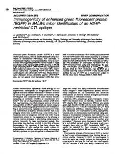
Fig. 1. Parasitaemic levels in different groups of mice. Values are expressed as mean ± standard deviation. ≠(p b 0.05) vs. nonpregnant-infected, *(p b 0.01) vs. nonpregnantinfected, #(p b 0.01) vs. pregnant-infected.
P
R O
O
F
2
2.3. Assessment of the first gestational day (GD)
98
Female mice were housed overnight with male mice of the same strain in the ratio of 2:1 and were monitored daily both in the morning and in the evening for the presence of vaginal plug. The mice showing presence of vaginal plug were marked as first gestational day [12,13,18].
99 100
U
N
C
O
R
R
E
C
T
were provided with standard pellet diet and water ad libitum. Care, use 95 and disposal of animals were done according to the guidelines of the 96 Institutional Animal Ethical Committee (44/99/CPCSEA). 97
Fig. 2. Percent reduction in parasitaemia on day 6 PI in P. berghei infected and sulphadoxine pyrimethamine (SP) or chloroquine (CQ) treated mice. Values are expressed as mean ± standard deviation.
Please cite this article as: Sharma L, Shukla G, Treatment of pregnant BALB/c mice with sulphadoxine pyrimethamine or chloroquine abrogates Plasmodium berghei induced placental pathology, Parasitology International (2013), http://dx.doi.org/10.1016/j.parint.2013.08.016
101 102 Q4
L. Sharma, G. Shukla / Parasitology International xxx (2013) xxx–xxx t1:1 t1:2
3
Table 1 Efficacy of sulphadoxine pyrimethamine and chloroquine treatment in P. berghei infected pregnant mice.
t1:3
Groups
No. of animals
No. of animals that survived to deliver
No. of pups delivered
Body weight of pups (g) a
% survival in pups after 30 days of delivery
t1:4 t1:5 t1:6
Pregnant Pregnant-infected Pregnant-infected sulphadoxine pyrimethamine treated Pregnant-infected chloroquine treated
8 8 8
8 2 8
36 5b 38
1.53 ± 0.13 1.32 ± 0.11 1.52 ± 0.13
100 0 100
8
8
33
1.49 ± 0.09
100
104
The animals were divided into seven groups. Group I (nonpregnantinfected, n = 8): These animals were inoculated intraperitoneally with 1 × 10⁶ iRBC [12,13]. Group II (nonpregnant-infected, CQ treated, n = 8): These mice were infected as in group I but were administered chloroquine (IPCA Laboratories, Mumbai, India) orally as 6.25 mg/kg body weight daily for a period of 4 days [19]. The CQ treatment was started, 2 h after malarial infection. Group III (nonpregnant-infected, SP treated, n = 8): These mice were also infected as in group I but were treated orally with single dose of sulphadoxine pyrimethamine (LUPIN Ltd. Aurangabad, India; 26 mg/kg body weight), 2 h after malarial infection [20]. Group IV (pregnant, n = 9): These pregnant animals were inoculated intraperitoneally with normal saline on GD 10 ± 2. Group V (pregnant-infected, n = 16): Pregnant mice were inoculated intraperitoneally with 1 × 10⁶ iRBC on GD10 ± 2. Group VI (pregnant-infected,
T
117
125 126
C
115 116
Percent parasitaemia in all infected groups of mice was monitored on every alternate day in Giemsa stained tail blood films by examining at least 500 cells for one month and percent survival was calculated. Mice belonging to groups IV, V and VI were sacrificed by cervical dislocation on day 6 post infection (PI) and placentae were removed for estimation of oxidants and antioxidants, DNA isolation and for ethidium bromide/acridine orange staining. For histopathological
E
113 114
124
R
111 112
2.5. Follow-up of the animals
R
109 110
N C O
107 108
118
U
105 106
CQ treated, n = 16): These mice were infected as in group V but were treated orally with CQ (6.25 mg/kg body weight daily for a period of 4 days), 2 h after malarial infection [19]. Group VII (pregnant-infected, SP treated, n = 16): These mice were also infected as in group V and were treated orally with single dose of SP, 2 h after malarial infection [20].
F
2.4. Experimental design
E
103
O
b
Values are expressed as mean ± standard deviation. All pups died after 2 h of delivery.
R O
a
P
t1:8 t1:9
D
t1:7
Fig. 4. MDA Levels (A) and catalase activity (B) in the placenta of mice belonging to various groups. Values are expressed as mean ± standard deviation. *(p b 0.05) vs. pregnant, # (p b 0.05) vs. pregnant-infected.
Please cite this article as: Sharma L, Shukla G, Treatment of pregnant BALB/c mice with sulphadoxine pyrimethamine or chloroquine abrogates Plasmodium berghei induced placental pathology, Parasitology International (2013), http://dx.doi.org/10.1016/j.parint.2013.08.016
119 120 121 122 123
127 128 129 130 131
L. Sharma, G. Shukla / Parasitology International xxx (2013) xxx–xxx
C
T
E
D
P
R O
O
F
4
135 136 137
R
R
134
studies placentae were fixed in 10% buffered formalin, processed for haematoxylin and eosin (H&E) staining and observed by light microscope.
142
2.7. Lipid peroxidation assay
143 144
The amount of malondialdehyde (MDA), a measure of lipid peroxidation, was quantitated according to the method of Wills [22]. In brief, 0.5 ml of Tris–HCl buffer (0.1 M, pH 7.4) was added to 0.5 ml of tissue homogenate and incubated at 37 °C for 2 h. After incubation, 1.0 ml of 10% (w/v) chilled trichloroacetic acid was added followed by cold centrifugation at 100 × g for 10 min. To 1.0 ml of supernatant, 1.0 ml of 0.67% (w/v) thiobarbituric acid was added and kept in boiling water bath for 10 min. After cooling the tubes, 1.0 ml of distilled water was added and absorbance was measured at 532 nm. The results were expressed as nanomoles of MDA per milligram of protein, using the molar extinction coefficient of chromophore (1.56 × 10⁵ M−1 cm−1).
145 146 147 148 149 150 151 152 153
U
N
C
140 141
154
The catalase (EC 1.11.1.6) activity in post mitochondrial supernatant 155 was assayed by the method of Luck [23]. To carry out the assay 100 ml 156
2.6. Preparation of placental homogenates and post mitochondrial supernatant
Placental homogenates were prepared in phosphate buffered saline using a Potter-Elvehjem homogenizer. Placental homogenates were cold centrifuged at 8000 × g for 10 min, supernatants were labelled as post mitochondrial supernatants (PMS) and stored at −20 °C till further use. Protein concentration was measured as per Lowry et al. [21].
138 Q5 139
2.8. Measurement of catalase activity
O
132 133
E
Fig. 5. GSH levels (A) and SOD activity (B) in the placenta of mice belonging to various groups. Values are expressed as mean ± standard deviation.
Fig. 6. DNA fragmentation assay in the placenta of P. berghei infected mice on day 6 PI. Pregnant-infected mice showing DNA fragmentation compared to intact DNA in mice belonging to pregnant-infected SP treated, pregnant-infected CQ treated and pregnant groups.
Please cite this article as: Sharma L, Shukla G, Treatment of pregnant BALB/c mice with sulphadoxine pyrimethamine or chloroquine abrogates Plasmodium berghei induced placental pathology, Parasitology International (2013), http://dx.doi.org/10.1016/j.parint.2013.08.016
L. Sharma, G. Shukla / Parasitology International xxx (2013) xxx–xxx
The levels of GSH were estimated as described by Ellman [24]. Briefly, 1 ml of placental homogenate was precipitated with 1.0 ml of 4% sulphosalicyclic acid, kept at 4 °C for at least 1 h and centrifuged at 100 ×g for 15 min at 4 °C. The assay mixture contained 0.1 ml of supernatant, 0.2 ml of 0.01 M dithionitro benzoic acid (DTNB) and 2.7 ml of phosphate buffer (0.1 M, pH 8.0). Absorbance was measured at 412 nm and results were expressed as micromoles of GSH per milligram of protein.
165 166 167 168 169 170 171
2.10. Assessment of super oxide dismutase (SOD) activity
173
SOD (EC 1.15.1.1) activity in post mitochondrial supernatant was assayed according to the method of Kono [25]. The reaction was initiated by the addition of 0.5 ml of hydroxylamine hydrochloride to the reaction mixture containing 2.0 ml nitroblue tetrazolium (NBT) and 0.1 ml PMS. SOD activity was expressed as units of SOD per milligram of protein where one unit activity is defined as the amount of SOD required to inhibit the rate of reduction of NBT by 50%.
189 190 191
193
202
196 197 198 199 200 201
Results were expressed as mean ± standard error (SE). The inter 203 group variation was assessed by two way analysis of variance 204 (ANOVA) and statistical significance was calculated at P b 0.05. 205
C E R
180
187 188
2.13. Statistical analysis
R
178 179
186
194 195
N C O
176 177
184 185
Cells were isolated by teasing placentas with frosted end slides. Dispersed cell suspension was filtered using a nylon mesh and centrifuged at 100 × g for 5 min at 25 °C. RBC in the cell pellet were lysed with chilled 2% saponin and centrifuged at 100 ×g for 10 min. The sediment containing placental cells were washed thrice and finally suspended in PBS. The number of cells in the suspension was counted using a haemocytometer and isolated placental cells were observed under a fluorescence microscope after staining with ethidium bromide/acridine orange stain.
U
174 175
192
T
172
2.12. Ethidium bromide/acridine orange staining for determination of apoptotic cells
F
164
182 183
O
2.9. Estimation of reduced glutathione (GSH)
DNA was isolated by the method described by Strauss [26] with minor modification. Briefly, 60–70 mg placental tissue was minced, suspended in 500 μl digestion buffer and kept at 50 °C overnight. An equal volume of the Tris-saturated phenol was added to the digested tissue and centrifuged at 8000 ×g for 15 min. The upper layer formed after centrifugation was subjected to phenol–chloroform–isoamylalcohol extraction procedure. Finally, DNA was precipitated with chilled ethanol, washed with 70% ethanol, dried and dissolved in Tris–EDTA buffer. Isolated DNA was electrophoresed on 1.2% agarose ethidium bromide gel and analysed by Gel Doc EZ Imager (Bio-Rad).
R O
163
181
P
161 162
2.11. DNA fragmentation assay
D
159 160
of phosphate buffer (0.05 M, pH 7.2) was made and 0.16 ml of H2O2 was added. The assay mixture consisted of 3 ml of the phosphate buffer and 5 μl of PMS. Change in absorbance was read at 240 nm. The results were expressed as millimoles of H2O2 decomposed per minute per milligram of proteins using the molar extinction coefficient of the chromophore (0.0394 mM−1 cm−1).
E
157 158
5
Fig. 7. Apoptotic cells in ethidium bromide/acridine orange (Etbr/Ar) stained placenta. Pregnant-infected mice showing higher number of apoptotic (orange nucleus) cells in the placenta compared with pregnant, pregnant-infected SP treated and pregnant-infected CQ treated groups.
Please cite this article as: Sharma L, Shukla G, Treatment of pregnant BALB/c mice with sulphadoxine pyrimethamine or chloroquine abrogates Plasmodium berghei induced placental pathology, Parasitology International (2013), http://dx.doi.org/10.1016/j.parint.2013.08.016
L. Sharma, G. Shukla / Parasitology International xxx (2013) xxx–xxx
208 209
223 224
The percent parasitaemia increased gradually both in nonpregnantinfected and pregnant-infected mice and attained peak parasitaemia on day 14 PI (62%) and day 10 PI (70%) respectively, but pregnant-infected mice had significantly higher (p b 0.05) percent parasitaemia at each point of observation beginning from day 2 PI compared with nonpregnant-infected mice. Interestingly, mice belonging to nonpregnant-infected CQ treated, nonpregnant-infected SP treated, pregnant-infected CQ treated and pregnant-infected SP treated had significantly lower (p b 0.01) percent parasitaemia compared with nonpregnant-infected and pregnant-infected mice. More specifically, all the treated animals had similar pattern of parasitaemia and became parasite free by day 24 PI (Figs. 1 & 2). However, when mice were treated with a reduced dose of CQ and SP (3.5 mg/kg body weight) for a period of 9 days, the maximum parasitaemia attained by mice was only 1.2% and parasitaemia was reduced to 0% by day 10 PI, which clearly indicated the effectiveness of these antimalarial drugs (Supplementary data, Fig. S1).
225
3.2. Percent survival
226 227
It was observed that all mice belonging to antimalarial treated groups survived compared with 100% mortality in nonpregnant-
3.4. Antioxidant levels
241
The catalase activity in the placenta of pregnant-infected CQ treated and pregnant-infected SP treated mice increased significantly (p b 0.05) on day 6 PI compared with pregnant-infected mice (Fig. 4B). However, levels of GSH and SOD were similar in the placentae of pregnant mice belonging to groups IV, V, VI and VII (Fig. 5A and B).
E T C E
221 222
236 237
R
219 220
Mice belonging to pregnant-infected CQ or SP treated groups had significantly reduced (p b 0.05) MDA levels in the placenta on day 6 PI compared with pregnant-infected mice. However, pregnantinfected mice had significantly higher (p b 0.05) MDA levels compared with pregnant mice (Fig. 4A).
R
217 218
235
O
215 216
3.3. Lipid peroxidation
C
213 214
N
211 212
U
210
F
3.1. Parasitaemia levels
228 229
O
207
infected and pregnant-infected mice (Fig. 3). Interestingly, all pregnant-infected CQ treated and pregnant-infected SP treated mice delivered normal pups as their weights were comparable to the weights of pups delivered by pregnant mice. There was 75% mortality in pregnant-infected mice before parturition and only 25% of the pregnant-infected mice survived to deliver pups and these pups died after 2 h of delivery (Table 1).
R O
3. Results
D
206
P
6
Fig. 8. (A) Number of apoptotic cells in the placenta of different groups of mice. Values are expressed as mean ± standard deviation. *(p b 0.05) vs. pregnant, #(p b 0.05) vs. pregnantinfected. (B) Bar diagram represents the percentage reduction in apoptotic cells in the placenta after CQ and SP treatment. Values are expressed as mean ± standard deviation.
Please cite this article as: Sharma L, Shukla G, Treatment of pregnant BALB/c mice with sulphadoxine pyrimethamine or chloroquine abrogates Plasmodium berghei induced placental pathology, Parasitology International (2013), http://dx.doi.org/10.1016/j.parint.2013.08.016
230 231 232 233 234
238 239 240
242 243 244 245 246 247
L. Sharma, G. Shukla / Parasitology International xxx (2013) xxx–xxx
255 256
Pregnant-infected SP or CQ treated mice had significantly (p b 0.05) less percent of apoptotic cells (67–70% reduction) compared with pregnant-infected mice (Figs. 7 & 8A & B).
257
3.7. Histopathological studies
258 259
264
Histopathologically, the placenta of pregnant-infected mice showed parasitized RBC in the intervillous space of placenta and deposition of malarial pigments compared with the normal cellular morphology of placenta belonging to pregnant mice. Interestingly, SP and CQ treated pregnant mice had normal cellular placental morphology, clearly demonstrating the abrogation of placental pathology by SP or CQ treatment (Fig. 9A, B, C, D).
265
4. Discussion
266
Despite all the efforts made to control malaria, it is still a major public health problem in underdeveloped and developing countries especially posing a high risk to children less than five years of age and pregnant women [4]. WHO and national health agencies recommend SP and CQ as the prophylaxis during pregnancy in malaria endemic areas, to reduce maternal death and successful pregnancy outcome
C E
271
R
269 270
R
267 268
N C O
262 263
U
260 261
F
254
O
3.6. Ethidium bromide/acridine orange staining
R O
253
P
251 252
Interestingly, no DNA fragmentation was observed in the placenta of mice belonging to pregnant, pregnant-infected CQ treated and pregnantinfected SP treated groups compared with faint DNA fragments in the placenta of pregnant-infected mice (Fig. 6).
D
249 250
[27,28]. Since the efficacy of SP and CQ in reducing malaria associated placental pathology with respect to oxidative stress and apoptosis has not been studied, the present study was designed. The study demonstrated that the treatment of pregnant and nonpregnant P. berghei infected mice either with SP or CQ resulted into 90% reduction in percent parasitaemia on day 6 PI and 100% survival compared with 100% mortality in pregnant-infected and nonpregnantinfected mice. The present observation is in accordance with earlier studies where 86.2% reduction in parasitaemia has been observed on day 5 PI and parasitaemia was reduced to 0% after antimalarial treatment in P. berghei infected mice [29,30]. Interestingly, all pregnant mice receiving antimalarial treatment after malarial infection delivered normally and survived till follow-up of the experiment. These observations clearly show that both SP and CQ are effective in attenuating the malarial infection which lead to normal delivery and can be used as the therapeutic agent in the late phase of pregnancy. Oxidative stress is the overabundance of reactive oxygen species (ROS) in the body which causes subsequent lipid peroxidation and has been found to be responsible for many human diseases, including malaria [31,32]. Recently, we have found that increased levels of MDA, a measure of lipid peroxidation, are responsible for the placental pathology in malarial infection [12,13]. Interestingly, in the present study we found that both SP and CQ treatments for pregnant-infected mice restored normal levels of MDA and catalase activity. However, results of catalase activity are contrary to earlier study, where a decreased activity of catalase was found in the blood of malaria patients (both male and female) treated with CQ [33]. The observed difference in catalase activity in this study may be either due to the murine model or material (placenta) used for the analysis. The antimalarial treatment further reduced the number of apoptotic cells in the placenta of infected mice as is evident by DNA ladder and ethidium bromide/acridine orange staining.
E
3.5. DNA fragmentation assay
T
248
7
Fig. 9. Photomicrograph of the placenta on day 6 post infection. Pregnant-infected mice showing plugging placental sinusoids with parasitized erythrocytes and malarial pigments compared with normal placental morphology in pregnant, pregnant-infected SP treated and pregnant-infected CQ treated mice (100×, H & E staining).
Please cite this article as: Sharma L, Shukla G, Treatment of pregnant BALB/c mice with sulphadoxine pyrimethamine or chloroquine abrogates Plasmodium berghei induced placental pathology, Parasitology International (2013), http://dx.doi.org/10.1016/j.parint.2013.08.016
272 273 274 275 276 277 278 279 280 281 282 283 284 285 286 287 288 289 290 291 292 293 294 295 Q6 296 297 298 299 300 301 302
323 324 325
The financial assistance provided by the University Grant Commission (UGC) New Delhi, India to carry out the present work is highly acknowledged.
326
Appendix A. Supplementary data
327
Supplementary data to this article can be found online at http://dx. doi.org/10.1016/j.parint.2013.08.016.
309 310 311 312 313 314 315 316 317 318 319
[13] [14] [15]
[16]
[17]
[18]
[19]
[20]
[21]
[23] [24] [25] [26]
E
328
[22]
[27]
References
330 331 332 333 334 335 336 337 338 339 340 341 342 343 344 345 346 347 348 349 350 351 352
[1] World Malaria Report. Geneva, Switzerland: World Health Organization; 2011. [2] Desai M, Ter Kuile FO, Nosten F, McGready R, Asamoa K, Brabin B. Epidemiology and burden of malaria in pregnancy. Lancet Infec Dis 2007;7:93–104. [3] Brabin BJ, Wasame M, Uddenfeldt-Wort U, Dellicour S, Hill J, Gies S. Monitoring and evaluation of malaria in pregnancy — developing a rational basis for control. Malar J 2008;7:S1–6. [4] WHO UNICEF. WHO/CDS/MAL/ , The Africa Malaria Report. Geneva, Switzerland: World Health Organization; 2003. p. 1093. [5] Steketee RW, Nahlen BL, Parise ME, Menendez C. The burden of malaria in pregnancy in malaria-endemic areas. Am J Trop Med Hyg 2001;64(S):28–35. [6] Saba N, Sultana A, Mahsud I. Outcome and complications of malaria in pregnancy. Gomal J Med Sci 2008;6:98–101. [7] Chawla SS, Manu SV. Malaria in pregnancy. MJAFI 2007;63:147–8. [8] Menendez C, Ordi J, Ismail MR, Ventura PJ, Aponte JJ, Kahigwa E. The impact of placental malaria on gestational age and birth weight. J Infect Dis 2000;181:1740–50. [9] Neres R, Marinho CRF, Goncalves LA, Catarino MB, Penha-Goncalves C. Pregnancy outcome and placenta pathology in Plasmodium berghei ANKA infected mice reproduce the pathogenesis of severe malaria in pregnant women. PLoS One 2008;3:e1608. [10] Poovassery J, Moore JM. Murine malaria infection induces fetal loss associated with accumulation of Plasmodium chabaudi AS-Infected erythrocytes in the placenta. Infect Immun 2006;74:2839–48. [11] Rogerson SJ, Pollina E, Getachew A, Tadesse E, Lema VM, Molyneux ME. Placental monocyte infiltrates in response to Plasmodium falciparum malaria infection and
N
C
O
R
R
E
C
[28] [29]
[30] [31]
[32] [33]
[34]
[35]
[36]
U
421
T
329
F
Acknowledgements
307 308
[12]
O
322
305 306
their association with adverse pregnancy outcomes. Am J Trop Med Hyg 2003;68:115–9. Sharma L, Kaur J, Rishi P, Shukla G. Plasmodium berghei: influence of infection on the oxidant and antioxidants levels in pregnant BALB/c mice. Exp Parasitol 2012;131:215–22. Sharma L, Kaur J, Shukla G. Role of oxidative stress and apoptosis in the placental pathology of Plasmodium berghei infected mice. PLoS One 2012;7:e32694. Nosten F, McGready R, Verhoeff F, Menendez C, Ana Bonell A, d'Alessandro U. Antimalarial drugs in pregnancy: a review. Curr Drug Saf 2006;1:1–15. WHO/AFRO. A Strategic Framework for Malaria Prevention and Control During Pregnancy in the African Region. World Health Organization Regional Office for Africa; 2004. NVBDCP. Directorate of National Vector Borne Disease Control Programme (Directorate General of Health Services) Ministry of Health & Family Welfare, Delhi. Malaria Drug Policy. NVBDCP; 2007 [http://www.nvbdcp.gov.in]. Ahmed A, Bararia D, Vinayak S, Yameen M, Biswas S, Dev V. Plasmodium falciparum isolates in India exhibit a progressive increase in mutations associated with sulfadoxine-pyrimethamine resistance. Antimicrob Agents Chemother 2004;48:879–89. Vinayak VK, Pathak G, Asnani PJ, Jain S, Malik AK. Influence of malaria infection on maternal foetal relationship in pregnant mice. Aust J Exp Biol Med Sci 1986;64:223–7. Peters W. The chemotherapy of rodent malaria. XXII. The value of drug resistance strains of Plasmodium berghei in screening for blood schizontocidal activity. Ann. Trop. Med Parasitol 1975;69:155–71. Uhegbu FO, Igwe CU, Oze GO, Ojiako AO. Comparative anti-malarial effects of sulphadoxine/pyrimethamine and aqueous leaf extract of Carica papaya, Magnifera indica in mice. Niger J Biochem Mol Boil 2009;24:29–31. Lowry OH, Rosenbrough NJ, Farr AL. Protein measurement with folin's phenol reagent. J Biol Chem 1951;193:265–75. Wills ED. Mechanism of lipid peroxidation formation in animal tissues. Biochem J 1996;99:667–76. Catalase Luck H. In: Bergmeyer HU, editor. Methods of Enzymatic Analysis. New York: Academic press; 1971. p. 885–94. Ellman GL. Tissue sulfhydryl groups. Arch Biochem 1959;82:70–7. Kono Y. Generation of superoxide radical during autooxidation of hydroxylamine and an assay of superoxide dismutase. Arch Biochem Biophys 1978;186:189–95. Strauss WM. Preparation of genomic DNA from mammalian tissue. In: Ausubel FK, Brent R, Kingston RE, et al, editors. Current Protocols in Molecular Biology. New York: Greene Publishing Associates & Wiley Interscience; 1987. p. 221–3. Aregawi M, Cibulskis R, Otten M, Williams R, Dye C. World Malaria Report; 2008. p. 1 [WHO/HTM/GMP/]. Kumar A, Valecha N, Jain T, Dash AP. Burden of malaria in India: retrospective and prospective view. Am J Trop Med Hyg 2007;77:69–78. Mohd Ridzuan MAR, Ruenruetai U, Noor RA, And Khozirah S, Zakiah I. Antimalarial properties of Goniothalamin in combination with chloroquine against Plasmodium yoelli and Plasmodium berghei growth in mice. Trop Biomed 2006;23(2):140–6. Chandel S, Bagai U. Antiplasmodial activity of Ajuga bracteosa against Plasmodium berghei infected BALB/C mice. Ind J Med Res 2010;131:440–4. Jauniaux E, Poston L, Burton GJ. Placental-related diseases of pregnancy: involvement of oxidative stress and implications in human evolution. Hum Reprod Update 2006;12:747–55. Hubel CA. Oxidative Stress in the Pathogenesis of Preeclampsia, 222PSEBM; 1999. Farombi EO, Shyntum YY, Emerole GO. Influence of chloroquine treatment and Plasmodium falciparum malaria infection on some enzymatic and non-enzymatic antioxidant defense indices in humans. Drug Chem Toxicol 2003;26:59–71. Leke RGF, Taylor DW. The use of intermittent preventive treatment with sulphadoxine- pyrimethamine for preventing malaria in pregnant women. Clin Infect Dis 2011;53:231–3. Rogerson SJ, Chaluluka E, Kanjala M, Mkundika P, Mhango C, Molyneux ME. Intermittent sulfadoxine–pyrimethamine in pregnancy: effectiveness against malaria morbidity in Blantyre, Malawi, in 1997–99. Trans R Soc Trop Med Hyg 2000;94:549–53. Parise ME, Ayisi JG, Nahlen BL, Schultz LJ, Roberts JM, Misore A. Efficacy of sulfadoxine-pyrimethamine for prevention of placental malaria in an area of Kenya with a high prevalence of malaria and human immunodeficiency virus infection. Am J Trop Med Hyg 1998;59:813–22.
R O
320 321
Histopathologically too, the placental cell morphology was normal in pregnant treated mice. These observations clearly indicate that SP and CQ are equally effective in reducing peripheral percent parasitaemia and malaria associated placental pathology in mice particularly when used in the later phase of pregnancy. The present study clearly highlights the protective mechanism offered by SP and CQ and corroborates with earlier studies, where intermittent preventive treatment with SP greatly reduced the malarial burden in pregnant women and associated low birth weight incidence in foetuses in malaria endemic areas [34–36]. This is the first ever study providing a strong rationale about the protective efficacy of sulphadoxine pyrimethamine and chloroquine treatment in preventing the placental pathology and associated foetus loss during malaria infection due to decreased percent parasitaemia, reduced oxidative stress and apoptosis in the placenta. However, present observations need to be investigated in humans as the results may vary due to biological differences in human and murine model and presence of sulphadoxine pyrimethamine and chloroquine resistant Plasmodium falciparum strains in the community.
P
303 304
L. Sharma, G. Shukla / Parasitology International xxx (2013) xxx–xxx
D
8
Please cite this article as: Sharma L, Shukla G, Treatment of pregnant BALB/c mice with sulphadoxine pyrimethamine or chloroquine abrogates Plasmodium berghei induced placental pathology, Parasitology International (2013), http://dx.doi.org/10.1016/j.parint.2013.08.016
353 354 355 356 357 358 359 360 361 362 363 364 365 366 367 368 369 370 371 372 373 374 375 376 377 378 379 380 381 382 383 384 385 386 387 388 389 390 391 392 393 394 395 396 397 398 399 400 401 402 403 404 405 406 407 408 409 410 411 412 413 414 415 416 417 418 419 420