and the infection spreads systemically (Samuel, 1931; Jockusch, 1966~). Fraser (1983) ..... Jager et al., 1977; Dawson and White, 1978, 1979; Hardy et al., 1979;.
ADVANCES IN VIRUS RESEARCH, VOL. 29
PLANT VIRUS-SPECIFIC TRANSPORT FUNCTION AND RESISTANCE OF PLANTS TO VIRUSES J.
G.Atabekov and Yu. 1. Dorokhov
Department of Virology and Laboratory of Molecular Biology and Bioorganic Chemistry Moscow State University Moscow, USSR
I. Introduction. . . . . . . . . . . . . . . . . . . . . . . . . . . . . 11. Transport of Virus Genome from Infected to Healthy Cells: An Active Virus-Coded Function . . . . . . . . . . . . . . . . . . . . . . . . A. The Phenomenon of Transport and the Role of Cell Plasmodesmata . . B. Ways of Determining the Efficiency of Transport . . . . . . . . . . C. The Structure of Tobacco Mosaic Virus Genome and the Virus-Coded Transport Function . . . . . . . . . . . . . . . . . . . . . . . 111. Resistance of Plants to Viruses as a Problem of Transport of the Virus Genome from Infected to Healthy Cells. . . . . . . . . . . . . . . . . A. Extreme and Facultative Resistance of Plants to Viruses . . . . . . . B. Role of Transport Function in Virus Host Range Control . . . . . . . C. Two Conceivable Means of Cell-to-Cell Transport: Modification of Plasmodesmata and Suppression of the Plant Defense Reactions. . . . D. The Subliminal Symptomless Infections . . . . . . . . . . . . . . IV. The Transport Form of Viral Infection . . . . . . . . . . . . . . . . . A. Mature Virions Do Not Appear to Participate in Infection Transport. . B. Detection and Properties of Virus-Specific Informosome-Like Ribonucleoprotein (vRNP) . . . . . . . . . . . . . . . . . . . . C. Structure of vRNP . . . . . . . . . . . . . . . . . . . . . . . . D. Functions of vRNP . . . . . . . . . . . . . . . . . . . . . . . V. Concluding Remarks . . . . . . . . . . . . . . . . . . . . . . . . . References . . . . . . . . . . . . . . . . . . . . . . . . . . . . .
313 315 315 319 322 337 337 341 344 345 348 348 349 350 352 358 359
I. INTRODUCTION At the moment of inoculation of a plant with a virus only a very small number of cells become infected. The virus replicates in these primarily infected cells and then moves to the neighboring healthy cells. It has been widely accepted that the transport of infection (the cell-to-cell movement and systemic spreading of the virus) is a passive process, i.e., that the infective material accumulates in the primarily infected cells and, as its concentration increases, migrates then to the surrounding healthy cells. Almost 30 years ago it had been assumed that “the rate of intercellular 313
Copyright 0 1984 by Academic Press, Inc. All rights of reproduction in any form reserved. ISBN 0-12-039829-X
314
J. G. ATABEKOV AND YU. L. DOROKHOV
virus spread may depend solely upon the rate of production of infectious units within a cell, the velocity of cyclosis and the number of available cell exits in the form of plasmodesmata” (Rappaport and Wildman, 1957). The notion on the transport of viral infection as a passive, i.e., not a virus-coded, process has persisted until recently (see, e.g., reviews by De Zoeten, 1981;Matthews, 1981). However, considerable evidence collected during the last years allows us to consider in the present review the transport process as a distinct virus-specific function- transport function (TF)-performed by virus-coded (or virus-induced) protein(s). Until not long ago the virus genome was held to be transported either in mature virion particles or in the form of free RNA. Recently special virus-specific ribonucleoprotein particles (vRNP) were found to be formed in the virus-infected plant. They differ from the virion in structure and contain substantial amounts of subgenomic and a relatively small quantity of genomic viral RNA(s); besides, vRNP contains virus-specific proteins (Dorokhov etal., 1983a). Evidence is available favoring that vRNP plays the part of the transport form of viral infection (Dorokhov etal., 1983b), and contains the subgenomic RNA coding for the transport protein as well as probably the transport protein itself. If the transport event does not take place (i.e., the virus cannot be transported to the surrounding healthy cells) the plant remains healthy as the portion of the primarily infected cells containing the virus progeny is infinitesimal. In those cases when the TF cannot be performed the plant behaves as resistance to the virus: virus propagation is restricted to the primarily infected cells owing to the blockage of the TF in the particular virus - host combination. In certain cases the virus infection does not spread systemically over the infected plant but is localized within (or near from) the site of entry-the virus replication is inhibited by the defense reactions of the host. Although considerable success has been reached in the study of different forms of resistance of plants to virus infection, it would be beyond the scope of this review to discuss the phenomena of hypersensitivity, acquired resistance, and cross-protection covered by other reviewers: the reader is referred to recent reviews of Kassanis (1981), Sela (1981), Fraser (1982), (1982), and Van Loon (1982, Gianinazzi (1982, 1983), Loebenstein etal. 1983). Only the aspects of the problem of localized resistance will be considered, which are related to the phenomenon of movement (transport) of the virus genetic material over the infected plant.
PLANT VIRUS-SPECIFIC TRANSPORT FUNCTION
315
11. TRANSPORT OF VIRUSGENOME FROM INFECTED TO HEALTHY CELLS: AN ACTIVE VIRUS-CODED FUNCTION A. The Phenomenon of Transport and the RoleofCell Plasmodesmata During the primary infection of the plant the virus particles penetrate through microinjuries into the cells of the epidermis and probably into occasional cells of the mesophyll (Sulzinski and Zaitlin, 1982). Further systemic spreading of infection takes place by two ways: (1)slow cell-tocell movement (short distance transport) in the parenchyma and (2) rapid migration over long distances via the conducting tissues (long-distance transport). The long-distance transport of plant viruses occurs usually in the phloem, and in a few cases in the xylem. The phenomenology of shortand long-distance transport has been extensively discussed in a number of reviews (Bennett, 1956;Esau, 1956; Schneider, 1965; Gibbs and Harrison, 1976; Zhuravlev, 1979; Matthews, 1981). The plant cell protoplasts are interconnected with strands of cytoplasm, thus forming a united system, the symplast. The cytoplasmic bridges connecting the adjacent cells are known as plasmodesmata. It is universally believed that it is the plasmodesmata that play the role of the transport channels through which the infective principle is transferred from cell to cell, although the virus particles have been visualized in sieve pores as well (for review, see Bennett, 1956; Matthews, 1981). The diameter of plasmodesmata is estimated differently by different authors and depends on a number of factors (see reviews by Robards, 1975;Zhuravlev, 1979). In some cases the plasmodesmata may be quite sizable, reaching 200 and even 500 nm. Their number also varies in different tissues and depends on the plant species. There have been numerous reports on the presence of the virions of plant viruses inside the plasmodesmata of infected tissues; these observations by no means signify that the virion is the obligate transport form of viral infection (see further), but give an idea about the relative dimensions of plasmodesmata. In recent years some information has been obtained on the structure of plasmodesmata. They turned out to be not simply homogeneous cytoplasmic strands joining adjacent cells; their composition includes a desmosoma1 element (desmotubules) resembling microtubules (Robards, 1975). By virtue of the existence of the system of plasmodesmata, a joint continuous meshwork is formed of the endoplasmic reticulum of neighboring cells in the symplast. This, however, does not mean that the first act of infection of a single cell would suffice for the subsequent systemic spreading of infection throughout the symplast.
316
J. G . ATABEKOV AND YU. L. DOROKHOV
The systemic cell-to-cell spreading is a discrete process in the sense that virus replication in the primarily infected cell and transport of its genome into the adjacent healthy cell are two separate events. Though the infective principle is transferred via the plasmodesmata, it nevertheless seems likely that the plasmodesmata of the originally infected cells are "closed" for the virus, i.e., do not permit the infection to pass into the adjoining healthy cells. The virus must act on the cells so as to modify them and thereby open the gates for the migration of the virus genetic material to the healthy cells. The rate of virus transport in the infected tissue appears to be determined by the number of plasmodesmata connecting the neighboring cells (Wieringa-Brants, 1981); the increase in the number or size or structural alterations of plasmodesmata may favor enhanced transport of infection (Shalla etal., 1982; Sulzinski and Zaitlin, 1982). In other words, it is possible that the transport function consists in a certain modification of the infected cell (e.g., proteolytic degradation of tubular elements, increase of the number or size of plasmodesmata). The latter suggestion is supported by some observations on the modification of plasmodesmata in virus-infected plants (Esau etal., 1967;Davison, 1969; Kitajima and Lauritis, 1969; Esau and Hoefert, 1972; Kim and Fulton, 1973; Chamberlain et aL, 1977) and is appealingly simple. Unfortunately, nothing is yet known about the molecular mechanism of the modification of plasmodesmata during the transport of infection. The above-mentioned speculation concerning the proteolytic degradation of the tubular elements of plasmodesmata by a virus-specific transport protein cannot be ruled out by the experimental data now a t hand. Several viruses were shown to code for proteolytic activities cleaving the precursor proteins, the translation products of the viral RNA. Cowpea mosaic virus (CPMV) has been reported to code for such protease(s) (Franssen etal., 1982; Goldbach and Krijt, 1982; Goldbach etal., 1983). The proteolytic activity is encoded in B-RNA of CPMV, which can replicate independently but needs expression of M-RNA for efficient cell-tocell transport (Rezelman etal., 1982). The results of Rezelman etal. (1982) allow one to suggest that the transport coded for by M-RNA of CPMV is performed by particular polypeptide(s) formed upon the cleavage of the primary translation product. Such polypeptide(s) can be thought to have proteolytic activity. As a result of a number of consecutive acts of short-distance transport the infection moves through the plasmodesmata from cell to cell within the parenchyma tissue. In each of the newly involved parenchymal cells the virus replicates to form a progeny of daughter particles. The systemic infection developing on the basis of short-distance transport at a certain
PLANT VIRUS-SPECIFIC TRANSPORT FUNCTION
317
stage affects the cells of phloem parenchyma, companion cells, occupies the sieve tubes, and further proceeds by the pathway of assimilate transport through the phloem. Penetration of the infective principle (the transport form of the virus) into the phloem cells signifies the transition from short- to long-distance transport of infection. In the long-distance transport the infective agent is carried passively in the flow of metabolites. The critical step in this process is not the long-distance transport through the vessels as such, but the release of the infective material into the conducting system, its transport from the mesophyll cells to the sieve tubes of the phloem. The transfer to the sieve tubes can be supposed to take place via the plasmodesmata connecting the cells of the parenchyma and the conducting bundle, i.e., to be the concluding act of short-distance transport. Further, the infective material is “ejected” into the conducting tissue and passively transported in the flow of metabolites. In other words, the short- and the long-distance transport are two intimately connected processes, and the virus genetic material most probably participates in both as a physically invariant “transport form” (see below). By the distance and rate of migration Schneider (1965) discerns a third mode of the transport of viral infection, intermediate-distance transport. In elongated cells of phloem parenchyma the virus is transported twice as fast as in the cells of the leaf mesophyll (Weintraub etal., 1976). There are also carrier cells positioned between the parenchyma cells and conducting elements (Hiruki etal., 1976). The carrier cells are modified sieve elements and have in their walls specific protrusions which enhance the efficiency of their exchange with the surrounding tissue (Esau, 1972,1978; Cronshaw, 1974). The carrier cells are connected by numerous plasmodesmata with sieve elements, parenchyma cells, and with each other. They appear to link the rapid and the slow transport. Obviously the phloem cells have a well-developed system of connections with the parenchyma cells, which ensures the virus migration from the vascular system to the mesophyll and back. The viruses appear to be distinctly specific in the manner in which they are transported within the plant: 1.Typical viruses of the so-called “mosaic group” are introduced into the host by mechanical inoculation and produce symptoms like a combination of yellow and green patches on leaves. Typical mosaic viruses are not restricted to any specific tissue, and the virus (e.g., tobacco mosaic virus, TMV) multiplies in all types of living tissues of the host and is distributed through all tissues including the phloem and even the xylem (Esau, 1956). These viruses multiply primarily in the parenchyma and then enter the vascular bundles of the inoculated leaf.
318
J. G . ATABEKOV AND YU. L. DOROKHOV
Thus the following transport events take place during the spreading of a mosaic virus over the host plant. First, the cell-to-cell transport occurs within the parenchyma of the originally infected leaf. Then the virus penetrates the vascular system, moving from the parenchyma into the phloem of the same leaf. The infection is then passively carried through the sieve elements of the stem downward to the roots and upward to the upper leaves. The final event is the penetration of the virus from the phloem into the parenchyma of a secondarily infectedleaf (and subsequent cell-to-cell transport within it). Thus, the typical mosaic viruses like TMV can effectively move in two directions: from parenchyma into phloem and back. On the other hand, they fail to cause systemic infection when experimentally introduced into the vascular system of the host plant (Caldwell, 1931; Matthews, 1981; Taliansky et al., 1982a,b) through the cut stem or petioles. It can be suggested that normally no movement of the virus occurs in the xylem vessels although TMV probably can penetrate the xylem (Esau and Cronshow, 1967). In any case, having been experimentally introduced into the xylem the mosaic virus is unable to leave the vessels and move into the parenchyma. A certain barrier can be assumed to operate on the way of transport of such viruses as TMV and turnip yellow mosaic virus (TYMV) from xylem to parenchyma. 2. In contrast to the first group, a certain group of viruses is distinguished by their ability to move from the tracheary elements into the mesophyll cells. Schneider and Worley (1959a,b) working with the southern bean mosaic virus (SBMV) obtained results that are best explained by the movement of the virus particles in the water stream in the xylem with their subsequent transport and replication in the surrounding living cells of the undamaged leaf. In this case the transport of the virus from the xylem to parenchyma cells seems not to be barred. 3. The third group may be represented by the beet yellows virus (BYV) which appears to primarily affect the phloem tissues; hence it is able to pass into the mesophyll (Esau, 1960a). Probably BYV does not multiply in the enucleated cells of mature sieve elements and its replication is confined to the young ones (Esau, 1960b). 4. The fourth group can be recognized as phloem-limited viruses. These species are associated with the phloem, i,e., multiply and move there and remain restricted to the phloem invariably. For example, geminiviruses and luteoviruses are confined to the phloem tissue, namely sieve tubes, phloem parenchyma, and companion cells (Kubo, 1981). The phloemlimited viruses are normally unable to move from the phloem into parenchyma cells, though they can multiply in the isolated mesophyll protoplasts (Imazumi and Kubo, 1980a) as well as in the primarily inoculated epidermal cells of the host plant (Imazumi and Kubo, 1980b).
PLANT VIRUS-SPECIFIC TRANSPORT FUNCTION
319
It has for a long time been known (for review, see Bennett, 1956; Esau, 1956) that some plant viruses are restricted to specific tissues of the infectedplant, but this phenomenon has never been discussed in terms of the blockage of an active virus-specific transport function which should be performed to release the virus beyond the tissue to which the virus is confined.
B. Ways ofDetermining the Eficiency ofTramport In the studies of the systemic spreading of viruses this phenomenon is usually assessed only qualitatively, without applying quantitative criteria of transport efficiency. Not infrequently the rate of virus expansion through the infected plant is determined by assaying in time the infectivity of extracts from leaves not subjected to mechanical inoculation and situated above (or below) the virus-inoculated leaf (for review, see Matthews, 1981). With such an approach, the observedoverall rate of transport is the resultant of the rates of slow cell-to-cell transport within the inoculated leaf, rapid long-distance transport through the vessels during the spreading of the infection beyond the inoculated leaf, and the final movement of the infection from the phloem again to the parenchyma. This approach does not seem to be always correct since the rates of the short- and longdistance transport may depend on different factors (see below). It is known that local lesions caused by different TMV strains in Ngene-carrying hosts may substantially vary in size. It seemed quite reasonable to use the kinetics of lesion growth for the comparative assessment of TF in different virus strains and mutants (Rappaport and Wildman, 1957;Siegel, 1960). Indeed, the TMV mutants Lsl and Ni2519 form small lesions on the necrotic host plant, unlike the normally spreading strains. It is, however, doubtful that this feature can be used as a universal criterion for detecting transport mutants. For instance, it is known that the bean TMV strain (dolichos enation mosaic virus, DEMV) produces small lesions in Nicotianaglutinosa L plants, which does not appear to prevent this virus from normally spreading in systemically infected bean plants. Some TMV mutants defective in the coat protein are also known to form small (as compared to uulgare) lesions on necrotic hosts (Sehgal, 1973); yet the decrease in the lesion size is not directly related to this property, as other defective mutants of this type produce large lesions (Kapitsa et at., 1969; Bhalla and Sehgal, 1973). The doubtfulness of assessing the virus-specific T F by the size of lesions becomes especially obvious on admitting that the lesion size and the virus transport depend not only on the virus itself but also on the strength of the defense reactions of the N-gene-carrying host. The necrotization of virus-infected cells is not a prerequisite for the
320
J. G . ATABEKOV AND YU. L. DOROKHOV
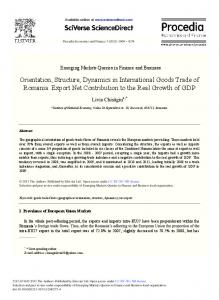
localization of virus infection (Cohen and Loebenstein, 1975; Roberts, 1982), i.e., other active antiviral reactions should be responsible for the virus localization (for reviews, see Van Loon, 1982, 1983; Loebenstein et al., 1982). It was concluded by Van Loon (1982) that “the localizing mechanism is operative in neighboring not yet infected cells rather than in the cells in which the virus is actively multiplying”; however, it is widely accepted that the resistance localizing the spreading of the virus is based on the inhibition of virus replication but not on the suppression of the TF as such. The lesion size is probably determined by several factors: (1)the efficiency of the virus-specific TF as the performance of the transport gene; (2) the efficiency of the virus in inducing the defense reactions of acquired resistance (AR) in the host plant; (3) the efficiency of the plant antiviral reactions; and (4) the rate of necrotization and callose accumulation in the infected cells. In this connection an obvious advantage is offered by the fact that AR and antiviral defense, as well as the very lesion formation in N-gene-carrying plants, are temperature-sensitive; no lesions are formed a t 32 -33°C and the infection spreads systemically (Samuel, 1931; Jockusch, 1966~). Fraser (1983) reported that, similarly to the N gene, the hypersensitive reaction of the N’ gene is temperature-sensitive. This circumstance makes it possible to estimate the efficiency of virus transport under switched-off reactions of AR and lesion formation (32-33°C) with subsequent transfer of the plant to 22- 24°C to restore the necrotic reaction (Jockusch, 1966a). The temperature-shift treatment (TST) permits, after the formation of the primary lesion (3 days at 22”C),necrotization to be suppressed (2 days a t 32-33°C) so as to allow the radial systemic spreading of the infection. Upon subsequent transfer of the plant back to 22°C the necrotization is again switched on, entailing an appreciable expansion of the lesion owing to the formation around the primary lesion of an aureole-an additional zone of cells taken over by the wave of virus transport a t 32-33°C and necrotized upon the return to 22°C. TST is a convenient means of testing the t s phenotype in transport. By the use of TST it is easy to establish, for instance, that the Nil18 TMV mutant ts in the coat protein is trin the transport function (active in spreading under switched-off necrotization at 32 -33°C). On the other hand, Ni2519 and Lsl behave in this test as ts in transport (lack of lesion growth after 32-33°C) (Figs. 1and 2). The efficiency of short-distance transport can be also assessed by immunofluorescence microscopy. In this case the spreading of the viral infection from cell to cell is visualized by detecting the viral antigen with fluorescent antibodies. Under blocked infection transport this method
PLANT VIRUS-SPECIFIC TRANSPORT FUNCTION
321
FIG.1. Lesions produced in an inoculated leaf of Xanthi-nc plants kept for 5 days in a greenhouse at 22 to 30°C. The right half was inoculated with Lsl(O.1 pg/ml) and the left half with L (0.1 pg/ml). Courtesy of Dr. M. Nishiguchi. FIG.2. Lesions produced in a leaf of a Xanthi-nc tobacco plant kept a t 22°C for 3 days, then at 32°C for 2 days, and finally a t 22°C for 1day. The upper half was inoculatedwith Lsl (0.1 pg/ml) and the lower half with L (0.1pg/ml).
reveals mainly individual infected (antigen-containing) cells; on the other hand, under conditions of active transport the fluorescent antibodies stain groups of infected cells (Nishiguchi etal., 1980). The most proper procedure to estimate the efficiency of short-distances (1978). The leaves transport in infected tissue is that of Nishiguchi etal., of a systemic host are inoculated with the virus and kept under conditions optimal for the onset of infection and initial transport. Then disks are cut out of the inoculated leaves and incubated for some more time; the controls are disks without such incubation. The efficiency of virus transport in infected tissues is judged from the increment in the percentage of virus-infected (antigen-containing) cells in a disk. The determination of the short-distance-transportrate of the virus on the basis of the increase in the
322
J. G. ATABEKOV AND YU. L. DOROKHOV
number of infected cells in the disk per unit of time is a quite proper technique, yet it is desirable to combine it with the assay of infectivity under the same conditions related to the number of infected cells. C. The Structure of Tobacco MosaicVirusGenome and theVirus-Coded Transport Function 1. Molecular Organization of TMV Genome
The structure and expression of the TMV genome have been discussed in detail in a number of reviews (Atabekov and Morozov, 1979; Davies, 1979;Hirth and Richards, 1981;Davies and Hall, 1982;Van Vloten-Doting and Neelman, 1982). This issue is considered below only in the aspect of the transport of the virus genetic material from infected to neighboring healthy cells as a function encoded in the genome of the virus itself. In addition to normal host products preexisting in the healthy cell, two types of products participating in virus replication arise owing to the viral infection and expression of the virus genetic material: (1)virus-specific proteins, i.e., the proteins coded by the viral genome, and (2) virus-induced proteins, i.e., those coded for by the cell genome but activated upon viral infection. It can be assumed that the synthesis of virus-induced proteins is either stimulated or derepressed by infection but they are invariably coded for by the host cell. Induction of this type of products can be exemplified with interferon formation in virus-infected animal cells and with the production of the so-called pathogenesis-related proteins in virus-infected plants (for review, see Van Loon, 1983). Unfortunately, it is difficult to discriminate experimentally between virus-coded and virusinduced proteins. The genomic TMV RNA directs in cell-free systems from wheat embryos or rabbit reticulocytes the synthesis of two polypeptides (MW 130 X lo3 and 165 X lo3) with overlapping amino acid sequences (Knowland etal., 1975; Beachy etal., 1976; Pelham and Jackson, 1976; Pelham, 1978). No synthesis of the TMV coat protein (MW 1978; Scalla etal., 17,500) is observed upon translation of the genomic RNA (Hunter etal., 1976;Zaitlin etal., 1976);its synthesis inuiuorequires formation of a short subgenomic RNA (LMC, low-molecular-weight component), which directs the coat protein synthesis in a cell-free system as well (Jackson etal., 1972; Siege1 etal., 1973, 1976; Hunter etal., 1976). In infected leaf tissues, besides LMC and genomic TMV RNA, a heterogeneous population of RNA molecules is synthesized with molecular weights ranging from 0.6 to 1.6 X lo6,which is at least partly present in the total RNA isolated from TMV virions (Whitfeldand Higgins, 1976;Zaitlin
PLANT VIRUS-SPECIFIC TRANSPORT FUNCTION
323
etal., 1976;Beachy and Zaitlin, 1977). The subgenomic RNA with molecular weight around 0.68 X lo6is designated as intermediate-size RNA 2 (I, RNA), and the set of RNAs with molecular weights of 0.9- 1.6 X lo6as I, RNA (Zaitlin etal., 1976; Beachy and Zaitlin, 1977). LMC, I,, and I, RNAs were shown to have a common 3’-terminal nucleotide sequence with the genomic TMV RNA (Zaitlin etal., 1976;Beachy and Zaitlin, 1977). In a cell-free protein-synthesizing system RNAs I, and I, direct the synthesis of a 30,000-MW polypeptide having no common 1976; amino acid sequence with the TMV coat protein (Beachy etal., 1976; Beachy and Zaitlin, 1977). This Bruening etal., 1976; Zaitlin etal., polypeptide is also revealed in the TMV-infected leaf tissue (Leonard and 1983) and protoplasts (Watanabe etaL,1983; Zaitlin, 1982; Joshi etal., Ooshika etal., 1983). Thus, TMV can produce in uiuoat least four polypeptides with molecular weights of 165,000 and 130,000, 30,000, and 17,500 (coat protein), whose synthesis is directed by genomic RNA, subgenomic I, and I, RNAs, and LMC, respectively (Fig. 2). It is not excluded that in virus-infected plant cells “chymeric” proteins may be synthesized, of which the amino acid sequence is coded partly by the viral and partly by the host-cell genome. This fantastic assumption stems from the analysis of the so-called H protein (MW 26,500) found in the TMV preparation in the amount of a single molecule per virion (Asselin and Zaitlin, 1978). Studies of the peptide structure of this protein testify to the presence in its composition of all the peptides of the coat protein and of another protein of host origin (Collmer and Zaitlin, 1983; Collmer etal., 1983). The mechanism giving rise to such a protein is unknown. The complete nucleotide sequence has been reported for the RNA of TMV uulgare (Goelet etal., 1982); considerable parts of RNA of cowpea have been TMV strain C, and tomato strain OM very similar to uulgare sequenced by Japanese workers (Meshi etaL,1982a,b,c). Examination of the nucleotide sequences of TMV RNA (common strain) showed the presence of three open reading frames in the virus genome (Fig. 3). The first open reading frame corresponds to the protein of molecular weight 125,941 (130K protein) which terminates at the UAG triplet at residue 3417 from the 5‘ end, and its readthrough product is a protein of molecular weight 183,253 (165K protein). The right-hand portion of the cistron coding for the 130K and 165K protein overlaps by five codons with a reading frame, in the second phase, that codes for a protein of molecular weight 29,987 (30K protein). The gene coding for the 30K protein is terminated by the UAA triplet two nontranslated nucleotides before the initiator codon of the coat protein (Guilley etal., 1979; Goelet etal., 1982). The nucleotide sequences
324
J. G . ATABEKOV AND YU. L. DOROKHOV amber terminator UA6
mG ’
p 130
I
r
PI60
T I I
P 87
7 r
RNA molecule =translation initiation m’G = cap I = translation termination t = aminoacylating n = protein p = MW x 10-3 CP= coat protein =
t
V I
.
;
P 83
I
.
I
. P46
,
.
’
’
’
P30
CP(P17)
0
FIG.3. A tentative map of the TMV (common strain) genome. For explanation see text.
coding for the 30K protein of common and cowpea C, strains have been compared by Meshi etal. (1982a,b). It turned out that the 30K protein of the C, strain is 15 residues longer than that of the common strain. In contrast to the common strain, the 30K cistron of the C, strain overlaps with the coat protein gene and is terminated within its initial region. Therefore, the 30K gene of the C, strain overlaps by its left portion with the 165K gene and by its right portion with the coat protein gene. In addition to LMC and subgenomic RNA coding for the 30K protein, a family of 3’-coterminal and colinear messengers (probably subgenomic RNAs) have been found by Goelet and Karn (1982). These RNAs can be
PLANT VIRUS-SPECIFIC TRANSPORT FUNCTION
325
encapsidated (since they contain the origin of assembly) and code for the proteins of molecular weights in the range 20,000-90,000. A set of proteins can be translated in uitro from the encapsidated subgenomic RNAs, including those of molecular weights 30,000,29,000 and 23,000 which have overlapping COOH-terminal sequences (data of T. Hunter, R. J. Jackson, and D. Zimmern, referred to by Goelet et al., 1982). The origin and significance of the population of 3'-colinear subgenomic TMV RNAs is obscure. It should be noted that there is no direct evidence that all the members of this population are functionally essential and code for functionally active TMV-specific proteins. It cannot be excluded that at least partly these RNAs are produced by inefficient termination of the transcription of the genomic RNA into negative strands with subsequent synthesis of the positive strands to yield double-stranded (ds) RNA. It is noteworthy that a set of discrete dsRNAs presumably serving as the replicative form (RF) for subgenomic RNAs (sub-RF) have been isolated from TMV-infected cells (Zelcer and Zaitlin, 1981; Dawson and Dodds, 1982), including the sub-RF for the 30K protein gene. Interestingly, no sub-RF corresponding to LMC has been found. A reasonable suggestion is that with TMV and some other plant viruses (Smit and Jaspars, 1982; Nassuth et al., 1983) the subgenomic RNAs coding for the coat protein are not produced by autonomous replication through sub-RF formation. 2. Analysis of T M V Mutants
Certain advance in studying the structure of TMV genome was achieved through analysis of mutants. It must, however, be admitted that though quite a number of TMV mutants have been described, in the overwhelming majority of cases it is difficult if at all possible to establish the connection between the phenotypic manifestation of mutation(s) and the virus-specific function affected. For instance, mutants selected by particular symptoms they produce on appropriate indicator plants (e.g., by their ability to cause lesions on N.siluestris Speg and Comes) may actually have different functions impaired, i.e., differentiating between these mutants requires other more specific tests. The properties and peculiarities of behavior of TMV mutants with mutations in different genes have been discussed in a number of reviews (see, e.g., Atabekov, 1977). Here TMV mutations are analyzed with respect t o their possible influence on the T F of the virus. a. Coat Protein Mutants. We still cannot definitely state that the coat protein has any other function(s) besides the structural one; mutation in this gene seems to have no dramatic effect on the expression of other viral genes. It is well known that in the absence of an active coat protein RNA of many viruses can replicate to form infective daughter RNAs that are not
326
J. G. ATABEKOV AND YU. L. DOROKHOV
protected from RNase action in the sap of the infected plant. Consequently, the infective principle is unstable without the coat protein (Jockusch, 1966a). Various forms have been described of “defective” mutants in the coat protein that are deficient in their ability to produce mature TMV particles. Siegel etal. (1962) isolated two unusual TMV mutants deficient in production of mature TMV particles. No virus-like nucleoprotein particles could be isolated from plants infected with these mutants; in the cells of such plants the viral RNA is not protected with the coat protein. These coat protein mutants were designated as PM1 and PM2. Defective mutants spread from cell to cell but usually remain within the inoculated leaves. The long-distance spreading of the infection from the lower inoculated leaves to the upper ones occurs extremely slowly (Siegelet aL, 1962;Sarkar, 1969;Kapitsa etaL,1969;Bhalla and Sehgal, 1973). The deep suppression of the long-distance transport is characteristic of all TMV mutants defective in the coat protein, which “tend to travel in vascular parenchyma, and do not enter the conducting elements or are inactivated there” (Siegel and Zaitlin, 1964). The causes of suppressed long-distance transport in the absence of the coat protein are discussed below (see Section IV). Recently a new interesting mutant (DT-1G) defective in the coat protein has been described which differs from all previously known by its complete inability to produce the coat protein (Sarkar and Smitamana, 1981a,b). Irrespective of the causes of the absence of the viral protein, the DT-1G mutant offers an attractive model for studying the influence of the coat protein on the transport function. It turned out that the DT-1G infection lacks symptoms and the infective principle is present in the green tissues of the infected leaf (Gfrom “green”). This prompts a conclusion that even mutation(s) causing the complete abolishment of the synthesis of the TMV structural protein do not preclude the short-distance transport of the virus, i.e., the coat protein is not indispensable for the expression of the TF. It cannot be excluded, however, that in this case the TF is also somewhat suppressed; this can be inferred only from the smaller lesions produced on leaves of Xanthi-nc tobacco by the DT-1G (as compared to vulgure) (Sarkar and Smitamana, 1981a). It is quite probable that the transport efficiency is lower simply because of the lack of mature virus particles which, albeit not obligate for the TF, nevertheless provide an additional possible form of near and long-distance transport of the infection. In the systemic spreading the virus may probably use different forms of particles to transfer the viral genome, which functionally complement each other. We should specially dwell on the effect of temperature-sensitive ( t s ) mutations in the coat protein gene on the expression of the TF.
PLANT VIRUS-SPECIFIC TRANSPORT FUNCTION
327
Jockusch (1966a) showed that various TMV ts mutants fall into two classes: 1. The tsmutants of class I are typically represented by the mutants Nil18 and flauumand possess the following characteristics. A t nonpermissive temperatures viral particles are formed in negligible amounts; the infectivity of the extracts at 30-33°C is low or entirely lacking. Under nonpermissive conditions, cells infected with tsImutants accumulate infectious viral RNA unprotected or partially protected by the protein, i.e., the situation is in principle similar to that reported for plants infected with defective mutants of the PM series (see above). The behavior of the ts1 mutants at high temperatures and of the constitutively defective PM mutants is also similar in respect of the T F their long-distance transport is suppressed but they retain the cell-to-cell transport, although it may also be partly inhibited. Hence provisionally both these types of mutants (PM series and tsI) can be regarded as defective in the coat protein synthesis but active in the short-distance transport and RNA replication. For several other plant viruses the coat protein gene has been shown to be not essential for viral RNA replication (for references, see Sarachy etaL, 1983). 2. TMV tsmutants of class I 1contain a mutation in a portion of the virus genome beyond the coat protein gene. Informaton concerning the properties and functions of nonstructural proteins encoded in the TMV genome is quite scanty. Class I 1 mutants are considered below.
b.Non-Coat-Protein Mutants.We do not know for certain how many other genes essential for the virus replication the TMV RNA contains besides the coat protein gene. Unfortunately, in those rare cases when the mutant can be assigned to the tsII class in Jockusch’s terminology, it is rather difficult to reliably identify the affected gene(s). Among the tsmutants deficient in the nonstructural protein function there are grounds for discerning at least two types of mutations whose phenotypic expression may affect the TF. i.Mutations which influence thesynthesis of genomic andlor subgenomic RNAs. According to the classical scheme, the parental plus-strand RNA molecule serves as a template for the synthesis of RNA chains through formation of dsRNA (RF) and partial dsRNA, or the replicative intermediate. Subsequently the minus-strand RNA direct the synthesis of complementary daughter RNA plus chains. It is assumed that a virus-specific enzyme, RNA-dependent RNA polymerase (or RNA replicase) is responsible for the replication of the virus genome. More exactly, viral RNA is replicated in the so-called replication complex consisting of viral RNA, virus-specific and/or cell-specific proteins. It can be concluded that an indispensable function in the replica-
328
J. G. ATABEKOV AND YU. L. DOROKHOV
tion of plant virus RNA is performed by some nonstructural virus-coded protein(s). This conclusion is supported by the existence of mutants conditionally lethal in this function (Dawson and White, 1978,1979;Dawson, 1983). Unfortunately, no definitive evidence has been obtained on the structure and even the origin of the RNA replicase activity in the virus-infected plant (for references, see Dorssers etal., 1983). A number of indirect facts accumulated in recent years argues in favor of the host (and not the virusspecific) origin of the enzymic system providing for the synthesis of viral RNA in the infected cell. Firstly, some (albeit rather low) RNA-dependent RNA polymerase activity was found in the healthy cells of some plants (for review, see Ikegami and Fraenkel-Conrat, 1979;Dorssers etal., 1983; Fraenkel-Conrat, 1983), and preparations of RNA polymerase isolated from healthy and from TMV-infected cells proved similar in several respects (Romaine and Zaitlin, 1978). Second, RNA polymerase preparations isolated from plants infected with different viruses did not show template specificity (Semal and Hamilton, 1968; May etal., 1970; Zaitlin et al., 1973; Weening and Bol, 1975; Le Roy etal., 1977), i.e., showed no significant preference for the RNA of the infecting virus as template. Third, the products of polymerase reaction, when analyzed, were shown to be small and heterogeneous (for review, see Miller and Hall, 1983). On the other hand, a considerable body of evidence suggests that viruscoded product(s) participate in the replication of plant virus RNA (De Jager etal., 1977; Dawson and White, 1978, 1979; Hardy etal., 1979; Goldbach et al., 1980;Robinson et al., 1980; Nassuth et at., 1981;Bujarski et al., 1981; Nassuth and Bol, 1983; Sarachy etal., 1983). Dorssers etal. (1983) showed that two different RNA polymerase activities are operative in CPMV-infected cowpea, one associated with replicating viral RNA and the other representing the host-coded RNA-dependent RNA polymerase system. Very important data of Hall and co-workers (Bujarskiet al., 1982; Miller and Hall, 1983) show that RNA 1 of brome mosaic virus (BMV) codes for the template-specific active viral replicase. The question of the coding of the TMV-specific polymerase remains obscure, most probably because of the peculiarities of the system and the difficulties of detecting minor TMV-coded protein( s) in RNA replicase preparations isolated from TMV-infected plants. The facts available at present allow of three possibilities: (1)The TMV RNA replicase is a virusinduced protein, i.e., its synthesis is directed by the cell genome but activated upon viral infection; (2) the RNA replicase is a virus-specific product, encoded in the viral genome; (3) the RNA replicase system is of mixed origin and contains, besides the cell components (which is characteristic of many RNA replicase systems), the virus-specific protein(s) as well, probably as minor constituents.
PLANT VIRUS-SPECIFIC TRANSPORT FUNCTION
329
It is not clear whether any connection exists between the mutations affecting the replication of viral RNA and the virus TF. No information is available in the literature on the dependence of the transport rate on the rate of synthesis of virus components (RNA, coat protein) in the infected cell. The lack of such information complicates the interpretation of a number of facts; in particular, it would have substantially clarified the central question whether the transport is an active virus-coded function. An adherent of the passive transport, De Zoeten (1981) claimed that a membrane-associated dsRNA (the replication complex) is the form in which viral RNA can be transported from cell to cell, and speculated that the “transport of infective entity must be achieved in a passive manner” following “the movement of the normal cell constituents.” Assuming that the transport of the infective principle (as free viral RNA or in another form) is passive, there should be a dependence between the efficiency of the synthesis of viral RNA and the rate of the cell-to-cell transport of infection. On the other hand, it is quite obvious that in any case the ts mutations affecting the replication of genomic RNA would inevitably manifest themselves as temperature-sensitive in the TF; mutations suppressing the synthesis of genomic RNA and mutations in transport would be phenotypically similar. Therefore the question can be rephrased: are there real mutants in TF, or does the decline in the transport rate merely reflect the decreased synthesis rate of genomic TMV RNA? If the TF is performed independently from RNA replication (i.e., by the protein product of a special transport gene), then mutants must exist that are ts in transport by trin RNA replication. TMV mutants temperaturesensitive only in transport have indeed been described; the genomic RNA of such ts mutants is normally replicated but the virus transport is inhibited at nonpermissive temperature (see further). It may be assumed that genomic TNA replication and the synthesis of subgenomic TMV RNAs are two independent enough processes, i.e., the synthesis of subgenomic RNAs is controlled by a particular virus-specific protein. If this suggestion is correct, one should expect mutations in the TMV genome affecting the synthesis of different subgenomic TMV RNAs (LMC and I-class RNAs) but not of the genomic RNA. Would such mutations tell on the transport function? Since the production of the TMV coat protein is not obligate for the virus transport, a mutation suppressing the synthesis of the subgenomic LMC RNA is hardly likely to have appreciable influence on the TF. On the other hand, mutations affecting the synthesis of the I-class subgenomic RNAs can be thought to have basically different phenotypic manifestations. Earlier we have suggested (Atabekov and Morozov, 1979) that the 30K protein, the product of translation of the intermediate-class
330
J. G. ATABEKOV AND YU. L. DOROKHOV
subgenomic RNAs, is the transport protein. If this is so, the transport function would be inhibited to an equal extent by (1)mutations causing the ts behavior of the transport gene product and (2) mutations suppressing the synthesis of subgenomic RNAs coding for this protein. From this it followsthat with the ts mutations exhibiting retarded transport there are at least three conceivable causes giving rise to this phenotype: (1)temperature sensitivity of the synthesis of genomic RNA, (2) temperature sensitivity of the synthesis of subgenomic RNA coding for the putative virus-specific transport protein, and (3) temperature sensitivity of the transport protein as such. The idea of the existence of a special transport gene independent of the RNA replicase gene(s) is compatible with the results of the studies on the expression of the multipartite genome of CPMV (see a review by Jaspars, 1974). The heavy (B) component of CPMV RNA was known to code for functions related to viral RNA replication (Goldbach et al., 1980). Recently Rezelman etal. (1982) showed that B-RNA is capable of infecting cells and replicating there; however, the infection is not transferred to adjacent cells since the TF is encoded in the other genomic component, the light CPMV RNA (M-RNA). Thus the replicase and the transport protein are coded for by B-RNA and M-RNA, respectively. It is of interest that in another bipartite virus, the tobacco rattle virus (TRV), both these functions (RNA replication and transport) are encoded in the heavy RNA. Inoculation of plants with the heavy TRV component results in RNA replication and systemic spreading of the infection which exists in an “unstable form” (Sanger and Brandenburg, 1961; Cadman, 1961). ii. Mutations in thetransport gene. At least three TMV mutants temperature-sensitive in the function of systemic spreading have been isolated Ni2519 (Jockusch, 1968), 11-27 (Peters and Murphy, 1975), and Lsl (Nishiguchi et aL, 1978). It seems reasonable to assume that acertaingene product nonfunctional at restrictive temperatures is responsible for this effect. Strain Lsl has been derived from the temperature-resistant tomato strain L (Nishiguchi et aL, 1978); its temperature sensitivity manifests itself as the block of cell-to-cell transport of infection at nonpermissive temperatures (32- 33“C). Under such conditions the virus replicates only in the primarily infected cells. It must be emphasized that Lsl is not ts in either RNA replication, coat protein synthesis, or virion assembly; the virus normally infects and reproduces in cultured protoplasts at 32’ C, the temperature nonpermissive for Lsl in the intact plant. Moreover, at restrictive temperature Lsl accumulates in infected cells in greater amounts than at permissive temperature (22”C),i.e., its genomic RNA is even more actively replicated at
PLANT VIRUS-SPECIFIC TRANSPORT FUNCTION
331
higher temperatures. Taking into account that the TF under such conditions is completely inhibited, one can suppose that the transport of the viral genome into neighboring cells does not directly depend on the amount of genomic RNA produced in the primarily infected cell but is controlled only by the transport gene(s). The rate of short-distance transport in tissues infected with a trstrain L was found to be the same at 22 and 32 C although at 32 C the virus accumulated in infected cells in substantially larger quantities than at 22 C (Nishiguchi et al., 1978). It is not excluded that the transport depends on RNA synthesis only at certain critical RNA concentrations in the infected cell. There may be a minimal threshold RNA concentration that must be attained for the transport to become possible. It remains utterly obscure, however, why after overcoming the threshold further increase in the viral RNA concentration within the cell does not accelerate the transport of strain L temperature-resistant in this function. If the lack of dependence between the rates of transport and RNA synthesis would be proved, the transport of infection from infected to healthy cells could be thought not to be a continuous process concurrent with RNA replication, but to take place probably in a single step at a moment when the total amount of RNA is relatively small; after this the virus transport from this particular cell stops though the synthesis of the viral RNA goes on. This is in accordance with the suggestion that the 30K protein is the most probable candidate for the transport protein (Atabekov and Morozov, 1979;Leonard and Zaitlin, 1982). Ooshika et al. (1983)and Watanabe et al. (1983)succeeded in detecting the 30K protein and its mRNA in TMV-infected protoplasts and studied their time course in viva The synthesis of the 30K protein was first detected in 3- 4 hours after inoculation (before infectivity appeared). However, the 30K protein and its mRNA were transiently synthesized in 2 to 3 hours after inoculation and gradually declined afterward. In contrast, the 130K,180K,and coat protein, as well as genomic RNA and LMC, were synthesized continuously. These data suggest first that the synthesis of subgenomic RNAs of different classes is controlled by different mechanisms and, second, that the 30K protein is unstable and degrades or becomes inaccessible for the extraction relatively soon after inoculation. If the 30K protein is indeed the transport protein the T F would cease to operate simultaneouslywith this event. The transport rate may be independent of the rate of genomic RNA replication for some other reasons. For instance, the transport of the viral RNA into adjoining cells at a certain stage of virus replication may compete with virion maturation: the “flux” of the newly synthesizedviral RNA changes its direction and is switched over from the transport to the assembly of the viral particles, which probably do not participate in transport.
332
J. G. ATABEKOV AND YU. L. DOROKHOV
It can also be assumed that the transport form of infection (see further) contains a certain host or virus-specific protein necessary for its functioning. Then the TF is controlled not by the RNA synthesis rate but by the availability and amount of this hypothetical protein. Unfortunately, we are practically ignorant of the properties and functions of the transport protein. Leonard and Zaitlin (1982) carried out a comparative peptide analysis of the 30K proteins directed in a cell-free protein-synthesizing system by RNAs from two TMV strains: L (wild type) and ts-transport mutant Lsl; a small difference was found in the peptide maps of the 30K protein coded for by these viruses. The nucleotide sequences of the 30K protein cistrons of L ( t rand ) Lsl (ts in TF) strains have been recently compared by Ohno etal.(1983). The authors found a single base substitution which caused the replacement of a proline in the L strain 30K protein by serine in the Lsl 30K protein. Detection of a single amino acid substitution in the 30K protein of Lsl compared to that of the L strain is not enough to state that this protein is the product of the transport gene. This result cannot yet be linked to the T F for several reasons. First, the 30K protein coded for by Lsl RNA was not shown to be temperature-sensitive and, second, and most importantly, there is yet no direct proof that the 30K protein performs the transport function. If nevertheless the 30K protein does perform the TF, it must be noted that different TMV strains differ dramatically in the amino acid sequences of their 30K proteins. The amino acid compositions of the 30K proteins of several TMV strains (uulgare, OM, C,, L) have been deduced from the 1982a,b;Goelet etal., nucleotide sequence data on TMV RNA (Meshi etal., 1982; Takamatsu etal., 1983; Watanabe etal., 1983). The 30K proteins coded for by different TMV strains may strongly differ in the content of particular amino acids (Watanabe etal., 1983). For example, the 30K of OM TMV contains 11methionines in the total 267 amino acid residues; in contrast, the 30K of the C, strain has only three in the total 282 (Watanabe etal., 1983). 3.Complementation of tsTransport Function by trHelperVirus
Various forms of complementation between plant viruses have long been known: during mixed infection the partner viruses provide each other with the gene products which can rescue the defective virus (for review, see Dodds and Hamilton, 1976). Pertinent examples are the phenomena of satellitism of viruses (for review, see Atabekov, 1977)and the replication of viruses with fragmented genomes (Jaspars, 1974). The possibility of complementation between plant virus mutants was first demonstrated with a number of TMV mutants in this group (Shas-
PLANT VIRUS-SPECIFIC TRANSPORT FUNCTION
333
kolskaya etal., 1968;Atabekov etal., 1970a,b,c) and then reproduced (Sarkar, 1969) and confirmed (Kassanis and Bastow, 1971a,b; Kassanis and Conti, 1971). Upon mixed infection of plants with two TMV strains one of which produces a tsand the other a tr coat protein, their complementation can take place under conditions nonpermissive for the ts mutant alone. This gives rise to virions with a so-called masked genome (mixed virus particles in which the RNA of the tsmutant is encapsidated with the coat protein of the trhelper). Later we used in the complementation analysis the ts TMV mutant Ni2519 (Taliansky etal., 1982a) that has no mutational defect in the coat protein gene. This non-coat mutant was derived from the natural TMV strain A14 and described as ts (Jockusch, 1968; Bosch and Jockusch, 1972). A more detailed characteristic of this mutant was given by Taliansky etd.(1982a,b) and Kaplan etal. (1982). At nonpermissive temperature (33°C) the assembly of Ni2519 particles is blocked (Bosch and Jockusch, 1972). It has been shown that in uitro the Ni2519 RNA can be reconstituted with the coat protein, yet infectious virions are formed only at permissive temperature; at restrictive temperature the assembly gives rise to defective particles. The aberrant assembly was found to result from the ts behavior of the Ni2519 RNA molecule itself, its conformational instability at 33°C. Under such conditions the assembly of the virus particles is initiated not from a single initiation site (as with trstrains or with Ni2519 RNA a t permissive temperature); at 33°C an additional assembly initiation site is demasked in the Ni2519 RNA molecule directly in the coat protein gene (Kaplan etal., 1982; Taliansky etal., 1982a,b). Thus, Ni2519 is a representative of a new peculiar class of ts virus mutants. Taking into account this peculiarity of Ni2519, it was logical to suppose that its assembly function would not be complemented by any helper virus. Indeed, complementation of the assembly of Ni2519 was shown to be impossible (Taliansky etal., 1982a). However, Ni2519 is ts not only in the virion assembly. Two distinct virus-specific functions, namely virus assembly and spreading of infection from cell to cell (TF),are t s in Ni2519 (Bosch and Jockusch, 1972). It has been recently shown by Taliansky etal. (1982a,b) that the ts TF of Ni2519 can be complemented by a helper virus. The experimental system for the complementation of virus spreading was as follows. The N'-gene-carrying plants used developed local lesions in response to Ni2519 (dependent ts virus) but formed a systemic reaction upon infection with a common TMV strain uulgare (helper tr virus). The leaves were jointly infected with a mixture of the viruses, and the plants were subjected to the TST. Since Ni2519 alone is incapable of spreading systemically a t 33"C, the local
334
J. G. ATABEKOV AND YU. L. DOROKHOV
lesions produced by Ni2519 could increase upon TST only by virtue of complementation by the helper virus which can spread at high temperatures. The appearance of lesions surrounded by a collapsed area testified to the complementation of the cell-to-cell transport of Ni2519 at 33°C. Obviously the Ni2519 infection spreads under these conditions in a form other than mature virions, as its assembly cannot be complemented by the helper virus (Taliansky etal., 1982a). In another work (Taliansky etal., 1982c) uae was made of the ts mutant Lsl which is temperature-sensitive only in TF but normally multiplies at nonpermissive temperature in primarily infected cells and protoplasts. Complementation of the cell-to-cell movement of Lsl in the presence of a trhelper was tested either by the TST of necrotic N'-gene-carrying plants (as above) or by immunofluorescence microscopy. The results in a general form are given in Table I. It is important that the role of the helper virus in transport can be played not only by a related TMV strain but also by a quite unrelated, foreign virus that can systemically infect the particular host. Thus potato virus X (PVX) could serve as a helper for TMV-Ls1 in the cell-to-cell movement. The next experimental system used for complementing the TF in Lsl TABLE I COMPLEMENTATION OF t s TRANSPORT FUNCTION BY tr HELPER VIRUS** Virus ts TMV
Lsl
strains 33
24
Ni2519
33
24
trhelper
TMV-UI Potato virus X
+
Transport of the ts T("C) virus from cell to cell
ts tr Lsl UI Lsl potato virus X Ni2519 UI
+ +
+
33
24 33
24 33 33 33
+ + + + + +
+
Summarized results of Taliansky etal.(1982a,d). (+) and ( - ) correspond to effective and blocked transport, respectively; the trhelper viruses spread normally at both temperatures used.
PLANT VIRUS-SPECIFIC TRANSPORT FUNCTION
335
was extremely simple; yet it served its purpose. It is well known that it is impossible to infect the upper leaves of a healthy plant when the virus is imbibed through the cut stem. The virus moves along the conducting tissues of the stem (probably through the xylem), accumulates there, but cannot move to the parenchyma and infect the leaf. It was assumed that the penetration of the virus from the conducting tissues into the leaf parenchyma is a transport event, which is blocked since the TF cannot be expressed without virus replication. Therefore the influence of preinfection of the upper leaves with a helper virus (which systemically spreads there) on the transport of the dependent virus from the conducting tissues to the leaf parenchyma was studied. Such preinfection before imbibing the dependent virus into the stem was found (Taliansksy etd,1982c,d) to promote its penetration into the mesophyll of the preinfected leaf and its spreading there. The systemic infection of the plant upper leaves with the helper virus appeared to “open the gates” for the transport of the dependent virus. The helper virus renders the leaf susceptible for systemic infection with the dependent virus that otherwise - when alone- would be confined to the conducting tissues (Table 11). As can be seen from Table 11, the TF of the dependent virus can be complemented in this system not only by strains of the same virus (TMV) TABLE I1 HELPER VIRUSPROMOTES THE TRANSPORT OF DEPENDENT VIRUS** Dependent virus imbibed through the stem
Helper virus: preinfection of upper leaves
TMV U2
TMV UI No helper Potato virus X No helper TMV Nil18 ( t s coat protein, trtransport) No helper TMV L (tr transport) No helper TMV LsI (ts transport)
TMV L TMV Lsl ( t s in transport) TMV Lsl TMV L
Transport and
T replication of ( C) dependent virus 24 24
33
+++ -
24 - 33
+
24 33
Summarized results of Taliansky etal. (1982a). and (-) correspond to effective and blocked transport function. a
* ( +)
336
J. G. ATABEKOV AND YU. L. DOROKHOV
but also by an unrelated helper (PVX). Thus, the T F is not very specific since an unrelated virus can assist the spreading of the dependent virus over the leaf. It would be interesting to identify and compare the transport proteins coded for by TMV and PVX; it is not excluded that the transport proteins of different viruses (capable of systemic spreading in the same host species) have basically different properties. Nevertheless such proteins must have some common functional specialties determining their transport faculty. It is evident from Table I1 that TMV Lsl can be used as a dependent virus, and its transport at nonpermissive temperature can be complemented with TMV Nil18 (ts coat protein, trtransport) or with its parental strain L. The Lsl was used not only as a dependent virus but also as a helper at restrictive and permissive temperature. As can be seen (Table 11),Lsl can serve as the transport helper for TMV L only at low temperature (24°C) but is ineffective at 33°C. It is noteworthy that when the upper leaves are preinfected with Lsl (helper) at 24°C and then the upper portion of the plant is cut off and transferred to 33' C, placing the stem into the suspension of TMV L (dependent virus), the ability of Lsl to assist the transport is lost not immediately but within several hours after the transfer from 24 to 33°C (Taliansky etal., 1982~).It seems likely that at restrictive temperature the t s transport protein of Lsl is rendered nonfunctional and its activity is gradually lost. 4.Interaction betweenViruses Possibly LeadingtoTF Complementation
Numerous different forms of nongenetic interaction between plant viruses under conditions of mixed infection have been repeatedly reviewed (see, e.g., Kassanis, 1963; Dodds and Hamilton, 1976; Matthews, 1981). The multiplication rate and/or distribution of one virus are often enhanced in the presence of another. A classical example of the synergism between plant viruses is the stimulation of PVX replication in double infection with different unrelated viruses potato virus Y (PVY), TMV, tobacco etch virus, cucumber mosaic virus (CMV). The increase in PVX concentration is attended by more severe symptoms (Rochow and Ross, 1954, 1955; Stoffer and Ross, 1961a,b). This phenomenon might be ascribed to the increased rate of PVX cell-to-cell transport in the presence of a helper virus; however, it has been found that (1)the rate of PVX transport remains practically the same in single and double infection, and (2) PVX is stimulated in the presence of TMV upon mixed infection of isolated protoplasts (M. E. Taliansky etal., unpublished data). And still, the interaction between PVX and PVY seems to involve complementation of the TF as well. According to Close (1962),at 31°C PVX replication was restricted to the inoculated leaves, i.e., the long-distance transport was
PLANT VIRUS-SPECIFIC TRANSPORT FUNCTION
337
inhibited. However, PVX spread systemically a t 31°C in plants doubly infected with PVX and PVY. It is of interest that the transport of PVX in plants kept at 31°C could also be assisted by TMV and some strains of tobacco ringspot virus and CMV (Close, 1962). The experiments of Rochow and Ross (1955) and Close (1962) support the suggestion that the phenomenon described by them represents the complementation of TF; the transport of PVX from parenchyma into phloem (which is inhibited in single infection, especially at high temperature) can be nonspecifically complemented by unrelated viruses that are active in transport at this temperature. Some examples of synergism between strains reviewed by Kassanis (1968) are probably based on the effects of TF complementation. Thus the necrotic strain of tomato spotted wilt virus (TSWV) acquired the ability to spread systemically over the potato plants in the presence of the ringspot TSWV strain (Norris, 1951) which appeared to act as a helper virus complementing the TF. One more example of interaction between viruses that can be regarded as TF complementation was reported by Benda (1957) who used two TMV strains for mixed inoculation of N. syluestris plants. The common TMV spread systemically while the yellow aucuba (YA) remained confined to the inoculated leaf, producing local lesions. The plants infected with both strains developed yellow spots produced by YA in the presence of the common strain. It seems reasonable to think that the common TMV helped the YA strain in the TF, providing for its systemic spreading. There are some variants of barley yellow dwarf virus (BYDV) confined to the phloem in oats. However, double infection with two different BYDV variants predisposed the xylem to infection (Gill and Chong, 1981). This unexpected phenomenon shows that interaction between two viruses each of which by itself is inable to penetrate the xylem can lead to the appearance of such ability, possibly as a result of the combined action of their transport proteins. 111. RESISTANCE OF PLANTS TO VIRUSES AS A PROBLEM OF TRANSPORT OF THE VIRUSGENOME FROM INFECTED TO HEALTHY CELLS
A. Extremeand Facultative Resistance of Plants toViruses The biochemical mechanisms determining the success or failure of the viral infection in the plant cell are not clear. Solution of this question is hampered by the lack of information on the functions performed by the virus and by the host plant in virus replication. It is quite probable that
338
J. G. ATABEKOV AND YU. L. DOROKHOV
the mechanisms controllingthe resistance of plants to viruses may operate at any level of virus replication, i.e., nonperformance of any function(s) may render the plant resistant to the virus. Normal development of infectionproceeds through a sequence of events: 1. First phases of infection: virus penetration, adsorption on the cell receptors, and deproteinization. 2. Replication of viral RNA and its translation into virus-specific proteins. 3. Systemic spreading of the infection from the primarily infected to healthy cells (transport). 4. Assembly of mature virions.
The first phases of interaction with the cell, which play such a prominent role in the restraint of infection and host-range control of animal viruses, turn out to be of lesser importance during plant infection by plant viruses (see reviews by Atabekov, 1975; De Zoeten, 1981). It can be stated that in most cases plant resistance to the particular virus is caused not by the blockage of the first phases of infection in the host cells but by other factors. Plant cells probably also have cell receptors specific for certain viruses (Novikovand Atabekov, 1970), however, the virus -receptor interaction does not seem to be so decisive for the development of the viral infection as with most animal viruses. It has more than once been demonstrated that the resistance of plants to viruses usually holds at the level of the whole plant but is lost at the single-cell level (for review, see Van Loon, 1983). Only in a few cases the resistance of a plant species to a particular virus is maintained in isolated protoplasts. Van Loon (1983) argued that “in general, individual plant cells are capable of replicating essentially any virus.’’ In spite of its apparent extremeness,this concept seems to be true for many host-virus combinations. The term “extreme resistance’’ may be conveniently adopted for those probably infrequent cases when the plant and isolated cells (protoplasts) prove resistant both to the whole virus and to its free RNA (ie., when the resistance is determined not by the first phases of virus replication). Accordingly,when the resistance is exhibited only at the level of the intact plant (or tissues) while the individual cell is susceptible to the virus an appropriate term would be “facultative resistance.” The same term can also be reasonably applied to those infrequent situations when the plant resistant to the native virus can be infected with a preparation of free viral RNA (see a review by Atabekov, 1975).
PLANT VIRUS-SPECIFIC TRANSPORT FUNCTION
339
Thus, the phenomenon of extreme resistance consists in the fact that the virus replication turns out to be in principle impossible in the cells of resistant plants. Facultative resistance implies that virus replication in the cells is not basically forbidden, though in the whole plant the infection is blocked by (1)a barrier for the first phases of infection, which can be overcome by infecting the resistant plants with free viral RNA; (2) impossibility of the transport of infection from the primarily infected cells where the virus can multiply (in this case the protoplasts isolated from the resistant plant are infected by the virus); and (3) the antiviral defense reactions operating in plant tissues but not in protoplasts or individual cells. Extreme resistance can be exemplified by the immunity of N . tabacurn plants and protoplasts to the wild-type strain of bromegrass mosaic virus (BMV) (Motoyoshi et al., 1974). It must be noted that even in this case some amount of BMV-susceptible protoplasts can be revealed (Sakai et al., 1983). Similar resistance is exhibited by the tomato line Tm-1 toward TMV L (strain of type 0)(Motoyoshi and Oshima, 1977) and by the Arlington line of Vigna sinensis unguiculata Walp. toward the SB strain of CPMV (Beier et al., 1977, 1979). Virus replication turns out to be impossible or profoundly suppressed in the cells and protoplasts of the plants having extreme resistance. Interestingly, extreme resistance may cover not all strains of a given virus. Thus, N . tabacum plants and protoplasts proved susceptible for another BMV strain, V5 (Motoyoshi et al., 1974),the TMV strain CH2 (a strain of type 1)can overcome the resistance of TM-1, and the plants or protoplasts of the V. sinensis unguiculata line resistant to CPMV SB are infected by CPMV strain DG (Beier et al., 1979). Extreme resistance appears to be underlied by the impossibility of carrying out the late phases of virus reproduction after the completion of the early phases. Most probably the barrier is put up at the level of replication of genomic or subgenomic viral RNAs. The obstacle can hardly be in translation since the results of numerous experiments testify to the lack of selectivity in mRNA translation by eukaryotic ribosomes (see, e.g., a review by Atabekov and Morozov, 1979). It was found that accumulation of infective TMV RNA was extremely inhibited in the presence of the Tm-1 gene in tomato, suggesting that this gene influenced the viral RNA replication (Motoyoshi, 1982). In the manifestation of the extreme resistance in protoplasts a prominent role is played by some cell substances suppressing viral replication. Thus it was observed that BMV, which is incapable of multiplying in radish and turnip mesophyll protoplasts, acquires this ability upon exposure of the protoplasts to ultraviolet light or actinomycin D (Maekawa et al., 1981).
340
J. G. ATABEKOV AND YU. L. DOROKHOV
Separate consideration should be given to those cases when the infected cell (and protoplast) isolated from a virus-resistant host nevertheless proves capable of replicating the virus, i.e., when immunity to the virus exists in tissues but not in individual cells. Quite a number of examples of this type have been described 1.TMV can infect cowpea protoplasts but not the whole plants (Huber et al., 1977). 2. BMV strain ATCC66 can infect protoplasts of Raphanus sativus L. while the plants are BMV-resistant (Furusawa and Okuno, 1978). 3. CMV can infect protoplasts of cowpea, the plant normally resistant to this virus (Kioke etal., 1977; Gonda and Symons, 1979; Maule etal., 1980). 4. Several lines of V. sinensis unguiculata were found to be resistant to CPMV-SB; upon inoculation of the resistant plants with the virus at a concentration of 250 pg/ml no symptoms develop and the virus is virtually not accumulated. The resistance is lost at the level of protoplasts: the protoplasts of all resistant lines (except Arlington) could be infected with CPMV-SB (Beier etal., 1977, 1979). 5. Tomato line TM-2 plants are resistant to TMV but their protoplasts are quite susceptible (Motoyoshi and Oshima, 1977).
All the above cases can arbitrarily be classed as examples of facultative plant resistance based on the block of systemic spreading (transport) of the virus from the primarily infected to the surrounding healthy cells in the resistant plant. It must, however, be noted that per se the ability of the virus to replicate in protoplasts isolated from the tissues where it cannot accumulate does not always mean that the host resistance is based on the block of just the virus-specific TF. The resistance in this virus-host system may be due to the inability of the virus to replicate in the host cells organized into leaf tissue (e.g., because these cells contain a certain substance impeding virus multiplication). During isolation of protoplasts the cell wall is removed and the plasma membrane undergoes fundamental changes, accompanied by partial exchange of the cell contents with the medium and probably loss of some cytoplasmic components. It is admissible that all these events alter the composition of the cytoplasm, enabling the virus to replicate in the protoplasts though this was impossible in the tissue. Rigorous proof of the fact that one is dealing with the resistance actually based on blocked infection transport for each particular virus - host system requires demonstration that the virus replicating in protoplasts can also accumulate in primarily infected cells of the intact facultatively resistant (1978) in the work plant. Such proof was presented by Nishiguchi etal.
PLANT VIRUS-SPECIFIC TRANSPORT FUNCTION
341
with the TMV ts mutant Lsl, and by Sulzinski and Zaitlin (1982)who used TMV to infect resistant plants of V.sinensis End1 and Gossipium hirsutum L. In both cases TMV replication was shown to take place only in the primarily infected cells, i.e., the plants behaved as facultatively resistant (see also Section 111,D).
B. Role of Transport Function in Virus Host Range Control The role of virus-coded TF in the host range control of a plant virus was studied by Taliansky et al. (1982d) using the experimental system described above: the dependent virus was imbibed into the conducting tissues and moved into the mesophyll cells of the upper leaves preinfected with the helper virus (Taliansky et al., 1982~). It was assumed that a certain plant species may be resistant to a given virus (be a nonhost for it) simply because the virus genetic material cannot move from cell to cell over this plant. As a result, the virus remains and multiplies only in the primarily infected cells. The plant looks like a nonhost only owing to blocked TF. Taliansky et al. (1982d) demonstrated that in several cases this type of resistance can be overcome, and the nonhost plant surrenders to infection in the presence of a helper -another virus that can normally spread in this plant. It can be seen from Table I11 that the BMV Russian strain acquires the ability to spread over the leaves of tomato (the plant facultatively resistant to BMV) in the presence of a helper virus-tomato strain of TMV. Similarly, BMV can infect beans (which are also resistant to BMV) when assisted by another helper TMV strain (bean strain, or dolichos enation mosaic virus, DEMV) which is able to systemically infect bean plants (Table 111). Dodds and Hamilton (1972) and Hamilton and Nichols (1977) demonstrated the complementation between barley stripe mosaic virus (BSMV) and TMV in barley plants (Hordeurn vulgare L. var Black Hulless) doubly infected with these viruses. A similar effect was observed when BMV was used in mixed infection with TMV (Hamilton and Nichols, 1977). As a result of this interaction, TMV systemically spread over barley plants coinfected with BSMV or BMV. Taliansky et al. (1982d) presumed that this phenomenon was based on the complementation of TMV TF by BSMV in barley where the transport of TMV was restricted. As can be seen from Table 111,BSMV served as a helper virus promoting the systemic spreading of TMV vulgare over wheat plants. It is important that similar results were obtained when the TMV mutant Lsl (ts in TF) was used as a dependent virus. Lsl was shown to be capable of systemically spreading at
342
J. G. ATABEKOV AND YU. L. DOROKHOV TABLE I11
HELPER VIRUSPROMOTES THE TRANSPORT OF DEPENDENT VIRUSOVER NONHOST PLANTS"* ~
Plant resistant to dependent virus
Dependent virus imbibed through the stem
Tomato Bean Wheat
Bromegrass mosaic virus Bromegrass mosaic virus TMV UI
Wheat Tomato Tm-2 (resistant to TMV)
Helper virus preinfection of upper leaves
Transport and T replication of ("C) dependent virus
TMV LsI ( t s in transport)
TMV L TMV bean strain Barley stripe mosaic virus Barley stripe mosaic virus
24 24 24 33
TMV L
Potato virus X
24
33
+
+ + + + +
Summarized results of Taliansky etal.(1982d). correspond to effective and blocked transport function.
* (+) and (-)
nonpermissive temperature (33"C) in the presence of a temperature-resistance helper (BSMV). These observations indicated that it was the TF that was complemented by BSMV under such conditions. Similar results were obtained when tomatoes of the Tm-2 line were used in such experiments. Table I11 shows that Tm-2 tomatoes can be systemically infected with TMV in the presence of another unrelated virus, PVX, used as a helper. These results led to the conclusion that a "nonhost" (facultatively resistant) plant, whose resistance to the virus was due only to the blockage of the TF, can be infected by this virus owing to complementation of transport by the unrelated helper virus. One could ask whether the dependent virus which in the presence of a helper overcomes the transport blockage and moves from the conducting system into parenchyma then continuously needs the assistance of the helper virus for spreading over the parenchyma or, having once moved from the vascular system into parenchyma with the help of the assisting virus, it can then spread alone, without the helper. To clarify this question a complementation experiment was performed where Lsl TMV was used as asystemically spreading helper virus and BMV as a dependent one. Three days after BMV was imbibed into the cut stem of Lsl-infected tomatoes kept at 24"C, the plants were transferred to 32°C. As can be seen in Table IV, the spreading of BMV within the tomato leaf parenchyma started during the 3 days of incubation at 24°C. In controls continuously incubated at 24"C the virus moved continuously. However, BMV transport within the tomato leaf was arrested as soon as the T F of the helper virus (Lsl) was switched off at 32°C (M. E. Taliansky etaL, unpublished).
PLANT VIRUS-SPECIFIC TRANSPORT FUNCTION
343
TABLE IV T F SHOULD BE CONSTANTLY COMPLEMENTED FOR SPREADING OF BMV OVER TOMATO LEAF PARENCHYMA
T
Helper virus used
("C)
Effectiveness of transport: percent of BMV-infected mesophyll protoplasts isolated from TMV-infected tomato leaf (days after incubation) 3
5 ~
Lsl(ts) Control' Lsl Control L (tr) Control
24 24 24-,32* 24-32 24 24 24-32 24-32
L
Control ~
10 0
8 0 13 0 9 0
7 1 0 ~
18 0 11
0 27 0 21 0
29 0 10 0 35 0 36 0
~~
52 0 11 0 58 0 53 0
~~~~~
No helper used. Three days after BMV was imbibed into the cut stem of TMV (Lsl or L)-infected tomatoes (at 24°C) the plants were transferred to 32°C and kept for the time indicated.
Another phenomenon that can provisorily be regarded as a manifestation of plant facultative resistance is the tropism of viruses to particular cell types. Multiplication of many viruses is known to be restricted to phloem cells (see above). Apparently in these cases we again encounter the suppression of the infection transport, but here the barrier arises at the stage of the transfer of infection from the phloem to the parenchyma cells. That this barrier has at its foundation the infection transport block is favored by the results of Kubo and Takanami (1979) for the tobacco necrotic dwarf virus (TNDV). Replication of TNDV is restricted to the phloem, but the authors have shown that this virus can infect tobacco mesophyll protoplasts in uitro. By analogy to the experiments on TF complementation described above, it seemed reasonable to use a systemically spreading virus as a helper compensating for the inability of a phloem-limited virus to be transported from the phloem to parenchyma. It was demonstrated by Atabekov etal. (1983) that in Datum stramonium L. plants mixedly infected with PVX
344
J. G . ATABEKOV AND YU. L. DOROKHOV
(systemically spreading helper virus) and potato leafroll virus, PLRV (dependent phloem-limited virus) the latter moves from the phloem into parenchyma cells and multiplies there. As a result of TF complementation PLRV accumulates in doubly infected plants in amounts exceeding that in the controls (PLRV alone). Like results were obtained by Carr and Kim (1983a,b) with another phloem-limited virus, bean golden mosaic virus (BGMV) which was shown to spread systemically in beans coinfected with BGMV and TMV (bean strain).
C. Two Conceivable Means ofCell-to-Cell Transport: Modification of Plusmodesmata and Suppression ofthePlantDefense Reactions At least two different mechanisms can be envisaged as the general means of cell-to-cell transport of the virus genome. The first one, already mentioned, postulates that a plant cell is normally prepared for the replication of a virus introduced into it but “not prepared” to let the virus out, to let it pass to the neighboring healthy cells. A virus-coded TF should be performed to “open the gates.” The plasmodesmata of healthy cells playing the role of such gates are closed for the transport of virus infection and should be modified by a virus-specific transport proteins to ensure virus spreading. Another quite different mechanism can be offered as well to explain the phenomena of transport and its blockage and complementation. It is based on the generally accepted notion than upon the primary inoculation of a plant certain defense mechanism(s) may be triggered, rendering the neighboring noninoculated cells and tissues resistant to challenge inoculation. Apparently, this phenomenon should be due to systemic movement of certain antiviral factor(s) from inoculated to noninoculated cells (for review, see Kassanis, 1963; Sela, 1981; Fraser, 1982; Gianinazzi, 1982, 1983; Van Loon, 1982, 1983; Loebenstein etal., 1982). Thus, it can be speculated that there is no need to modify the plasmodesmata since they are always opened for transport, but the surrounding healthy cells may happen to be resistant to the infection. The virus-coded TF probably consists in overcoming this defense in the cells at or around the point of entry of the virus, making them susceptible to the virus infection. This function can be thought to be performed only in a certain host-virus combination, the transport protein coded by a given virus being effective in suppressing the defense reaction in surrounding cells only in a certain plant species (in which the virus spreads systemically). If it is so, the mechanism of T F complementation consists in overcoming the plant antiviral defense reactions by the transport protein of the helper virus, and not in modification of plasmodesmata.
PLANT VIRUS-SPECIFIC TRANSPORT FUNCTION
345
The question arises why the virus replicates in the primarily infected cells. It can be speculated that in this case the defense reaction is induced only after the virus replication has begun, i.e., later than necessary for the cell to achieve a fully “antiviral” state. On the other hand, the blockage of the virus transport may be due not to the virus-induced but to some constitutive resistance to a particular virus in aparticular host. This constitutive defense mechanism may be inactive when isolated protoplasts or primarily inoculated cells are infected, but operates against secondary infection to cause complete inhibition of virus replication or some kind of abortive infection. Thus, the term “transport function” may mean not an active mechanism for transporting the virus genome from infected to healthy cells but a mechanism that serves to break the defense of healthy cells against virus replication.
D. The Subliminal Symptomless Infections Several forms of localized virus infection have been listed by Loebenstein etal. (1982) including different types of necrotic reactions, starch lesions produced by TMV in cucumber cotyledons, and the so-called subliminal symptomless infection. The last phenomenon is considered below, being the less studied and the one directly connected with the problem of the cell-to-cell transport of virus infection. The term “subliminal infection” was offered to designate the kind of virus - host interactions when no necrotization or any visible symptoms develop and extremely little infective virus progeny can be recovered from the inoculated leaves (Cheo, 1970; Cheo and Gerard, 1971). The phenomenon of subliminal infection basically differs from tolerance (in a tolerant plant the replication and transport of the virus are not prevented although the infection is symptomless) and from immunity (an immune plant is totally unable to support the virus replication in individual cells and tissues). In the subliminally infected host plant the virus infection can be localized by a mechanism similar to that operating upon localization at nonpermissive temperature of TMV mutant Lsl temperature-sensitive in the TF (see above). In these cases the localization mechanism does not operate through inhibiting virus multiplication: the virus can replicate but cannot move to the surrounding cells (Koike etal., 1976; Wieringa-Brant etal., 1978; Huber etal., 1981; Sulzinski and Zaitlin, 1982). It can be assumed that the virus-specific transport protein is nonfunctional in the subliminally infected host. The restriction of virus spreading also takes place in the hypersensitive host reaction upon production of ringspot-type local lesions or some other
346
J. G. ATABEKOV AND YU. L. DOROKHOV
symptoms. However, at least in some of these cases the localization reactions are temperature-sensitive and can be switched off at 30-33°C; as a result, the virus moves at these temperatures (see above). On the contrary, the cell-to-cell virus transport seems to be invariably blocked at either temperature in subliminally infected plants. Therefore, it is possible to discriminate between the localization of a virus owing to suppressed TF and that due to the inhibition of virus replication upon the development of the hypersensitive reaction. The evidence is accumulating that the phenomenon of subliminal infection represents a separate form of localizing resistance which is a common system of plant defense against viruses. Several examples of “facultative” resistance of plants to viruses have been mentioned above, including the resistance to TMV of barley plants and tomatoes homozygous for the TM-2 gene. The host plant mechanically inoculated by the phloem-limited viruses can be regarded as subliminally infected since some phloemlimited viruses were shown to multiply in the primarily inoculated epidermal cells. The phloem-limited viruses normally transferred by the vector are restricted to the phloem and cannot be transported into parenchyma cells, i.e., such host -virus combinations in a certain sense can also exemplify the subliminal form of virus infection. It can be assumed that more than one mechanism may be responsible for the phenomenon of subliminal infection in different host -virus combinations. It is important to know whether the resistance developed in the subliminally infected host is constitutive or induced by the virus; in other words, whether the blocking of the TF in the subliminally infected plant is an intrinsic capacity of a given plant species with a given virus, or it is induced by this virus as an active defense reaction against the virus infection. If the blockage of the TF is a virus-induced reaction of the host cell, there are at least two possibilities: (1) the defense reaction(s) involved in the localization of the virus in the primarily infected cells develop in noninfected neighboring cells in response to a signal from primarily infected cells; or (2) the defense reaction(s) develop only directly in the primarily infected cells owing to a certain modification of the latter. It was shown by Shalla etal. (1982) that the number of plasmodesmata between the cells in LsI-infected tissues is significantly smaller at nonpermissive than at normal temperature. One can suggest that a similar reduction of the number of plasmodesmata between the primarily infected and neighboring healthy cells occurs in the subliminally infected host. However, the reduction in the number of plasmodesmata can hardly explain the phenomenon of complete transport blockage. The observations of Nishiguchi etal. (1980) allow a suggestion that the cell-to-cell translo-
PLANT VIRUS-SPECIFIC TRANSPORT FUNCTION
347
cation of Lsl at nonpermissive temperature is stopped completely although the reduction in the number of plasmodesmata is rather a quantitative than a qualitative event: no differences in the structure of plasmodesmata were observed upon blocking of the cell-to-cell virus trans1982). It is not excluded, however, that the reduction of port (Shalla etal., the number of plasmodesmata connecting the primarily infected cells with the neighboring healthy ones represents a universal defense reaction of a plant against the virus infection, and the function of a virus-specific transport protein consists in overcoming this reaction. It can be speculated that the transport protein coded for by a certain virus is functionally active not in any plant species, hence a subliminal infection develops in a host plant where the transport protein is inactive. Apparently, the transport-blocking mechanism operating in a subliminally infected plant (as well as in a plant infected with TMV ts transport mutant Lsl at nonpermissive temperature) is specific, i.e., it is directed only against the definite virus (e.g., against TMV in cowpea and cotton, or against Lsl TMV a t 33°C) although it does not prevent the infection of the same plant by other viruses and their systemic spreading. It would be of interest to know whether the physiological changes upon subliminal infection and blockage of virus transport are localized in the primarily infected cells, i.e., the neighboring cells are not involved in the process. To our knowledge there is no information about the replication of viral RNA in subliminally infected plants. Unexpectedly it turned out that in some cases when the transport of infection is apparently blocked, replication and transport of at least some species of viral RNA are not suppressed. Appreciable synthesis of short (MW 0.7 X lo6 to 1.2 X lo6) “subgenomic” RNAs with very little if any replication of genomic RNA was demonstrated in wheat infected with TMV or PVX. It is important that newly synthesized “subgenomic” RNAs are detected in the noninoculated parts of TMV-inoculated wheat leaves although no virus progeny and no coat protein (or subgenomic RNA coding for it) can be found in noninoculated tissue (Dorokhov etal., 1983a,b). Hence it can be concluded that not only the originally infected cells are involved in the subliminal infection in TMV-infected wheat plants; secondarily infected cells do not produce the virus progeny probably because abortive infection develops. Similar results were obtained when another experimental system of this kind was analyzed. Different authors (Caldwell, 1931; Matthews, 1981) reported that plants cannot be infected with TMV, TYMV, or their RNA through the cut stem. It might have been thought that the virus introduced into the plant vascular system through the cut stem and entering the upper leaves is confined there and remains
348
J. G . ATABEKOV AND YU. L. DOROKHOV
inert, i.e., the TMV genome is not expressed under these conditions. However, the observations of Dorokhov etal. (198313) indicate that TMV introduced into the plant through the cut stem induces in the upper leaves the synthesis of TMV-specific RNAs shorter than genomic TMV RNA. The synthesis of these RNAs was localized in conducting and possibly in parenchymal cells closely associated with the vessels. The synthesis of genomic RNA does not occur or is deeply suppressed under such conditions. In other words, TMV introduced through the cut stem induces abortive infection in the upper leaves, marked by the lack of infectivity growth probably resulting from the block on genomic RNA synthesis. Tobacco leaves inoculated with the TMV ts-transport mutant Lsl at 33"C represent the third experimental system demonstrating that the virus infection is abortive when its transport is blocked. Dorokhov etal. (1983b) showed that in all three experimental systems (TMV in wheat, TMV in vascular system, and Lsl TMV in tobacco at 33"C)the blockage of the transport function was accompanied by the development of an unusual abortive form of infection; synthesized and transported are TMV-specific RNAs shorter than the genomic one whose synthesis is suppressed. In these cases the transport of the normal virus infection was indeed blocked. Still, an abortive form of TMV infection developed and seemed to spread at least in two of the three cases listed. IV. THETRANSPORT FORM OF VIRALINFECTION
A. MatureVirions Do NotAppearto Participate inInfection Transport Though the identity of the transport form (i.e., structures carrying the virus genetic material during the moving of infection) was obscure, it was assumed that this part could be played by mature virions, free plus RNA, or the replicative complex associated with cell membranes (De Zoeten, 1981). Mature virions are observable in the plasmodesmata of infected tissues; hence they were supposed to participate in the cell-to-cell transport of infection (Weintraub etal., 1976). However, there are several lines of evidence that virus particles are not used as the transport form or play only an ancillary role in the virus transport. 1.It is clear that the mere presence of virions in the primarily infected cell (even though daughter virions may have accumulated there through active replication) does not suffice for infection transport to healthy cells. This is illustrated by the results of Nishiguchi etal. (1978) with the TMV ts-transport mutant Lsl at nonpermissive temperature.
PLANT VIRUS-SPECIFIC TRANSPORT FUNCTION
349
2. Infection transport is a relatively early function and takes place not after but before the formation of mature virions. This is proved by experiments on the determination of the rate of TMV movement from the leaf epidermis to the underlying mesophyll (Matthews, 1981). The epidermal cells acquire the ability to infect mesophyll3 hours before the appearance of mature viral particles in the epidermis. 3. Evidence against the role of mature virions as the obligate form of infection spreading was obtained in studies of some viruses defective in the coat protein and TMV tsmutants in the coat protein. It is also obvious that the coat protein as well is not indispensable for the transport of infection from the infected cell. 4. Finally, the transport role of mature virions is contradicted by the results obtained by Dawson etal. (1975) by differential temperature treatment (DTT). In the course of DTT the lower leaves of a mechanically TMV-inoculated plant are kept at a temperature optimal for virus multiplication (25°C) while the upper leaves are placed into a cold chamber and kept at a temperature (5°C) precluding virus replication. Under such conditions a certain potentially infective material (the transport from of the virus) penetrates the upper noninfected leaves and accumulates there in substantial amounts. However, it is important to note that the infective entity gets there not as mature virions but in some other form. This follows from the fact that the upper leaves in the DTT conditions (several days at 5°C) contain no infective viral particles. On the other hand, there is no doubt that genomic TMV RNA reaches the upper leaves during DTT, since subsequent incubation at normal temperature (25°C) leads to intensive virus replication in these leaves, and the infection is even noticeably synchronized (Dawson etal., 1975; Dorokhov etal., 1981).
B. Detection and Properties of Virus-Specific Informosome-Like Ribonucleoprotein (vRNP) Taking into account that in eukaryotic cells the messenger RNA is contained in informosomes -cytoplasmic RNP particles built of cell proteins in complex with mRNA (for review, see Spirin, 1969,1972; Preobrazhensky and Spirin, 1978)-the viral RNA could also be thought to be able to form such structures. A convenient object to study this question is the TMV coat-protein ts mutant Nil18 (see above). Despite the absence of mature particles the Nil 18 infection can systemically spread a t nonpermissive temperature, i.e., this model is quite convenient for studying the identity of the transport form of virus infection. Dorokhov etal.(1980a, 1983a) found that the cells of plants infected
350
J. G . ATABEKOV AND YU. L. DOROKHOV
with Nil18 at nonpermissive temperature contain new types of ribonucleoprotein particles differing from mature virus in structure and buoyant density. These RNP are composed of genomic and subgenomic TMV RNAs and TMV-specific and cell proteins. Henceforth these structures are referred to as virus-specific RNP, or vRNP. It has been demonstrated that vRNP are present in cells infected with different TMV strains (including vulgare at 25°C) and with PVX (Dorokhov etal., 1983a,b). Formation of vRNP can be thought to be characteristic of the replication of various plant viruses. The properties of vRNP distinguishing them from mature virions have been elucidated (Dorokhov etal., 1983a): 1. The buoyant density of vRNP is considerably higher and corresponds to that of plant and animal cell informosomes. In cesium chloride or cesium sulfate density gradients vRNP particles are found in the zone of buoyant density 1.36-1.45 g/cm3 (Fig. 4). 2. The RNA of vRNP, unlike that of mature virions, is susceptible to RNase. 3. In the electron microscope TMV vRNP appear as filamentous structures differing from the rodlike TMV virions (Dorokhov et al., 1983a). 4. The results of pulse-chase experiments showed that vRNP does not serve as a precursor for the mature virion (Dorokhov etal., 1983b).
C. Structure of vRNP
In TMV vRNP, besides the genomic RNA, two types of RNA were found (Dorokhovet al., 1983a)which appear to be intermediate-class subgenomic RNA. This suggestion agrees first, with the results of hybridization of these RNAs with DNA of the recombinant plasmid pBR322-TMV containing nucleotide sequences complementary to genomic TMV RNA; second, RNA isolated from TMV vRNP stimulates in a rabbit reticulocyte cell-free protein-synthesizing system, among other TMV-specific proteins, the synthesis of the 30K protein (Dorokhov etd., 198313) which is known to be coded for by the I-class subgenomic TMV RNA (Beachy and Zaitlin, 1977). It should be noted that PVX vRNP contain genomic PVX RNA and a considerable amount of shorter (probably subgenomic) RNAs capable of coding for PVX-specific proteins in a cell-free system (Miroshnichenko etal., 1983). Polypeptide analysis revealed in TMV (vulgare) vRNP several proteins, the main components being 39K, 37K, 31K, and the coat protein (17.5K) (Fig. 5).
351
PLANT VIRUS-SPECIFIC TRANSPORT FUNCTION N i l 18 (ts-coat protein; tr-transport)
A
D
-
-1
25'C, 6h
1.42
-
6
x
25OC.6h.
F
33"C,6h,
uninfected
>
0
- 4
1.3
v 2
20
10
20
-i
1.4
k
10
c
1.5
X
2
v)
-6 1.6
P
4
.-c
uninfFcted
1 6
P
z
33"C,20h
33"CZ6h
c
L 10
20
Fraction number
FIG. 4. CsCl gradient diagrams of vRNP synthesized in Nill8-infected plants. (A,B) vRNP revealed after labeling with [3H]uridineat 25°C for6 and20 hours, respectively. (D,E) vRNP revealed after labelingwith [3H]uridineat 33°C for 6 and 20 hours, respectively. (C,F) Healthy leaf tissue labeling for 6 hours at 25 and 33"C, respectively. Buoyant densities are given above the zones.
The virus-specific nature of the 31k, 37K, and 39K polypeptides is evidenced by the following facts: (1)They are synthesized in the presence of actinomycin D when the host protein synthesis is suppressed. (2) They have overlapping amino acid sequences (Fig. 6 ) .This phenomenon is widely encountered in viruses: overlapping amino acid sequences are found in the TMV-specific proteins (Pelham, 1978) and several nonstructural proteins of other plant viruses (for review, see Davies and Hall, 1982). (3) It is important that the 31K, 37K, and 39K proteins are not observed in vRNP isolated from tobacco leaves infected with another virus, PVX. The PVX vRNP, besides the coat protein (23K), contain polypeptides 55K, 78K, 95K, 120K, and 145K (Dorokhov etal., 1983a). It is of interest that the 55K, 120K, and 145K polypeptides are found among the in vitro
352
J. G. ATABEKOV AND YU. L. DOROKHOV
A
B
x 1o3
-95.2 69
46
FIG. 5. Slab polyacrylamide gel electrophoresis of polypeptides of vRNP of TMV uulLane B, 14Cgare.Lane A represents the polypeptide pattern of vRNP of TMV uulgare. methylated marker proteins.
translation products of RNA isolated from PVX vRNP (Miroshnichenko et al., 1983). On the other hand, vRNP obtained from different plant tabacum, N .glutinosa, Lycopersicon esculentum) infected with species (N. TMV contain identical sets of polypeptides labeled in the presence of actinomycin D (Dorokhov et aL,1983b).
D. Functions of vRNP As already mentioned above, vRNP is not a precursor to the mature virion, and it can be supposed that one of its functions is to take part in the FIG.6. Peptide analysis of “C-labeled polypeptides of molecular weights 31,000 (A),
37,000 (B), and 39,000 ( C ) isolated from vRNP of TMV uulgare. (A,B,C) Autoradiograms of
peptide maps; (D,E,F) a scheme reflecting the positions of ninhydrin-stained spots of the TMV coat protein (not hatched) and radioactive spots of vRNP proteins (hatched).
354
J. G. ATABEKOV AND YU. L. DOROKHOV
synthesis of virus-specific proteins. This point of view is supported by a number of facts. 1. In the cells vRNP are found not only in the free state but also in the composition of polyribosomes synthesizing virus-specific proteins (Dorokhov etal., 1983b). 2. As already noted, in the cell-free protein-sythesizing system from rabbit reticulocytes the RNA from TMV vRNP can direct the synthesis of TMV-specific proteins, among which the 30K protein coded for by the I-class subgenomic TMV RNA is distinguished. Fingerprinting of the 30K species translated from vRNP RNA and from short I-class RNAs isolated from virions showed an identical set of peptides (Dorokhov etal., 1983b).
Evidence for the probable involvement of vRNP in transport was obtained in the work with two TMV mutants ts in transport, Ni2519 and Lsl. These mutants were shown to be able to form vRNP only at permissive but not at restrictive temperature when the transport is inhibited (Dorokhov etal., 1980a, 198313). The ways of spreading of the TMV transport form and its location in the upper noninoculated tobacco leaves were studied with the use of a modified DTT system (Dorokhov etal., 1981). The object was Samsun NN tobacco which, as mentioned above, develops lesions at 25°C but systemically responds to TMV at 33°C. The lower leaves of plants were infected with TMV and kept at 33°C for systemic spreading of the infection while the upper leaves of the same plants were kept at 5°C. As a result, lesions developedd neither in the lower (33°C) nor in the upper (5°C) leaves, but the transport form of infection moved from the lower to the upper noninfected leaves of the experimental plant. After 7- 10 days the upper leaves were transferred to 25°C to switch on the necrotic reaction (Fig. 7). The results obtained revealed the way of spreading and made it possible to locate the transport form of TMV in the DTT system. It is shown that when the N gene is switched off the transport form of the infection moves from the lowest mechanically inoculated leaves first to the basal regions of the top noninfected leaves and then to their distal parts and to the basal regions of the leaves of the next lower circle, spreading through the vascular system. Direct proof of the participation of vRNP in the transport of viral infection was obtained by Dorokhov etal. (1980b, 1983b) with the DTT technique. A study was made of the nature of the infective principle getting into the upper noninfected leaves (5°C) from the lower TMV-inoculated leaves (25°C) of tobacco plants. Labeling with [3H]uridine and 14C-la-
355
PLANT VIRUS-SPECIFIC TRANSPORT FUNCTION 1
2
8 crn 10 crn
FIG.7. Distribution and localization of infection in the upper leaves of TMV-infected tobacco Samsun NN plants after DTT. The upper leaves of tobacco Samsun NN were placed in a cold (5°C)chamber after inoculation of the lower leaves with TMV. The lower leaves were maintained at 33°C. Afterbeing kept in the chamber for aweek, the top oftheplantwas cut off and maintained at 25°C. Two days later lesions appeared on the leaves; the pattern of their localization is shown. Numbers correspond to the numeration and size of the leaves.
beled amino acids through the stem in DTT conditions in the presence of the inhibitors of host RNA synthesis and further analysis of extracts from the upper leaves revealed vRNP (Fig. 8). The absence of such structures from controls gave grounds for concluding that vRNP synthesized in the lower (infected, 25"C) leaves are transported to the upper (noninoculated, 5°C) ones under DTT conditions, i.e., that vRNP plays the role of the potentially infective material (the transport form) in the systemic spreading of viral infection. The fact that vRNP particles contain subgenomic I-class RNAs may reflect the functional role of vRNP. It has been suggested by Atabekov and Morozov (1979) and by Leonard and Zaitlin (1982) that the 30K protein coded for by the I, subgenomic RNA in vitro (Beachy and Zaitlin, 1977) acts as the transport protein essential for the systemic spreading of TMV infection in plant tissue. It is admissible that after vRNP containing the I, subgenomic RNA have moved from TMV-infected to healthy cells, the RNA can be translated to yield the 30K transport protein. Further, it is not excluded that this takes place even before the genomic
356
J. G. ATABEKOV AND YU. L. DOROKHOV
4
-1.6 -1.5 -1.4 -1.3
3
2 1
i
I
1
-
10
Y ?
20
30
;
10
20
30
X
14
g C
d
7
*
12
: m -. z 4._ tn
ID
1.40
-
1.6 1.5 2 1.4 1.3
10 8 6 4
2
10
20
30
10
Fraction number
20
30
FIG.8. Detection of vRNP in the upper noninoculated tobacco leaves infected with TMV under the DTT conditions. Healthy plants and those infected with TMV were kept for 72 hours under DTT conditions, then the plants were cut and the lower part of the stem was placed into a solution with AMD for 4 hours and then into a solution with [3H]uridine(A,B) or 14C-labeledamino acid mixture (C,D) in the presence of AMD and rifampicin. Plants under DTT conditions were labeled through the stems for 24 hours. Extracts from the upper noninoculated leaves were examined for vRNP presence by density gradient fractionation in CsCl. (A) TMV-infected plant; (B) healthy plant labeled in the presence of AMD and rifampicin; (C) TMV-infected plant, ‘“-labeled amino acid; (D) healthy control, “C-labeled amino acid.
TMV RNA penetrates the cell; vRNP translation and 30K production could be essential for subsequent modification of the cell which may precede the penetration and/or replication of full-length genomic RNA. It was already mentioned that if a systemic host is infected with a TMV strain effective in the coat protein the infection is confined to the mechanically inoculated leaves, i.e., the unstable form can perform only the shortdistance transport but is not involved in the long-distance transport along the vascular system. The movement of the virus is restricted to the vascu-
PLANT VIRUS-SPECIFIC TRANSPORT FUNCTION
357
lar parenchyma. It was supposed that the long-distance transport of the unstable form is impossible because the viral RNA is more readily degraded in the vascular system than in the parenchymal cells (Siege1etal., 1962). The situation changes radically on assuming that the transport form is not free RNA but vRNP. It has been mentioned above that one of the main vRNP components is the coat protein (Fig. 5 ) which was found in vRNP formed by TMV vulgare and by TMV ts mutants at permissive temperature. On the other hand, the coat protein was shown to be absent from the vRNP of TMV class I ts mutants at nonpermissive temperature (Dorokhov etal., 1983b). It is important that the lack of the coat protein in vRNP is not accompanied by a loss of short-distance transport of these mutants, i.e., the presence of the coat protein is not crucial for the near transport. Conversely, there are grounds for believing that the coat protein in the transport form (vRNP) is needed to protect viral RNAs during the long-distance transport. To test this suggestion, experiments were run on the complementation of the long-distance transport of viral infection. To this end, plants were simultaneously infected with two TMV mutants: Lsl (ts in transport but trin the coat protein) and Nil18 ( t s in the coat protein and in long-distance transport but trin short-distance transport). It was expected that during mixed infection Lsl will make the tr coat protein which will be incorporated into the vRNP of the ts mutant Nil18 instead of its homologous coat protein at 33°C. As a result, the Nil18 RNA will be protected from RNase attack in mixed vRNP and will therefore be able to participate in long-distance transport. It should be borne in mind that under such conditions the complementation could be reciprocal: first, it was expected that the mixed Nil18 vRNP having acquired the Lsl coat protein would be involved in long-distance transport, and second, it was less possible but not excluded that Lsl itself could spread in the presence of Ni118. In agreement with this suggestion, Dorokhov etal. (1983b)found considerable amounts of 3H-labeled vRNP in the upper leaves of tobacco plants infected with both Nil18 and Lsl; no such material was seen in controls singly infected with Nil18 or Lsl (Fig. 9). It should be noted that the viral RNA was considerably more sensitive to RNase in the coat-protein-free vRNP of Nil18 than in vRNP of vulgare containing the coat protein. The coat protein can be supposed to indirectly serve for T F expression by protecting the viral RNA in the vRNP particles, thereby being essential for the long-distance transport. In this connection the mechanism of transport of viroid infection appears enigmatic. It is quite clear that the viroid RNA does not code for any viroid-specific proteins in the infected plant (for review, see Dickson, 1979), but nevertheless the infection spreads. Hence the transport of
358
J. G. ATABEKOV AND YU. L. DOROKHOV
- 12
i
2
C
k 10
30
10
30
/.
10
30
10
30
u 10
10
30
30
Fraction number
.
m
10
g
30
2.
.-c In
1.5 X 1.3
10
30
FIG.9. Biochemical testing of complementation between TMV mutants Lsl and Ni118. (A-C) CsCl gradient diagrams of RNP synthesized in leaves mechanically inoculated with Lsl (A), Nil18 (B),Lsl Nil18 (C). (E-G) CsCl gradient diagrams of RNP synthesized in noninoculated upper leaves systemically infected by L s l (E),Nil18 (B),and Lsl Nil18 (G); D,H, healthy controls. (From Dorokhov et aL,1983b.)
+
+
viroids does not require any new proteins or may involve some viroid-induced proteins of host origin. The molecular form in which viroid RNA is transported (free RNA or some form of RNP) is also unknown.
V. CONCLUDING REMARKS Systemic spreading of viral infection over the plant, i.e., the transfer of the virus genome from infected to healthy cells, is regarded not as a passive process but as an active function (TF) performed by virus-coded protein (s). The TF can be complemented in mixed infection with a helper virus: a temperature-resistant helper assists systemic spreading of the virus temperature-sensitive in TF. Complementation can be achieved with some unrelated viruses, i.e., the TF is nonspecific. Resistance of nonhost plants to viruses can be due to the impossibility of
PLANT VIRUS-SPECIFIC TRANSPORT FUNCTION
359
the infection transport (subliminal infection) and the virus is confined to the primarily infected cells. In these cases the TF can be compensated by a heterologous helper so that in mixed infection the plant surrenders to both viruses. In other words, a TF-complementing helper expands the host range of some other viruses, making them infectious for normally resistant plants. The virus genetic material is transported through the infected plant in the form of special transport particles -virus-specific informosome-like ribonucleoproteins (vRNP) differing from mature virions. These vRNP contain viral genomic and subgenomic RNAs and virus-specific and cell proteins. Some hypothetical possibilities are considered as to why the virus genetic material may or may not be actively transported in a particular virus - host combination.
ACKNOWLEDGMENTS We wish to thank Drs. J. F. Bol, R. S. S. Fraser, S. Gianinazzi, T. C. Hall, G. Loebenstein, T. Meshi, M. Nichiguchi, I. Sela, S. Sarkar, A. Van Kammen, and L. C. Van Loon for sending us their reprints andpreprints. Thanks are due to Dr. A. V. Galkin for his help in translating this text into English.
REFERENCES Asselin, A., and Zaitlin, M. (1978). Virology 91,173. Atabekov, J. G. (1975). Annu. Rev. Phytopathol. 13,127. Atabekov, J. G. (1977). Compr. Virol. 1 1 , 143-216. Atabekov, J. G., and Morozov, Yu. S. (1979). Adv. Virus Res.25, 1. Atabekov, J. G., Novikov, V. K., Vishnichenko, V. K., and Javakhia, V. V. G. (1970a). Virology 4 1 , 108. Atabekov, J. G., Schaskolskaya, N. D., Atabekova, T. I., and Sacharovskaya, G. A. (1970b). Virology 4 1, 397. Atabekov, J. G., Novikov,V. K., Vishnichenko, V. K., andgaftanova, A. S. (1970~).Virology 4 1 , 519.
Atabekov, J. G., Drampyan, A. H., and Taliansky, M. E. (1983). In preparation. Beachy, R. N., and Zaitlin, M. (1977). Virology 81, 160. Beachy, R. N., Zaitlin, M., Bruening, G., and Israel, H. W. (1976). Virology 73, 498. Beier, H., Siler, D. J., Russell, M. L., and Bruening, G .(1977). Phytopathology 6 7 , 917. Beier, H., Bruening, G., Russell, M. L., and Tucker, C. L. (1979). Virology 96, 165. Benda, G. T. A. (1957). Virology 3 , 601. Bennett, C. W. (1956). Annu. Rev. Plant Physiol. 7, 143. Bhalla, R. B., and Sehgal, 0. P. (1973). Phytopathology 63,906. Bosch, F. X., and Jockusch, H. (1972). Mol. Gen. Genet. 116,95. Bruening, G., Beachy, R. N., Scalla, R., and Zaitlin, M. (1976). Virology 71,498. Bujarski, J. J., Hardy, S. F., Miller, W. A,, and Hall, T. C. (1982). Virology 1 1 9 , 465.
360
J. G. ATABEKOV AND YU. L. DOROKHOV
Cadman, C. H. (1961). Nature (London) 193,49. Caldwell, J. (1931). Ann. Appl. Biol. 1 8 , 19. Carr, R. J., and Kim, K. S. (1983a). Phytopathology 73,499. Carr, R. J., and Kim, K. S. (198313). J.Gen.Virol. 64, 2489-2492. Chamberlain, J. A., Catherall, P. L., and Jellings, A. J. (1977). J. Gen.Virol. 3 6 , 297. Cheo, P. C. (1970). Phytopathology 60,41. Cheo, P. C., and Gerard, J. S. (1971). Phytopathology 6 1 , 1010. Close, R. (1962). Referred to by Kassanis (1963). Cohen, J., and Loebenstein, G. (1975). Phytoputhology 6 5 , 3 2 . Collmer, C. W., and Zaitlin, M. (1983). Virology 126,449. Collmer, C. W., Vogt, V. M., and Zaitlin, M. (1983). Virology 126,429. Cronshaw, J. (1974). In “Dynamic Aspects of Plant Ultrastructure” (A. W. Robards, ed.), pp. 391-413. McGraw-Hill, New York. Da Graca, J. V., and Martin, M. M. (1975). Physiol. Plant Pathol. 7,287. Davies, J. W. (1979). In “Nucleic Acids in Plants” (T. C. Hall and J. W. Davies, eds.), Vol. 2, Chapter 10. CRC Press, Boca Raton, Florida. Davies, J. W., and Hall, R. (1982). J. Gen. Virol. 6 1 , l . Davison, E. M. (1969). Virology 37,694. Dawson, W. 0. (1983). Virology 1 2 5 , 314. Dawson, W. O., andDodds, J. A. (1982). Bwchem.Biophys. Res.Commun. 107,1230. Dawson. W . O., and White, J. L. (1978). Virology90, 209. Dawson, W. O., and White, J. L. (1979). Virology 93,104. Dawson, W. O., Schlegel, D. E., and Lung, M. C. Y. (1975). Virology 65,565. De Jager, C. P., Zabel, P., van der Beek, C. P., andVan Kammen, A. (1977). Virology76,164. De Zoeten, G. A. (1981). In “Plant Diseases and Vectors: Ecology and Epidemiology” (K. Maramorosch and K. F. Harris, eds.), p. 221. Academic Press, New York. Dickson, E. (1979). In “Nucleic Acids in Plants” (T. C. Hall and J. W. Davies, eds.), Vol. 2, pp. 153-193. CRC Press, Boca Raton, Florida. Dodds, J. A., and Hamilton, R. I. (1972). Virology 50,404. Dodds, J. A., and Hamilton, R. I. (1976). Adu. Virus Res.2 0 , 3 3 . Dorokhov, Yu. L., Alexandrova, N. M., Miroshnichenko, N. A., and Atabekov, J. G. (1980a). Biol. Nauki (Moscow) No. 6, p. 22. Dorokhov, Yu. L., Alexandrova, N. M., Miroshnickenko, N. A., and Atabekov, J. G. (1980b). Biol. Nauki (Moscow) No. 9, p. 23. Dorokhov, Yu. L., Miroshnichenko, N. A., Alexandrova, N. M., and Atabekov, J. G. (1981). Virology 108, 507. Dorokhov, Yu. L., Alexandrova, N. M., Miroshnichenko, N. A., and Atabekov, J. G. (1983a). Virology 127, 237. Dorokhov, Yu. L., Alexandrova, N. M., Miroshnichenko, N. A.,andAtabekov, J. G. (1983b). Virology (in press). Dorssers, L., Zabel, P., van der Meer, J., and Van Kammen, A. (1982). Virology 116,236. Dorssers, L., van der Meer, J., Van Kammen, A., and Zabel, P. (1983). Virology 125,155. Esau, K. (1956). Am. J. Bot. 4 3 , 739. Esau, K. (1960a). Virology 10, 73. Esau, K. (1960b). Virology 11, 317. Esau, K. (1972). New Phytol. 71,161. Esau, K. (1978). J. Ultrustruct.Res. 6 3 , 224. Esau, K., and Cronshaw, J. (1967). J. Cell Biol. 33,665. Esau, K., and Hoefert, L. L. (1972). Virology48, 724. Esau, K., Cronshaw, J., and Hoefert, L. L. (1967). J.CellBiol. 3 2 , 71.
PLANT VIRUS-SPECIFIC TRANSPORT FUNCTION
361
Fraenkel-Conrat, H. (1983). Proc. Natl. Acad.Sci. U.S.A. 80, 422. Franssen, H., Goldbach, R., Broekhuysen, M., Moerman, M., andVan Kammen, A. (1982) J. Gen.Virol. 41, 8. Fraser, R. S. S. (1982). ActaHortic. 127, 101. Fraser, R. S. S. (1983). Physiol. PlantPathol. 22, 109. Fraser, R. S. S., and Clay, C. M. (1983). Neth.J. PlantPathol. (in press). Fraser, R. S. S., and Loughlin, S. A. R. (1980). J . Gen.Virol. 48,87. Fraser, R. S. S., and Loughlin, S. A. R. (1982). Physiol. PlantPathol. 20,109. Furusawa, J., and Okuno, T. (1978). J. Gen.Virol. 40, 489. Gianinazzi, S. (1982). In “Active Defence Mechanisms in Plants” fR. K. S. Wood, ed.), pp. 275 - 298. Plenum, New York. Gianinazzi, S. (1983). In “Plant-Microbe Interactions, Molecular and Genetic Perspectives” (E. W. Nester and T. Kosuge, eds.), Vol. 1. Macmillan, New York (in press). Gianinazzi, S., and Kassanis, B. (1974). J. Gen.Virol. 23, 1. Gianinazzi, S., Vallee, J. G., and Martin, C. (1969). C.R. Hebd.Seances Acad.Sci. 282,800. Gianinazzi, S.,Martin, C., andVallee, J. C. (1970). C. R.Hebd.SeancesAcad. Sci. 270,2383. Gibbs, A,, and Harrison, B. (1976). “Plant Virology. The Principles.” Edward Arnold, London. Gill, C. C., and Chong, J. (1981). Virology 114, 405. Goelet, P., and Karn, J. (1982). J.Mot. Biol. 164, 541. Goelet, P., Lomonosoff, G. P., Butler, P. J. P., Akam, M. E., Gait, M. J., and Karn, J. (1982). Proc. Natl. Acad.Sci. U.S.A. 79, 5818. Goldbach, R., and Krijt, Y. (1982). J. Virol. 43, 1151. Goldbach, R., Rezelman, G., and Van Kammen, A. (1980). Nature(London) 286,297. Goldbach, R., Frenssen, H., Rezelman, G., Van Kammen, A., Verver, J., and Wezenbeek, P. (1983). ColdSpring HarborSymp.Quant. Biol. (in press). Gonda, T. J., and Symons, R. T. (1979). J.Gen.Virol. 45, 723. Guilley, H., Jonard, G., Kukla, B., and Richards, K. E. (1979). Nucleic AcidsRes.6, 1287. Hamilton, R. I., and Nichols, C. (1977). Phytopathology 67,484. Hardy, S. F., German, T. L., Loesch-Fries, S. L., and Hall, T. C. (1979). Proc. Natl. Acad.Sci. U.S.A. 76,4956. Hirth, L., and Richards, K. E. (1981). Adv.VirusRes.26, 145. Hiruki, C., Shukla, P., and Rao, D. V. (1976). Phytopathology 66,594. Huber, R., Rezelman, G., Hibi, T., and Van Kammen, A. (1977). J. Gen.Virol. 34, 315. Huber, R., Hontelez, J., and Van Kammen, A. (1981). J. Gen.Virol. 5 5 , 241. Hunter, T. R., Hunt, T., Knowland, J., and Zimmern, D. (1976). Nature(London)260,759. Ikegami, M., and Fraenkel-Conrat, H. (1979). J. Biol. Chem.264,149. Imazumi, I., and Kubo, S. (19BOa). Ann.Phytopathol. Soc.Jpn.46, 54. Imazumi, I., andKubo, S. (1980b). Ann.Phytopathol. Soc. Jpn.46, 422. Jackson, A. O., Zaitlin, M., Siegel, A., and Francki, R. I. B. (1972). Virology 48,655. Jaspars, E. M. J. (1974). Adu.Virus Res.19, 37. Jockusch, H. (1966a). 2. Vererbungsl. 98, 320. Jockusch, H. (1966b). 2. Vererbungsl. 98,344. Jockusch, H. (1966~). Phytopathol. 2.55, 185. Jockusch, H. (1968). Virology 35,94. Joshi, S., Pleij, C. W., Haenni, A. L., Chapeville, F., andBosch, L. (1983). Virology 127,100. Kapitsa, 0. S., Vostrova, N. G., and Andreeva, E. N. (1969). Vopr.Virusol. 5,623. Kaplan, I. B., Kozlov, Yu. V., Pshennikova, E. S., Taliansky, M. E., and Atabekov, J. G. (1982). Virology 118,317. Res.10,219. Kassanis, B. (1963). Adv.Virus
362
J. G. ATABEKOV AND YU. L. DOROKHOV
Kassanis, B. (1968). Adu. Virus Res.13,147. Kassanis, B. (1981). Phytoputhol. 2.1 0 2 , 277. Kassanis, B., and Bastow, C. (1971a). J. Gen. Virol. 1 0 , 95. Kassanis, B., and Bastow, C. (1971b). J.Gen.Virol. 1 1 , 171. Kassanis, B., and Conti, M. (1971). J. Gen. Virol. 1 3 , 361. Kassanis, B., Gianinazzi, S., and White, R. F. (1974). J . Gen. Virol. 2 3 , 11. Kim, K. S., and Fulton, J. P. (1973). J. Ultrastruct. Res. 45,328. Kitajima, E. W., and Lauritis, J. A. (1969). Virology 3 7 , 681. Knowland, J., Hunter, T., Hunt, T., and Zimmern, D. (1975). Colloq. -Zmt. Nutl. Sunte Rech.Med. 47,211. Koike, M., Hibi, T., and Yora, K. (1976). Ann. Phytoputhol. SOC.Jpn.42, 105. Koike, M., Hibi, T., and Yora, K. (1977). Virology 8 3 , 413. Kubo, S. (1981). “SMI/AAB Descriptions of Plant Viruses,” No. 234. Kubo, S., and Takanami, Y. (1979). J. Gen.Virol. 4 2 , 387. Leonard, D. A., and Zaitlin, M. (1982). Virology 117,416. Le Roy, C., Stussi-Garaud, C., and Hirth, L. (1977). Virology 8 2 , 4 8 . Loebenstein, G., Spiegel, S., and Gera, A. (1982). 1n“Active Defense Mechanisms in Plants” (R. K. S. Wood, ed.), pp. 211-230. Plenum, New York. Maekawa, E., Furusawa, I., and Okuno, T. (1981). J. Gen.Virol. 5 3 , 353. Matthews, R. E. F. (1980). Compr. Virol. 16,297-359. Matthews, R. E. F. (1981). “Plant Virology,” 2nd ed., Academic Press, New York. Maule, A. J., Boulton, M. I., and Wood, K. R. (1980). J . Gen. Virol. 6 1 , 27. May, J. T., Gilliland, J. M., and Symons, R. H. (1970). Virology 4 1 , 653. Meshi, T., Ohno, T., and Okada, Y. (1982a). Nuckic Acids Res.10,611. Meshi, T., Ohno, T., and Okada, Y. (1982b). J. Biochern. (Tokyo) 9 1 , 1441. Meshi, T., Takanami, N., Ohno, T., and Okada, Y. (1982~). Virology 118,64. Miller, W. A., and Hall, T. C. (1983). Virology 1 2 5 , 236. Miroshnichenko, N. A., Dorokhov, Yu. L., Alexandrova, N. M., and Atabekov, J. G. (1983). In preparation. Motoyoshi, F. (1982). Proc. Int. Congr.Tissue Cell Cult. 5th,1982 pp. 659-660. Motoyoshi, F., and Oshima, N. (1977). J. Gen.Virol. 34,499. Motoyoshi, F., and Oshima, N. (1979). J. Gen.Virol. 44,801. Motoyoshi, F., Bancroft, J. B., and Watts, J. W. (1974). J. Gen.Virol. 25, 31. Nassuth, A., and Bol, J. F. (1983). Virology 1 2 4 , 75. Nassuth, A., Alblas, F., and Bol, J. F. (1981). J. Gen.Virol. 63,207. Nassuth, A., ten Bruggencate, G., and Bol, J. F. (1983). Virology 1 2 6 , 75. Nishiguchi, M., Motoyoshi, F., and Oshima, N. (1978). J. Gen.Virol. 3 9 , 5 3 . Nishiguchi, M., Motoyoshi, F., and Oshima, N. (1980). J. Gen.Virol. 46,497. Norris, D. 0. (1951). Aust. J. Agric. Res.2 , 221. Novikov, V. K., and Atabekov, J. G. (1970). Virology 4 1 , 101. Ohno,T., Takamatsu, T., Meshi, T., Okada, J., Nishiguchi, M., andKiho, Y. (1983). Virology 131,255.
Ooshika, I., Watanabe, Y., Meshi, T., Okada, Y., Igano, K., Inouye, K., and Yoshida, N. (1983). Virology 132, 71. Pelham, H. R. B. (1978). Nature (London) 272,469. Pelham, H. R. B., and Jackson, R. J . (1976). Eur.J. Biochem. 6 7 , 247. Peters, D. L., and Murphy, T. M. (1975). Virology 6 5 , 595. Preobrazhensky, A., and Spirin, A. (1978). Prog. Nucleic Acid Res.Mol. Biol.2 1 , 1. Rappaport, I., and Wildman, S.G. (1957). Virology 4 , 265. Rezelman, G., Franssen, H. J., Goldbach, R. W., Ie, T. S., and Van Kammen, A. (1982). J. Gen.Virol. 60, 335.
PLANT VIRUS-SPECIFIC TRANSPORT FUNCTION
363
Robards, A. W. (1975). Annu. Rev. Plant Physiol. 26, 13. Roberts, D. A. (1982). Virology 122,207. Robinson, D. J., Barker, H., Harrison, B. D., andMayo, M. A. (1980). J .Gen. Virol. 51,317. Rochow, W. F., and Ross, A. F. (1954). Phytoputhology 44, 504. Rochow, W. F.,and Ross, A. F. (1955). Virology 1, 10. Romaine, C. P., and Zaitlin, M. (1978). Virology 86,241. Rottier, R. J. M., Rezelman, C., and Van Kammen, A. (1979). Virology 92,299. Sakai, F.,Dawson, J. R. O., and Watts, J. W.(1983). J. Gen. Virol. 64, 1347. Samuel, G.(1931). Ann. Appl. Biol. 18, 494. Sanger, H.L., and Brandenburg, E. (1961). Naturwissenschajten 48,391. Sarachy, A. N., Nassuth, A., Roosien, J., Van Vloten-Doting, L., and Bol, J. F. (1983). Virology 125,64. Sarkar, S. (1969). Mol. Gen. Genet. 105, 87. Sarkar, S.,and Smitamana, P. (1981a). Naturwissenschajten 68, 145. Sarkar, S.,and Smitamana, P. (1981b). Mol. Gen. Genet. 184,158. Scalla, R., Romaine, C. P., Asselin, A.,Rigaud, J., and Zaitlin, M. (1978). Virology 91,182. Schneider, I. R. (1965). Adv. Virus Res.11,163. Schneider, I. R., and Worley, J. F. (1959a). Virology 8, 230. Schneider, I. R.,and Worley, J. F. (1959b). Virology 8,243. Sehgal, 0.P.(1973). Mol.Gen. Genet. 121, 15. Sela, I. (1981). Adu. Virus Res. 26,201. Semal, J., and Hamilton, R. (1968). Virology 36,293. Shalla, T. A., Peterson, L. J., and Zaitlin, M. (1982). Virology 60,355. Shaskolskaya,N. D., Atabekov, J. G., Sacharovekaya, G. N.,andJavachia,V. G. (1968). Biol. Nauki USSR 8,101. Siegel, A. (1960). Virology 11, 156. Siegel, A., and Zaitlin, M. (1964). Annu. Reu.Phytopathol. 2,179. Siegel, A., Zaitlin, M., and Sehgal, 0. P. (1962). Proc. Nutl. Acad. Sci. U.S.A. 48,1845. Siegel, A., Zaitlin, M., and Duda, C. T. (1973). Virology 53, 75. Siegel, A., Hari, V., Montgomery, I., and Kolacz, K. (1976). Virology 73, 363. Smit, C. H., and Jaspars, E. M. J. (1982). Virology 117,271. Spirin, A. S.(1969). Eur.J. Biochern. 10, 20. Spirin, A. S. (1972). I n “Mechanism of Protein Biosynthesis and its Regulation (L. Boscb, ed.), pp. 515-537. North-Holland Publ. Amsterdam. Stoffer, R. F., and Ross, A. F. (1961a). Phytopathology 51,5. Stoffer, R. F.,and Ross, A. F. (1961b). Phytopathology 51, 740. Sulzinski, M.A., and Zaitlin, M.(1982). Virology 121,lZ. Takamatsu, N., Ohno, T., Meshi, T., and Okada, Y. (1983). Nuckic Acids Res.11,3767. Taliansky, M.E.,Atabekova, T. I., Kaplan, I. B., Morozov, S. Yu., Maeyshenko, S. I., and Atabekov, J. G. (1982a). Virology 118,301. Taliansky, M. E., Kaplan, I. B., Yarvekulg, L. V., Atabekova, T. I., Agranovsky, A. A., and Atabekov, J. G.(1982b). Virology 116,309. Taliansky, M. E., Malyshenko, S. I., Pshennikova, E, S., Kaplan, I. B., Ulanova, E. F., and Atabekov, J. G.(1982~).Virology 122,318. Taliansky, M. E., Malyshenko, S. I., Pshennikova, E. S., and Atabekov, J. G. (1982d). Virology 122,327. Van Loon, L.S. (1976). J. Gen. Virol. 30,375. Van Loon, L. C. (1982). I n “Active Defense Mechanisms in Plants” (R. K. S. Wood, ed.), p. 247. Plenum, New York. Van Loon, L. S. (1983). I n “The Dynamics of Host Defense” (J. A. Bailey, ed.). Academic Press, New York (in press).
364
J. G. ATABEKOV AND YU. L. DOROKHOV
Van Vloten-Doting, L., and Neelman, L. (1982). Encycl. Plant Physiol., New Ser.16. Watanabe, Y., Emori, Y., Ooshika, I., Meshi, T., Ohno, T., and Okada, Y. (1983). Virology 133,18. Weening, C. J., and Bol, J. F. (1975). Virology63, 77. Weintraub, M.,Ragetli, H. W. J., and Leung, E. (1976). J. Ultrastruct. Res. 66,351. Whitfeld, P.R.,and Higgins, T. J. V. (1976). Virology 71,471. Wieringa-Brants, D. H.(1981). J.Gen.Virol. 64, 209. Wieringa-Brants, D. H., Timmer, F. A., and Rouweler, H. C. (1978). Neth. J.Plant Puthol. 84,239. Zaitlin, M., Duda, C. T., and Petti, M. A. (1973). Virology 53,300. Zaitlin, M., Beachy, R. N., Bruening, G., Romaine, C. P., and Scalla, R. (1976). I n “Animal Virology” (D. Baltimore, A. S. Huang, andF. C. Fox, eds.),pp. 567-581. Academic Press, New York. Zelcer, A., and Zaitlin, M. (1981). Virology 113,417. Zhuravlev, Yu.N.(1979). “Phytoviruses in Plants and Model Systems.” Nauka, Moscow.