www.nature.com/scientificreports
OPEN
received: 09 December 2015 accepted: 07 March 2016 Published: 23 March 2016
Ca2+ monitoring in Plasmodium falciparum using the yellow cameleon-Nano biosensor Kishor Pandey1,2,*, Pedro E. Ferreira1,3,4,*, Takeshi Ishikawa5, Takeharu Nagai6, Osamu Kaneko1 & Kazuhide Yahata1,* Calcium (Ca2+)-mediated signaling is a conserved mechanism in eukaryotes, including the human malaria parasite, Plasmodium falciparum. Due to its small size (300 nM). We determined that the mammalian SERCA inhibitor thapsigargin and antimalarial dihydroartemisinin did not perturb SERCA activity. The change of the cytosolic Ca2+ level in P. falciparum was additionally detectable by flow cytometry. Thus, we propose that the developed YC-Nano-based system is useful to study Ca2+ signaling in P. falciparum and is applicable for drug screening. Malaria is caused by the intracellular protozoan parasite Plasmodium, and remains a major global public health problem1. The malaria parasite life cycle is complex and involves several cellular transformation events and multiplication stages, within both the vertebrate host and mosquito vector. The means by which the parasite recognizes and responds to its environment, and the cell signaling mechanisms which regulate progression through the life cycle, are not well understood. Because these signaling mechanisms and downstream pathways likely involve parasite-specific components, they are of interest for characterization in order to identify drug targets. The importance of new drug development is highlighted by the lack of an effective vaccine, and the observed development of parasite resistance against the present regimens of antimalarial drugs. Thus the development and deployment of antimalarial drugs remains an important strategy to target the liver, blood and transmission stages of the malaria parasite. Malaria parasite infection begins with the inoculation of sporozoite stage parasites by the mosquito, followed by a period of parasite replication in the liver, and then release of merozoite stages into the blood to begin the intraerythrocytic cycle. It is the asexual repetition of red blood cell (RBC) invasion, development, multiplication via schizogony, and RBC rupture to release new merozoites which amplifies the blood stream parasite burden and provokes malarial symptoms and pathology. Some of the intra-erythrocytic parasites transform to non-replicative macro- and microgametocytes, which are the only stages transmissible to mosquitoes following the taking of a blood meal and which mediate sexual stage development in the mosquito midgut. The life cycle completes by the formation of sporozoites with midgut oocysts, and accumulation of these invasive sporozoites in the mosquito salivary gland. Herein we focus on the intraerythrocytic asexual cycle and gametocytes to characterize the storage and flow of calcium (Ca2+). Calcium is a universal secondary messenger for intracellular signaling in eukaryotic cells and regulates a variety of cellular functions through fluctuation of cytosolic free Ca2+ 2. In malaria parasites Ca2+ has been implicated 1
Department of Protozoology, Institute of Tropical Medicine (NEKKEN), Nagasaki University, 1-12-4 Sakamoto, Nagasaki 852-8523, Japan. 2Nepal Academy of Science and Technology (NAST), GPO Box: 3323, Khumaltar, Lalitpur, Nepal. 3School of Biological Science, Nanyang Technological University, Singapore. 4Life and Health Sciences Research Institute (ICVS), School of Health Sciences, University of Minho, Braga, Portugal. 5Department of Molecular Microbiology and Immunology, Graduate School of Biomedical Sciences, Nagasaki University, 1-12-4 Sakamoto, Nagasaki 852-8523, Japan. 6The Institute of Scientific and Industrial Research, Osaka University, 8-1 Mihogaoka, Ibaraki, Osaka 567-0047, Japan. *These authors contributed equally to this work. Correspondence and requests for materials should be addressed to O.K. (
[email protected]) or K.Y. (
[email protected]) Scientific Reports | 6:23454 | DOI: 10.1038/srep23454
1
www.nature.com/scientificreports/ as a key second messenger and is maintained at a low level in the cytosol3. Ca2+ signaling has an essential role in Plasmodium cell differentiation, motility, egress from and invasion into the RBC in the blood stage parasites, as well as predicted roles in other lifecycle stages4. However, the mechanism of Ca2+ signaling in malaria parasite is not well understood5. Calcium signaling has been shown to trigger the activation of calcium-dependent protein kinases (CDPKs) and proteinases, which in turn stimulate parasite egress from the infected RBC (iRBC)6,7. During the cell invasion step, the secretion of adhesins from the apical microneme organelles is stimulated by Ca2+ through a phospholipase C (PLC) pathway in both Plasmodium falciparum and the distantly related apicomplexan parasite, Toxoplasma gondii8,9. Among several putative intracellular Ca2+ storage compartments in P. falciparum10, the endoplasmic reticulum (ER) is known to regulate cytosolic Ca2+ through the sarco/endoplasmic reticulum Ca2+-ATPase (PfSERCA or PfATP6) pump11,12. Because SERCA has been studied as a target of therapeutic intervention in cancer13, and since P. falciparum genome contains only one SERCA gene14, targeting this essential pathway related to Ca2+ homeostasis in P. falciparum is an appealing approach to antimalarial drug development. The cell biology of intracellular Ca2+ has been studied using synthetic chemical fluorescent Ca2+ indicators; however, these types of fluorochromes have drawbacks15. Specifically, because the fluorochromes occupy not only the cytosol, but also intracellular compartments such as ER, mitochondria and digestive food vacuole (DV), they are not suitable to evaluate Ca2+ concentration in a specific compartmentalized space in the eukaryotic cells including malaria parasites16. Malaria parasites possess numerous transporters which presumably localize on the membrane of such different intracellular compartments to actively transport chemical compounds across membranes17. For example, Fluo 4-AM is accumulated in the DV of P. falciparum, and the degree of accumulation appears to differ among parasite strains depending on the gene copy number encoding the transporter located on this membrane18. These fluorochromes exhibit a high dissociation constant value not only in mammalian cells but also in malaria parasites, and are therefore not suitable to evaluate the low static Ca2+ concentration of less than 100 nM which is proposed for P. falciparum19. Thus, the investigation of a cytosolic free Ca2+ concentration using chemical Ca2+ indicators must be approached with caution. To overcome these limitations, we consider here a recently designed genetically encoded Ca2+ indicator, yellow cameleon-Nano (YC-Nano) which is ultrasensitive and able to detect nanomolar changes of Ca2+ concentration in mammalian cell lines20. The YC-Nano Ca2+ biosensors were based on cyan fluorescent protein (CFP) fused with the Ca2+ binding protein, calmodulin (CaM), and yellow fluorescent protein (YFP) fused with M13 peptide. The CaM domain binds to M13 peptide in the presence of Ca2+, which in turn shortens the distance and thereby increases fluorescence resonance energy transfer (FRET) efficiency between the two fluorescent proteins. The advantage of the biosensor compared with fluorochromes is the nature of the output, which is the ratio of the emitted YFP signal to the emitted CFP signal. This ratio metric output reduces the artifacts introduced by unstable focusing due to parasite movement during live cell imaging process, which may occur with non-ratiometric indicators such as fluorochromes. Here we report for P. falciparum the application of YC-Nano and the successful measurement of changes of cytosolic Ca2+. Live cell confocal microscopy revealed that cytosolic Ca2+ was maintained at a low concentration only at the trophozoite stage, and increased as intraerythrocytic development progressed. We show that the mammalian SERCA pump inhibitor thapsigargin (TG) and dihydroartemisinin (dART), a current first-line antimalarial, did not change the cytosolic Ca2+ concentration, indicating that these compounds do not inhibit P. falciparum SERCA pump activity. Docking analysis supported the insensitivity of the parasite to TG and dART. We also demonstrate detection of the FRET signal by a flow cytometry method, indicating that the transgenic reporter parasite is applicable for the high-throughput screening of compounds targeting P. falciparum Ca2+ homeostasis.
Results
Establishment and calibration of the Ca2+ biosensor YC-Nano in the malaria parasite P. falciparum. To monitor the changes of cytosolic free Ca2+ in P. falciparum, we generated transgenic lines express-
ing fluorescent protein-based Ca2+ biosensors YC-Nano15 or YC-Nano50 driven by the P. falciparum heat shock protein 86 (PfHSP86) constitutive promoter (Fig. 1a). The difference in the sensitivity to Ca2+ between these biosensors results from different lengths of the linker peptide between CaM domain and M13 peptide. The generated transgenic lines showed strong fluorescence signals in the parasite cytosol (Fig. 1b). In order to evaluate Ca2+ sensing capacity, we generated calibration curves using live trophozoite stages of these transgenic parasites. To exclude the indirect influence of Ca2+ in the iRBC cytosol and PV space, iRBC were treated with saponin to permeabilize the RBC and parasitophorous vacuole membrane (PVM) surrounding the parasite. The ratio of YFP and CFP was determined by confocal microscopy with Tyrode’s buffer containing different concentrations of Ca2+ (0–500 nM). The obtained calibration curves revealed a dissociation constant value of 15.5 nM and 45.8 nM for YC-Nano15 and YC-Nano50, respectively (Fig. 1c). These values are in agreement with the observed values by in vitro Ca2+ titration for those biosensors expressed in Escherichia coli; 15.8 nM and 52.5 nM, respectively20. Thus, our results indicate that malaria parasites efficiently express functional YC-Nano biosensors in the parasite cytosol. Because reports showed that the concentration of free Ca2+ in the parasite cytosol was roughly 40–100 nM by using synthetic chemical fluorescent Ca2+ indicator Fura 221, consistent with our preliminary data using generated transgenic parasites, we selected YC-Nano50 for further experiments to monitor Ca2+ in P. falciparum cytosol, which has a more suitable dynamic range than YC-Nano15 for this purpose.
Resting cytosolic Ca2+ concentration of intraerythrocytic P. falciparum. To describe the consti-
tutive expression of the fluorescent proteins, we examined the YC-Nano50 fluorescence throughout the malaria parasite life cycle in RBC in addition to the trophozoite stage. We detected fluorescent signals from all blood stages (amoeboid ring, schizont and merozoite stages) and gametocytes, at higher FRET signals than in the trophozoite stage (Fig. 2a). To estimate the resting Ca2+ concentration of the parasite cytosol from FRET signals, we used the in situ Ca2+ calibration method with the Grynkiewicz equation22 (Supplementary Fig. S1). Addition of Scientific Reports | 6:23454 | DOI: 10.1038/srep23454
2
www.nature.com/scientificreports/
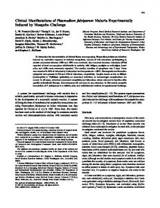
Figure 1. Design and properties of YC-Nano Ca2+ biosensors in P. falciparum. (a) Graphic representation of plasmid construction. Amino acid sequences of the linker between calmodulin (CaM) and myosin light chain (M13) peptide for YC-Nano15 and YC-Nano50 are shown. (b) Live cell images of the trophozoite stage parasite. Bright field (BF), CFP, YFP, and FRET (YFP/CFP) signals and merged image (BF-CFP-YFP). Purple to red color scale in the YFP/CFP panel represents low to high FRET efficiency (0 to 2.5). Scale bar, 2.5 μm. (c) The normalized fractional changes of the FRET signals (ΔR/R0) are plotted against the different Ca2+ concentration (0, 20, 40, 60, 80, and 100 nM). The curves represent the averaged data of ten parasites from 3 independent experiments.
10 mM Ca2+ containing the calcium ionophore A23187 to the Ca2+ free parasite culture increased YFP/CFP ratio from 1.36 (Ca2+ = 2.93 nM (median); minimum between 0–30 sec) to 2.66 (Ca2+ = 895.2 nM; maximum between 120–180 sec) in trophozoite stage parasites, confirming that YC-Nano50 has a large dynamic range in P. falciparum. The calculated median cytosolic Ca2+ concentration in the trophozoite stage was 30.0 (interquartile range: 5.6–55.0) nM. We found calculated Ca2+ concentrations in other stages of the parasite were much higher; 372.5 (253.0–483.0) nM at ring, 310.0 (256.2–514.9) nM at schizont, 949.6 (785.1–995.2) nM at merozoite, 131.3 (70.1– 185.1) nM at gametocyte (stage III) and 521.8 (387.2–942.2) nM at gametocyte (stage IV–V) stages (Fig. 2b).
P. falciparum cytosolic Ca2+ level is not modulated by thapsigargin, a mammalian SERCA inhibitor. The endoplasmic reticulum is an important Ca2+ storage compartment to maintain and regulate the cyto-
solic Ca2+ concentration in eukaryotic cells, and uptake of Ca2+ from cytosol to ER is regulated by SERCA. In Plasmodium conflicting reports describe the responses of malaria parasites against SERCA inhibitors, specifically thapsigargin (TG)21,23. We therefore revisited the effect of TG for parasite cytosolic Ca2+ homeostasis, and found that 15 μM CPA, a SERCA specific inhibitor consistently reported to inhibit P. falciparum SERCA (PfSERCA)24, increased the cytosolic Ca2+ (Fig. 3a); whereas 7.6 μM TG, a concentration reported to inhibit PfSERCA pump activity23, did not change the cytosolic Ca2+ concentration (Fig. 3b). The effect of TG on the cytosolic Ca2+ level was not observed even when 76 μM TG was applied (Supplementary Fig. 2). The positive control calcium ionophore A23187 increased the cytosolic Ca2+, and a solvent control DMSO showed no effect (Fig. 3c,d). Because the parasite is surrounded by Ca2+ rich environments in the human body and in the culture - for example, 45–86 nM in the RBC cytosol, ~40 μM in the PV space, and ~1 mM in the human plasma25,26 - we further evaluated the effect of CPA and TG in Ca2+-free medium after selective membrane permeabilization. Firstly, iRBCs were treated with streptolysin O (SLO) to selectively permeabilize the RBC membrane, but not the PVM and parasite plasma membrane (PPM). When TG was added to SLO-treated iRBC, no effect was observed, but
Scientific Reports | 6:23454 | DOI: 10.1038/srep23454
3
www.nature.com/scientificreports/
Figure 2. Cytosolic Ca2+ concentration in the different developmental stages of P. falciparum. (a) FRET signals from amoeboid ring (n = 11), trophozoite (n = 18), schizont (n = 10), merozoite (n = 10), and gametocyte (stage III (n = 6) and stage IV-V (n = 7)) stages of the parasite. Bright field (BF), merged image of BF, merged image (BF-CFP-YFP), FRET (YFP/CFP) signals, and calculated Ca2+ concentration with pseudo color are shown. Purple to red color scale in FRET (YFP/CFP) signals and calculated Ca2+ concentration represent low to high FRET efficiency (0 to 2.5) and 0 to 1000 nM Ca2+, respectively. Scale bar, 4 μm. (b) Calculated cytosolic Ca2+ concentrations of parasites with median and interquartile range are shown for each stage. **p = 0.0012, ***p