Epigenetics
ISSN: 1559-2294 (Print) 1559-2308 (Online) Journal homepage: http://www.tandfonline.com/loi/kepi20
Cadmium exposure and the epigenome: Exposureassociated patterns of DNA methylation in leukocytes from mother-baby pairs Alison Sanders, Lisa Smeester, Daniel Rojas, Tristan DeBussycher, Michael Wu, Fred Wright, Yi-Hui Zhou, Jessica Laine, Julia Rager, Geeta Swamy, Alison Ashley-Koch, Marie Lynn Miranda & Rebecca Fry To cite this article: Alison Sanders, Lisa Smeester, Daniel Rojas, Tristan DeBussycher, Michael Wu, Fred Wright, Yi-Hui Zhou, Jessica Laine, Julia Rager, Geeta Swamy, Alison Ashley-Koch, Marie Lynn Miranda & Rebecca Fry (2014) Cadmium exposure and the epigenome: Exposureassociated patterns of DNA methylation in leukocytes from mother-baby pairs, Epigenetics, 9:2, 212-221, DOI: 10.4161/epi.26798 To link to this article: http://dx.doi.org/10.4161/epi.26798
View supplementary material
Published online: 28 Oct 2013.
Submit your article to this journal
Article views: 210
View related articles
View Crossmark data
Citing articles: 27 View citing articles
Full Terms & Conditions of access and use can be found at http://www.tandfonline.com/action/journalInformation?journalCode=kepi20 Download by: [72.229.243.158]
Date: 23 August 2016, At: 06:59
Research paper
Epigenetics 9:2, 212–221; February 2014; © 2014 Landes Bioscience
Cadmium exposure and the epigenome
Exposure-associated patterns of DNA methylation in leukocytes from mother-baby pairs Alison P Sanders1,†,‡, Lisa Smeester1,‡, Daniel Rojas2, Tristan DeBussycher3, Michael C Wu4, Fred A Wright4, Yi-Hui Zhou4, Jessica E Laine5, Julia E Rager1, Geeta K Swamy6, Allison Ashley-Koch7, Marie Lynn Miranda8, and Rebecca C Fry1,2 Department of Environmental Sciences and Engineering; Gillings School of Global Public Health; University of North Carolina; Chapel Hill, NC USA; 2Curriculum in Toxicology; School of Medicine; University of North Carolina; Chapel Hill, NC USA; 3Center for Bioinformatics; University of North Carolina; Chapel Hill, NC USA; 4Department of Biostatistics; Gillings School of Global Public Health; University of North Carolina; Chapel Hill, NC USA; 5Department of Epidemiology; Gillings School of Global Public Health; University of North Carolina; Chapel Hill, NC USA; 6Department of Obstetrics and Gynecology; Duke University; Durham, NC USA; 7Center for Human Genetics; Duke University Medical Center; Durham, NC USA; 8School of Natural Resources and Environment; Department of Pediatrics; University of Michigan; Ann Arbor, MI USA †
Current affiliation: Department of Preventive Medicine; Icahn School of Medicine at Mount Sinai; New York, NY USA These authors contributed equally to this work.
‡
Keywords: cadmium, cigarette smoke, cord blood, epigenetics, heavy metal, maternal blood, methylation, pregnancy Abbreviations:5mC, 5-methylcytosine; Cd, Cadmium; CpG, Cytosine-phosphate-guanine; CEHI, Children’s Environmental Health Initiative; DAVID, Database for Annotation, Visualization and Integrated Discovery; DL, Detection limit; DNA, Deoxyribonucleic acid; FDR, false discovery rate; ICP-MS, Inductively Coupled Plasma-Mass Spectrometry; MEME, Multiple EM for Motif Elicitation; MTF-1, Metal-responsive transcription factor-1; MIRA, methylation CpG island recovery assay; NHANES IV, Fourth National Report on Human Exposure to Environmental Chemicals; PRR13, Proline rich 13; qPCR, Quantitative Polymerase Chain Reaction; SCEDDBO, Southern Center on Environmentally-Driven Disparities in Birth Outcomes; SRF, Serum response factor; TCF7L1, Transcription factor 7-like 1 (also known as TCF3); TCFAP2E, Transcription factor AP-2-eplison;
Cadmium (Cd) is prevalent in the environment yet understudied as a developmental toxicant. Cd partially crosses the placental barrier from mother to fetus and is linked to detrimental effects in newborns. Here we examine the relationship between levels of Cd during pregnancy and 5-methylcytosine (5mC) levels in leukocyte DNA collected from 17 mothernewborn pairs. The methylation of cytosines is an epigenetic mechanism known to impact transcriptional signaling and influence health endpoints. A methylated cytosine-guanine (CpG) island recovery assay was used to assess over 4.6 million sites spanning 16 421 CpG islands. Exposure to Cd was classified for each mother-newborn pair according to maternal blood levels and compared with levels of cotinine. Subsets of genes were identified that showed altered DNA methylation levels in their promoter regions in fetal DNA associated with levels of Cd (n = 61), cotinine (n = 366), or both (n = 30). Likewise, in maternal DNA, differentially methylated genes were identified that were associated with Cd (n = 92) or cotinine (n = 134) levels. While the gene sets were largely distinct between maternal and fetal DNA, functional similarities at the biological pathway level were identified including an enrichment of genes that encode for proteins that control transcriptional regulation and apoptosis. Furthermore, conserved DNA motifs with sequence similarity to specific transcription factor binding sites were identified within the CpG islands of the gene sets. This study provides evidence for distinct patterns of DNA methylation or “footprints” in fetal and maternal DNA associated with exposure to Cd.
Introduction Cadmium (Cd) is a heavy metal that ranks among the top ten chemicals in the Agency for Toxic Substances and Disease Registry priority list of hazardous substances.1 It is widespread in the environment, found in byproducts of industrial processes, contaminated water or soil, certain foods, and tobacco products.2 Cd is a known lung carcinogen and a
putative carcinogen in other tissues including the liver, prostate, kidney, bladder, stomach, and pancreas.3-5 In addition to its role as a carcinogen, Cd has also been associated with other health endpoints, including developmental effects early in life. For example, prenatal Cd exposure has been inversely associated with fetal growth parameters such as newborn length, weight, height, and head circumference,6-13 as well as adverse cognitive developmental effects later in life.12,14
*Correspondence to: Rebecca C Fry; Email:
[email protected] Submitted: 01/18/2013; Revised: 09/23/2013; Accepted: 10/14/2013; Published Online: 10/28/2013 http://dx.doi.org/10.4161/epi.26798 212 Epigenetics
Volume 9 Issue 2
©2014 Landes Bioscience. Do not distribute.
1
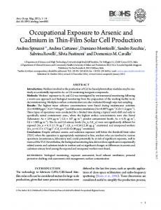
Figure 1. Heat map of genes with differential DNA methylation levels in fetal and maternal DNA associated with Cd (A), or cotinine (B). Venn diagram representing the total number of Cd-or cotinine-associated genes in fetal DNA (C) or maternal DNA (D). Heat maps represent average DNA methylation levels of exposure-associated gene sets. Data are z-score normalized for each gene. Individuals are ordered from left to right based on increasing level of exposure. Red indicates a relative increase in average DNA methylation and blue represents a relative decrease in average DNA methylation. In the Venn diagrams, the number in parentheses indicates the number of hypomethylated genes contained in each set. LE, lower exposed; HE, higher exposed; UE, unexposed; E, exposed.
Health effects associated with moderate to low levels of Cd exposure are of growing concern, particularly among susceptible populations such as pregnant women and children.15 Highlighting this issue, among pregnant women studied in the Fourth National Report on Human Exposure to Environmental Chemicals (NHANES IV), 66% had detectable blood Cd levels with an average level of 0.22 μg/L.16 The presence of Cd in the blood is particularly concerning as it has a half-life ranging up to 10 y.17 Cd is a component of cigarette smoke and concurrent exposure occurs. In utero exposure to both Cd and cigarette smoke is associated with lower newborn birth weight6,9,18 where Cd has been suggested to be the component of cigarette smoke that affects fetal skeletal growth.19 Cotinine, the primary metabolite of nicotine and biomarker of cigarette smoke exposure, is a reliable measure of actual dose received with a half-life of less than 1 d.20,21 Thus, cotinine is a general measure of recent exposure to tobacco products, whereas Cd can represent longterm and/or historic tobacco exposure. For non-smokers, diet is the major source of Cd exposure.17 The mechanism(s) of action of the detrimental health effects related to prenatal Cd exposure are not well established. An epigenetic mechanism has been hypothesized,22 but is understudied. The addition or removal of methyl groups from cytosine at the 5′ position (5mC) is an epigenetic mechanism that plays a key role in mediating gene expression and subsequent biological
processes,23 possibly contributing to subsequent health effects resulting from environmental toxicant exposure. Indeed there is evidence from animal and cell culture studies that Cd alters DNA methyltransferase activity and subsequently DNA methylation patterns.3,22,24-29 In addition, prenatal tobacco smoke exposure is a known modifier of DNA methylation patterns,30-32 and is therefore an important consideration in this study. In an effort to understand potential impacts of prenatal Cd exposure, in the present study we assess DNA methylation in blood leukocytes and distinguish between Cd- and cotinineassociated changes in a cohort of mother-baby pairs from Durham County, North Carolina. Maternal cotinine and Cd levels were used to compare and contrast the DNA methylation levels associated with either contaminant, enabling the differentiation between Cd-specific patterns in DNA methylation from those associated with cotinine. DNA methylation changes of more than 16 000 promoter-based cytosine-phosphate-guanine (CpG) islands were assessed in fetal and maternal DNA associated with maternal and thus in utero Cd and cotinine levels.
Results This study consisted of 34 subjects, 17 mother–newborn pairs, selected as a nested cohort from the CEHI Healthy
www.landesbioscience.com Epigenetics 213
©2014 Landes Bioscience. Do not distribute.
Research paper
Research paper
Pregnancy, Healthy Baby study in Durham, North Carolina. The pairs were selected from the larger cohort based on stratified maternal blood Cd levels. Maternal and infant characteristics are presented in Table 1. The average maternal age was 28 y. Most of the women had more than one child (n = 13; 76.5%), inclusive of the infants described in this study. There were similar proportions of male (n = 9; 52.9%) and female children (n = 8; 47.1%). All newborns, with one exception, had a birth weight greater than 2500 g (range: 2495–3740 g). Levels of maternal Cd ranged from below the detection limit to 1.05 μg/L with an average maternal blood concentration of 0.44 μg/L. Ten women in the study had Cd levels above the NHANES median level in pregnant women and were classified as the “higher cadmium-exposed” group (See Materials and Methods). Maternal cotinine levels ranged from below the detection limit to 166.96 μg/L with an average of 14.5 μg/L. While 11 women had detectable blood levels of cotinine, only two had levels above 10 μg/L, a level associated with active or passive smoking activity.20 The two individuals with elevated cotinine reported active smoking during pregnancy. As we
were also interested in the effect of exposure due to secondhand or inactive smoking, cotinine exposure was classified as any detectable level vs. none. Eleven women had a detectable level of cotinine and were classified as “cotinine-exposed.” The exposure data for each of the 17 mother-baby pairs is provided (Tables S1 and S2). Regression analysis revealed that maternal Cd or cotinine levels did not vary significantly with respect to maternal age, race, parity, child’s sex, or birth weight. Exclusion of the low birth weight infant from the data set did not significantly affect the results (data not shown). Generally, younger women had higher levels of Cd, but this finding was not statistically significant (P > 0.05). Spearman rank correlation revealed a positive relationship between maternal serum cotinine and Cd (r = 0.45, P = 0.07). Cd-associated gene-specific DNA methylation Methylated maternal and newborn DNA was isolated using a methyl-CpG-binding domain protein complex (MBD2b/ MBD3L1) and hybridized onto Affymetrix Human Promoter 1.0R arrays. These arrays contain more than 4.6 million probes
214 Epigenetics
Volume 9 Issue 2
©2014 Landes Bioscience. Do not distribute.
Figure 2. Significantly enriched DNA motifs identified within the Cd and cotinine-associated gene lists in fetal DNA (A) and maternal DNA (B) as identified by MEME. The motifs shown have standard orientation 5′ to 3′. Estimates of sequence conservation at each nucleotide position within the motif are shown where a higher bit score is represented by increased font size.
Mean ± SD (range)/N (%) Maternal cadmium (μg/L)
0.44 ± 0.31 (0–1.05)
Maternal cotinine (μg/L)
14.5 ± 42.23 (0–166.96)
Maternal age (years)
28 ± 7 (19–42)
Maternal race* NHB
/12 (70.6)
NHW
/4 (23.5)
Other
/1 (5.8)
Parity First
/4 (23.5)
Second
/5 (29.4)
Third or higher
/8 (47.1)
Child’s Sex M
/9 (52.9)
F
/8 (47.1)
Birth weight (g)
3210 ± 377 (2495–3740)
*This study intentionally over-sampled NHB women.
that cover more than 25 500 human gene promoter regions. Computational methods were used to summarize the DNA methylation levels at a gene-specific CpG island level annotated to the reference Human Genome 18 (HG18) as in our recent publications33,34 (see Materials and Methods). More than 16 000 CpG islands were included in this study. Prior to analysis for the exposures of interest, differences in average DNA methylation levels associated with maternal age, race, and infant sex were identified. Analysis of the maternal DNA revealed there was a significant difference in gene-specific methylation levels associated with maternal age (n = 596 genes) and race (n = 83 genes). In fetal DNA, there was a significant difference in gene-specific DNA methylation levels associated with maternal age (n = 39 genes), race (n = 949 genes), and infant sex (n = 176 genes) (Table S3). In relation to newborn environmental exposures of Cd and cotinine, two gene sets were identified with significantly different average DNA methylation abundances in fetal DNA including a Cd-associated gene set (n = 61) and a cotinine-associated gene set (n = 366) (Fig. 1A and B; Table S4). Of these, one gene showed hypomethylation with increasing Cd, and five genes showed hypomethylation with respect to increasing cotinine (Fig. 1C). A total of 30 genes overlapped between the Cd and cotinine gene sets and all were hypermethylated (Fig. 1C; Table S4). In maternal DNA, distinct sets of Cd-associated (n = 92) or cotinine-associated genes (n = 134) were identified (Fig. 1A and B; Table S5). Of these, 11 of the 92 Cd-associated differentially methylated genes were hypomethylated, whereas four of 134 cotinine-associated genes were hypomethylated. There were no overlapping genes between the Cd- or cotinineassociated genes in maternal DNA (Fig. 1D). A comparison of the Cd-associated genes between maternal and fetal DNA showed no overlap. Very few (n = 12) of the
cotinine-associated genes were differentially methylated in both fetal and maternal DNA (data not shown). The majority of differentially methylated genes showed increasing promoter methylation with increasing Cd or cotinine level for both fetal and maternal DNA (Fig. 1A and B). In addition to identifying genes that are differentially methylated and associated with Cd or cotinine levels, a comparison was also performed between the DNA methylomes of the women and their newborns. Regardless of environmental exposure, this comparison of the 34 individuals’ methylomes showed that there were 12 820 genes significantly differentially methylated between fetal and maternal DNA that were not due to differences in maternal age, race, or infant sex (Fig. S1). All of the 12 820 genes showed lower average methylation abundance in maternal DNA when compared with fetal DNA. Differential methylation does not reflect shifts in leukocyte cell types The Cd- and cotinine-associated gene sets were compared with known differentially methylated regions (DMRs) that correspond to shifts in the abundance of white blood cell types.35 Within a list of 500 genes with DMRs known to predict cell type in an adult population, 227 of these genes were contained on the MIRA array. No blood cell type-associated genes were contained in the maternal Cd- or cotinine-associated genes lists in this study. A single gene, UNC84 domain containing 1 (UNC84A), was present in the Cd-associated fetal DNA gene set and 4 genes: neutrophil cytosolic factor 4 (NCF4), CBP80/20dependent translation initiation factor (KIAA0427), sorting nexin 8 (SNX8), and scavenger receptor class F (SCARF) were present in the cotinine-associated gene set in fetal DNA. Biological functions are enriched among differentially methylated genes The identified Cd- and cotinine-associated gene sets in mother–baby pairs were analyzed to determine whether they encode proteins with similar functionality in the cell. Each of the gene sets was analyzed for enriched biological functions using two independent methods (see Materials and Methods). The most significantly enriched biological functional categories were gene expression, cell cycle, cell death, and nervous system development (Table 2). Interestingly, while there was no overlap between the individual gene sets, genes that encode proteins that play a role in regulation of transcription were enriched among Cd-associated genes in both fetal and maternal DNA (P < 0.001). Apoptosis was an enriched biological process in the Cd and cotinine-associated gene sets identified as differentially methylated within both maternal and fetal DNA (P < 0.05). Specifically, there were 14 apoptosis-associated genes including proline rich 13 (PRR13) in the fetal gene set and 20 additional genes among the maternal gene set (Table S6). Common DNA motifs identified in gene sets We hypothesized that the identified differentially methylated genes may contain common underlying sequences or motifs. To examine this, the promoter regions of the CpG islands for each of the differentially methylated genes were analyzed for statistically enriched common sequence patterns (e.g., motifs). The motifs were then compared with known transcription
www.landesbioscience.com Epigenetics 215
©2014 Landes Bioscience. Do not distribute.
Table 1. Characteristics of mother-newborn pairs (n = 34 subjects)
Table 2. Enriched biological functions within the differentially methylated gene sets Maternal DNA (P values)
Category
Function Annotation
Cd
Cotinine
Cda
Cotininea
Gene expression
Regulation of transcription
0.001* [0.01]
–
0.001* [0.001]
–
Tissue morphology
Adipose tissue quantity
0.004
–
–
–
Cancer
Hyperplasia
0.004
–
–
–
a
Lipid metabolism
Lipid accumulation
0.005
–
–
–
Cell death
Apoptosis
0.02 [0.05]
0.001*
1.2 × 10–4* [0.04]
0.05
Cell cycle
Delay in G1, interphase
–
3.0 × 10–5* [0.003]
–
–
Neuronal quantity
–
0.002 [0.03]
–5
5.2 × 10 * [0.03]
–
b
Nervous system
c
Cell proliferation
Tumor proliferation
–
0.002
–
0.03
Protein degradation
Proteolysis
–
0.003 [0.003]
–
0.01
Infectious disease
Replication of virus
–
–
–
0.01
Neurological disease
Movement disorder
–
–
–
0.02
*P < 0.001; [ ] = P value of gene ontology terms in DAVID. Associated contaminant exposure in fetal or maternal DNA. Lipid metabolism, molecular transport, small molecule biochemistry. cNervous system development and function. a
factor binding sites. Among the Cd- and cotinine-associated gene sets, significantly enriched motifs representing conserved DNA sequences were identified (Fig. 2). The conserved motifs showed sequence similarity to binding sites for several transcription factors including: transcription factor 7-like 1 (TCF7L1 also known as TCF3), metal-responsive transcription factor-1 (MTF-1), transcription factor AP-2-eplison (TCFAP2E), and serum response factor (SRF) (Table 3). Notably, for the Cd-associated gene sets identified both in maternal and fetal DNA, the enriched motifs had significant sequence similarity to the binding sites of MTF-1 and TCF7L1. Validation of MIRA DNA methylation results Validation of the MIRA-based methylation results was performed using two methodologies for both fetal and maternal DNA. First using a gene-specific analysis, the promoter region of the apoptosis-associated PRR13 gene was targeted for methylationspecific quantitative polymerase chain reaction (qPCR)-based analysis. Primers were designed to amplify the promoter region comparable to the MIRA-assessed site (see Materials and Methods). DNA collected from 15 newborns representing a range of Cd levels was bisulfite converted and assessed. The data show a significant Spearman’s rank correlation (r = 0.53, P < 0.05) between the MIRA-based methylation assessment and the qPCRbased methylation for PRR13 (Fig. S2). Second, the results from the MIRA platform were compared with data obtained using the Illumina 450K platform using 2 maternal DNA samples. While the Spearman’s rank correlation for the genome-wide (n = 11, 347) assessment was not statistically significant (r = 0.01, P = 0.1), the methylation levels for 83 of the 92 Cd-specific genes that could be compared between platforms were significantly correlated (r = 0.35, P = 0.0009). A majority of the Cd-associated CpG islands (82%) had concordant relative hyper- or hypo-methylation on the 450K platform as compared with the MIRA assay (Table S5). Of the 20 Cd-associated genes enriched for apoptosis, 17 could be compared on both platforms and these had significant Spearman’s rank correlation (r = 0.41, P = 0.07).
b
Discussion There is growing evidence that the prenatal environment may influence the burden of disease in adult life, and that this relationship is associated with epigenetic modifications altered during the prenatal period.23,36 The suggested impacts of Cd exposure include enzyme inhibition, generation of reactive oxygen species, and perturbation of apoptosis or cell cycle;37 however, the ability to induce genomic instability without genotoxic action has implicated a possible epigenetic mechanism of Cd toxicity.22 Cd-associated genome-wide DNA methylation has not been previously assessed in human samples. Given the relationship of Cd with birth outcomes in humans 6,9 and its known role as a mediator of DNA methyltransferase activity,22 we set out to identify whether patterns of differential promoter DNA methylation in fetal and maternal leukocyte DNA associated with exposure to Cd. The data were compared with cotinine-associated patterns of differential DNA methylation. Here differences in gene-specific levels of DNA methylation were observed and linked to Cd exposure in utero. These genes were largely independent of those associated with cotinine. Overall, the majority of the differentially methylated genes showed increased or hypermethylation associated with Cd exposure. The Cd-associated genes were classified into their known ontologies, and transcription regulatory processes emerged as significantly enriched in both fetal and maternal DNA. Cell death, specifically apoptosis, was a significantly enriched function for each gene list. The altered Cd-associated DNA methylation in maternal DNA was validated using an alternate genome-wide approach, as well as for a subset of apoptosis-enriched genes. In addition, a gene-specific approach was used for PRR13 in fetal DNA, known to play a role in apoptosis.38 Cd has been shown in vitro to perturb pathways involved in inflammatory response, cell survival, apoptosis, tumorigenesis, and oxidative stress.22,39-42 In animal models, in utero exposure to Cd is associated with a wide range of
216 Epigenetics
Volume 9 Issue 2
©2014 Landes Bioscience. Do not distribute.
Fetal DNA (P values) a
Table 3. Transcription factors with response element sequence similarity to the identified motifs within the Cd- or cotinine-associated gene sets Fetal DNA (P values) Transcription Factor
Cda
TCFAP2E
Maternal DNA (P values)
Cotininea
Cda
2.3 × 10–5*
9.2 × 10–4*
–
TCF7L1
6.7 × 10 *
2.0 × 10 *
5.1 × 10 *
1.5 × 10–4*
SRF
2.2 × 10–4*
3.2 × 10–4*
1.5 × 10–3
5.0 × 10⁻³
MTF1
3.1 × 10–4*
1.6 × 10–4*
3.5 × 10–4*
2.0 × 10–3
–5
–5
Cotininea 4.9 × 10⁻³ –4
cell cycle and proliferative genomic responses.43 The data presented here may support an epigenetic mechanism, namely DNA methylation, by which genes involved in transcriptional regulation and apoptosis could be influenced by Cd exposure. As RNA is not available for the specific study subjects analyzed here, gene expression analysis cannot be performed. It is important to note that it is not anticipated that all of the changes in DNA methylation will have functional consequence and impact gene expression. Relative to Cd, cotinine was associated with a greater number of genes with differential DNA methylation in both maternal DNA and fetal DNA. Three previous studies of prenatal cigarette smoke exposure examined gene-specific DNA methylation.30-32 One study reported global hypomethylation with gene-specific hypermethylation of eight genes investigated30 while the other found largely gene-specific hypo-methylation among 38 total genes.31 The third study reported 26 significant CpGs mapped to 10 genes with both hyper- and hypomethylation associated with plasma cotinine levels.32 Interestingly, none of the genes identified were common between the three studies and none are similarly reported in the present study. There are several factors that could potentially influence differences in observed DNA methylation patterns that include exposure type, exposure duration, tissue type (i.e., buccal vs. placental vs. cord blood), subject’s age at sampling, type of assay used, or other unaccounted for co-exposures. It is important to mention that a woman’s current Cd levels may reflect prior exposure to cigarette smoke. Although we have accounted for cotinine exposure (a measure of recent and active smoking) in the analysis, it cannot be ruled out that some of the Cd-associated changes may be due to historic cigarette smoke exposure. Our data are supported by previous in vitro and ex vivo studies showing that Cd exposure led to hypermethylation of DNA after prolonged chronic exposures, whereas hypomethylation was present after acute Cd exposure.22,25,26 Our data highlight that regardless of exposure, there were significant differences in DNA methylation profiles between maternal and fetal samples that were not due to differences in maternal age, race, or infant sex. This is supported by a recent study that demonstrated that relative to newborn DNA, there is less methylation is observed among CpG island promoters in older individuals.15 The increased methylation levels in fetal DNA may have relevance for developmental biology. Further evaluation of these basic processes may increase our understanding of how early life exposures resulting in epigenetic shifts can have long-term health effects.
Of interest, many of the genes contained in the Cd and cotinine gene sets showed common sequences in their CpG islands within promoters. Among these conserved motif regions, binding sites for a common set of transcription factors were identified. The specific binding of transcription factors to target sites is a mechanism that protects CpG islands from methylation.44,45 Very recently studies have demonstrated that transcription factor binding results in local regions of low methylation and in contrast, absence of DNA-binding factors triggers the remethylation of local promoter regions,46,47 however this phenomenon has not been described related to environmental exposures. Here we identify conserved motifs with sequence similarity to binding sites for transcription factors including MTF-1. Notably, MTF-1 is known to respond to changes in cellular concentrations of multiple metals and coordinate expression of genes protective against metal toxicity.48,49 All of the identified transcription factors (e.g., TCF7L1, TCFAP2E, MTF-1, SRF) are known to regulate developmental processes within cells and represent targets for future investigation.50-53 Taken together, we hypothesize that these results suggest that patterns of DNA methylation that are associated with Cd may represent “footprints” indicating transcription factor presence or absence that occur during periods of DNA methylation. As a limitation, the cord blood sampling done here is representative of newborn leukocyte DNA rather than potential target organ systems such as the kidney, liver, or bone. There are obvious ethical and technical reasons to use leukocyte DNA as a proxy for target tissue analysis. Moreover, the use of circulating white blood cells as proxies for disease has been shown.54,55 While patterns of DNA methylation can differ between white blood cell types,32,35,56 our data support that the Cd and cotinine gene sets are not simply due to shifts in blood cell types. There is increasing evidence that there are contaminant-specific changes to leukocyte DNA methylation associated with various environmental contaminant exposures.33,57,58 Future studies should aim to compare the Cd-associated changes here to tissuespecific changes. In summary, the data from the present study provide evidence of Cd- and cotinine-associated patterns in DNA methylation present in the leukocyte DNA of newborns and their mothers. We identify gene-specific changes in DNA methylation levels associated with in utero Cd exposure, and distinguish these from methylation changes attributable to cotinine exposure, a general proxy measure for exposure to tobacco products. These distinct patterns of environmentally-associated DNA
www.landesbioscience.com Epigenetics 217
©2014 Landes Bioscience. Do not distribute.
*P < 0.001. aAssociated contaminant exposure in fetal or maternal DNA.
Materials and Methods Study participants The Children’s Environmental Health Initiative (CEHI) conducted a prospective cohort study of pregnant women living in Durham County, North Carolina from 2005–2011. This study is a key component of the Southern Center on EnvironmentallyDriven Disparities in Birth Outcomes (SCEDDBO), an interdisciplinary center aimed at understanding how environmental, social, and host factors jointly contribute to health disparities (http://cehi.snre.umich.edu/projects/sceddbo/). The CEHI Healthy Pregnancy, Healthy Baby study was reviewed and approved by the Institutional Review Boards at Duke University (Pro00007633) and the University of North Carolina (#09-0866). All women participating in this study consented for maternal venous and newborn cord blood collection for chemical and genetic analysis. Women receiving prenatal care at either the Duke Obstetrics Clinic or the Durham County Health Department Prenatal Clinic were eligible to participate if they planned to deliver at Duke University Medical Center, were at least 18 y of age, were Englishliterate, lived in Durham County, and did not have a multi-fetal gestation or any known fetal genetic or congenital anomalies. Additional methods on subject recruitment, enrollment, and data collection have been described previously.59 Women were enrolled between 18 and 28 weeks of pregnancy, and demographic data were collected including maternal age, race, and parity, as well as child’s sex and birth weight. At delivery, biological samples including maternal venous blood and newborn cord blood were collected. From the CEHI study, we selected a nested subcohort of 17 mother-infant pairs stratified as above or below a maternal Cd level of 0.2 μg/L (see Supplemental Materials). Methylated CpG island recovery assay (MIRA) Venous maternal blood and newborn cord blood samples were obtained at delivery. DNA was extracted using Qiagen’s PAXgene Blood DNA kit (Qiagen) according to manufacturer’s protocol. DNA was re-suspended in nuclease-free water and stored at –80 °C prior to DNA methylation assessment. CpG methylated DNA was collected using the MethylCollector Ultra Kit (Active Motif) and enriched DNA was amplified using the WGA3 kit (Sigma) according to manufacturer instructions with the following modification: 10 mM dATP, 10 mM dCTP, 10 mM dGTP, 8 mM dTTP, and 2 mM dUTP. Amplified DNA was then hybridized to the Affymetrix Human Promoter 1.0R arrays (Affymetrix) which assess over 4.6 million sites. Statistical Analyses Linear regression analyses were performed using the statistical package SAS 9.3 (SAS Institute Inc.) to examine relationships
between maternal blood Cd levels, serum cotinine levels and demographic characteristics for women and children. Maternal demographics included age, race, and parity, and children’s demographics included sex and birth weight. The relationship between Cd and cotinine was assessed with linear regression as well as Spearman rank correlation. Cd and cotinine levels below detect were treated as zero. For each of the study subjects, the DNA methylation abundance data obtained through the MIRA assay assessing 4.6 million probes were normalized using robust multi-chip average.60 The DNA methylation levels were then summarized at a gene-specific CpG island level annotated to the reference HG18 where islands were defined as in Davies et al.61-63 The resulting average methylation abundances for 16 421 CpG islands were compared using ANOVA (Partek Genomic Suite 6.4) where differential DNA methylation levels were assessed for each island and statistically defined as: (1) average island promoter methylation with a minimum absolute change of 30%; and (2) a P value < 0.05. Additionally, a false discovery rate (FDR) corrected q-value estimate was calculated and is reported. After identifying covariate-associated DNA methylation patterns within CpG islands (see Supplemental Materials), differential methylation was also examined according to sample type (maternal vs. newborn), higher vs. lower Cd-exposed, and cotinine-exposed vs. cotinine-unexposed (see Supplemental Materials). The same statistical requirements were applied to all analyses. Gene ontology/pathway enrichment analysis Gene ontology/pathway enrichment analysis was performed using two independent methodologies. The differentially methylated genes were analyzed in the context of interacting networks using Ingenuity Pathway Analysis Software (Ingenuity Systems, Inc.) and functional clustering using the Database for Annotation, Visualization and Integrated Discovery (DAVID) (available online: http://david.abcc.ncifcrf.gov/tools.jsp). Enriched DNA motif identification CpG island sequences were retrieved from the UCSC genome browser website64 for each exposure-associated gene set. These sequences are representative of CpG islands within promoter regions of the genome. Position-specific letter probability matrices, also known as motifs, were identified using Multiple EM for Motif Elicitation (MEME) version 4.8.1.65 A first order Hidden Markov Model and negative position specific priors were calculated from a background set of 200 randomly selected CpG island sequences that were not differentially methylated. Additional parameter specifications are reported in the Supplemental Materials. The motif with the highest statistical significance (P value) for each group of differentially methylated genes was compared with known transcription factor binding sites using TOMTOM.65 Validation of MIRA results For gene-specific analysis, DNA from 15 newborn leukocyte samples was selected. DNA was bisulfite converted using the Zymo EZ DNA Methylation-Lightening kit (Zymo Research) according to the manufacturer’s instructions. Methylation was assessed using the EpiTect MethyLight Assay (Qiagen). Methylationindependent sequence-specific primers were designed for the
218 Epigenetics
Volume 9 Issue 2
©2014 Landes Bioscience. Do not distribute.
methylation alterations or “footprints” in fetal and maternal DNA may have functional consequences in the cell and warrant further research. Metal exposure continues to be an important area of public health concern for both maternal and child health. Increased education about the potential risks of environmental contaminants including Cd and cigarette smoke will be key to reducing and preventing harm to infants.
References 1. ATSDR. Priority List of Hazardous Substances. Online at: http://wwwatsdrcdcgov/spl/ 2011. 2. ATSDR. Toxicological Profile for Cadmium. Atlanta, GA: Centers for Diseaes Control, 2008. 3. Arita A, Costa M. Epigenetics in metal carcinogenesis: nickel, arsenic, chromium and cadmium. Metallomics 2009; 1:222-8; PMID:20461219; http://dx.doi. org/10.1039/b903049b 4. Waalkes MP. Cadmium carcinogenesis in review. J Inorg Biochem 2000; 79:241-4; PMID:10830873; http://dx.doi.org/10.1016/S0162-0134(00)00009-X 5. IARC. Volume 58. Cadmium. 1993:119-238 6. Fréry N, Nessmann C, Girard F, Lafond J, Moreau T, Blot P, Lellouch J, Huel G. Environmental exposure to cadmium and human birthweight. Toxicology 1993; 79:109-18; PMID:8497864; http://dx.doi. org/10.1016/0300-483X(93)90124-B 7. Lin CM, Doyle P, Wang DL, Hwang YH, Chen PC. Does prenatal cadmium exposure affect fetal and child growth? Occup Environ Med 2011; 68:6416; PMID:21186202; http://dx.doi.org/10.1136/ oem.2010.059758 8. Nishijo M, Tawara K, Honda R, Nakagawa H, Tanebe K, Saito S. Relationship between newborn size and mother’s blood cadmium levels, Toyama, Japan. Arch Environ Health 2004; 59:22-5; PMID:16053205; http://dx.doi.org/10.3200/AEOH.59.1.22-25 9. Salpietro CD, Gangemi S, Minciullo PL, Briuglia S, Merlino MV, Stelitano A, Cristani M, Trombetta D, Saija A. Cadmium concentration in maternal and cord blood and infant birth weight: a study on healthy non-smoking women. J Perinat Med 2002; 30:3959; PMID:12442603; http://dx.doi.org/10.1515/ JPM.2002.061
did not meet this minimum threshold for detection. The data were further filtered for probes positioned within CpG islands for comparison with the MIRA assay. For statistical comparison, average β values were calculated and a ratio of β Cd-exposed/ β Cd-unexposed determined for each gene. A Spearman rank correlation was calculated to compare the β ratio to the MIRA FC for all comparable genes (n = 11 347), the focused set of 92 Cd-associated genes in maternal DNA, and the subset of 20 Cd-associated apoptosis-related genes. Disclosure of Potential Conflicts of Interest
No potential conflicts of interest were disclosed. Acknowledgments
This research was funded in part by grants from the NIEHS (T32-ES007018, P42-ES005948, ES019315, and P30-ES010126), the USEPA (RD83329301), and in part by the North Carolina Translation and Clinical Sciences Institute (NC TRaCS) grants UL1RR025747, KL2RR025746, and TLRR025745 from the NIH National Center for Research Resources and the National Center for Advancing Translational Sciences, National Institutes of Health. We acknowledge researchers at Expression Analysis in Durham, NC for their assistance with the processing of the Illumina 450K microarrays. Supplemental Materials
Supplemental materials may be found here: www.landesbioscience.com/journals/epigenetics/article/26798
10. Ronco AM, Arguello G, Muñoz L, Gras N, Llanos M. Metals content in placentas from moderate cigarette consumers: correlation with newborn birth weight. Biometals 2005; 18:233-41; PMID:15984568; http://dx.doi.org/10.1007/s10534-005-0583-2 11. Llanos MN, Ronco AM. Fetal growth restriction is related to placental levels of cadmium, lead and arsenic but not with antioxidant activities. Reprod Toxicol 2009; 27:88-92; PMID:19103280; http:// dx.doi.org/10.1016/j.reprotox.2008.11.057 12. Tian LL, Zhao YC, Wang XC, Gu JL, Sun ZJ, Zhang YL, Wang JX. Effects of gestational cadmium exposure on pregnancy outcome and development in the offspring at age 4.5 years. Biol Trace Elem Res 2009; 132:51-9; PMID:19404590; http://dx.doi. org/10.1007/s12011-009-8391-0 13. Shirai S, Suzuki Y, Yoshinaga J, Mizumoto Y. Maternal exposure to low-level heavy metals during pregnancy and birth size. J Environ Sci Health A Tox Hazard Subst Environ Eng 2010; 45:1468-74; PMID:20694885; http://dx.doi.org/10.1080/10934 529.2010.500942 14. Bonithon-Kopp C, Huel G, Moreau T, Wendling R. Prenatal exposure to lead and cadmium and psychomotor development of the child at 6 years. Neurobehav Toxicol Teratol 1986; 8:307-10; PMID:3736760 15. Heyn H, Li N, Ferreira HJ, Moran S, Pisano DG, Gomez A, Diez J, Sanchez-Mut JV, Setien F, Carmona FJ, et al. Distinct DNA methylomes of newborns and centenarians. Proc Natl Acad Sci U S A 2012; 109:10522-7; PMID:22689993; http://dx.doi. org/10.1073/pnas.1120658109
16. Woodruff TJ, Zota AR, Schwartz JM. Environmental chemicals in pregnant women in the United States: NHANES 2003-2004. Environ Health Perspect 2011; 119:878-85; PMID:21233055; http://dx.doi. org/10.1289/ehp.1002727 17. Järup L, Akesson A. Current status of cadmium as an environmental health problem. Toxicol Appl Pharmacol 2009; 238:201-8; PMID:19409405; http://dx.doi.org/10.1016/j.taap.2009.04.020 18. Yu XD, Yan CH, Shen XM, Tian Y, Cao LL, Yu XG, Zhao L, Liu JX. Prenatal exposure to multiple toxic heavy metals and neonatal neurobehavioral development in Shanghai, China. Neurotoxicol Teratol 2011; 33:437-43; PMID:21664460; http:// dx.doi.org/10.1016/j.ntt.2011.05.010 19. Godfrey K, Walker-Bone K, Robinson S, Taylor P, Shore S, Wheeler T, Cooper C. Neonatal bone mass: influence of parental birthweight, maternal smoking, body composition, and activity during pregnancy. J Bone Miner Res 2001; 16:1694-703; PMID:11547840; http://dx.doi.org/10.1359/ jbmr.2001.16.9.1694 20. Klebanoff MA, Levine RJ, Clemens JD, DerSimonian R, Wilkins DG. Serum cotinine concentration and self-reported smoking during pregnancy. Am J Epidemiol 1998; 148:259-62; PMID:9690362; ht tp : //d x.doi.org /10.1093 /ox fordjou rna ls.aje. a009633 21. Jarvis MJ, Russell MA, Benowitz NL, Feyerabend C. Elimination of cotinine from body fluids: implications for noninvasive measurement of tobacco smoke exposure. Am J Public Health 1988; 78:6968; PMID:3369603; http://dx.doi.org/10.2105/ AJPH.78.6.696
www.landesbioscience.com Epigenetics 219
©2014 Landes Bioscience. Do not distribute.
apoptosis-associated gene PRR13 with forward primer sequence: (5′ GGTTTGGGTG ATTAGGAAGAGT 3′) and reverse primer sequence: (5′ AAAATCCAAA TACCCCATCA C 3′). A methyl-specific reporter probe for quantification was also designed (5′ 6FAM-GCCGCCTAAA CTTACTACGT 3′). The amplified region of PRR13 represents promoter region on chromosome 12, position 52121562–52121835, based upon HG18. Amplicons were assessed using qPCR in technical triplicate. qPCR methylation abundance was determined as 100/(1 + cycle threshold [CT]) for each sample, averaged across triplicates. A Spearman rank correlation and corresponding P value were calculated to compare the methylation abundances between the qPCR and MIRA assays. Genome-wide comparison was performed on two maternal DNA samples stratified by maternal blood Cd levels using the 0.2 μg/L exposure cutoff and matched on maternal race, age, and insurance status. Bisulfite conversion was performed using the Zymo EZ DNA Methylation kit (Zymo Research) according to the manufacturer’s instructions. Methylation was assessed at 485 577 CpGs in maternal DNA using the Illumina Infinium HumanMethylation450 BeadChip (Illumina Inc.). BeadChip processing was performed at Expression Analysis Inc. (www.expressionanalysis.com) and processed with Illumina’s GenomeStudio Methylation module Version 1.8 (Illumina Inc.). The proportion of methylation (β) for each CpG was calculated as the ratio of methylated signal intensity divided by the sum of both methylated and unmethylated signals. For quality control, probes with detection P value < 0.0001 were required. β values were excluded from analysis for probes which
34. Bailey KA, Wu MC, Ward WO, Smeester L, Rager JE, García-Vargas G, Del Razo LM, Drobná Z, Stýblo M, Fry RC. Arsenic and the epigenome: interindividual differences in arsenic metabolism related to distinct patterns of DNA methylation. J Biochem Mol Toxicol 2013; 27:106-15; PMID:23315758; http://dx.doi. org/10.1002/jbt.21462 35. Houseman EA, Accomando WP, Koestler DC, Christensen BC, Marsit CJ, Nelson HH, Wiencke JK, Kelsey KT. DNA methylation arrays as surrogate measures of cell mixture distribution. BMC Bioinformatics 2012; 13:86; PMID:22568884; http://dx.doi.org/10.1186/1471-2105-13-86 36. Bollati V, Baccarelli A. Environmental epigenetics. Heredity (Edinb) 2010; 105:105-12; PMID:20179736; http://dx.doi.org/10.1038/ hdy.2010.2 37. Waisberg M, Joseph P, Hale B, Beyersmann D. Molecular and cellular mechanisms of cadmium carcinogenesis. Toxicology 2003; 192:95-117; PMID:14580780; http://dx.doi.org/10.1016/ S0300-483X(03)00305-6 38. Lih CJ, Wei W, Cohen SN. Txr1: a transcriptional regulator of thrombospondin-1 that modulates cellular sensitivity to taxanes. Genes Dev 2006; 20:2082-95; PMID:16847352; http://dx.doi. org/10.1101/gad.1441306 39. Benton MA, Rager JE, Smeester L, Fry RC. Comparative genomic analyses identify common molecular pathways modulated upon exposure to low doses of arsenic and cadmium. BMC Genomics 2011; 12:173; PMID:21457566; http://dx.doi. org/10.1186/1471-2164-12-173 40. Andrews GK. Regulation of metallothionein gene expression by oxidative stress and metal ions. Biochem Pharmacol 2000; 59:95-104; PMID:10605938; http://dx.doi.org/10.1016/S0006-2952(99)00301-9 41. Shin HJ, Park KK, Lee BH, Moon CK, Lee MO. Identification of genes that are induced after cadmium exposure by suppression subtractive hybridization. Toxicology 2003; 191:121-31; PMID:12965115; http://dx.doi.org/10.1016/S0300-483X(03)00210-5 42. Robinson JF, Yu XZ, Moreira EG, Hong SW, Faustman EM. Arsenic- and cadmium-induced toxicogenomic response in mouse embryos undergoing neurulation. Toxicol Appl Pharmacol 2011; 250:117-29; PMID:20883709; http://dx.doi. org/10.1016/j.taap.2010.09.018 43. Robinson JF, Yu X, Hong S, Griffith WC, Beyer R, Kim E, Faustman EM. Cadmium-induced differential toxicogenomic response in resistant and sensitive mouse strains undergoing neurulation. Toxicol Sci 2009; 107:206-19; PMID:18974090; http://dx.doi.org/10.1093/toxsci/kfn221 44. Brandeis M, Frank D, Keshet I, Siegfried Z, Mendelsohn M, Nemes A, Temper V, Razin A, Cedar H. Sp1 elements protect a CpG island from de novo methylation. Nature 1994; 371:435-8; PMID:8090226; http://dx.doi. org/10.1038/371435a0 45. Macleod D, Charlton J, Mullins J, Bird AP. Sp1 sites in the mouse aprt gene promoter are required to prevent methylation of the CpG island. Genes Dev 1994; 8:2282-92; PMID:7958895; http://dx.doi. org/10.1101/gad.8.19.2282 46. Lienert F, Wirbelauer C, Som I, Dean A, Mohn F, Schübeler D. Identification of genetic elements that autonomously determine DNA methylation states. Nat Genet 2011; 43:1091-7; PMID:21964573; http://dx.doi.org/10.1038/ng.946 47. Stadler MB, Murr R, Burger L, Ivanek R, Lienert F, Schöler A, van Nimwegen E, Wirbelauer C, Oakeley EJ, Gaidatzis D, et al. DNA-binding factors shape the mouse methylome at distal regulatory regions. Nature 2011; 480:490-5; PMID:22170606
220 Epigenetics
48. Selvaraj A, Balamurugan K, Yepiskoposyan H, Zhou H, Egli D, Georgiev O, Thiele DJ, Schaffner W. Metal-responsive transcription factor (MTF1) handles both extremes, copper load and copper starvation, by activating different genes. Genes Dev 2005; 19:891-6; PMID:15833915; http://dx.doi. org/10.1101/gad.1301805 49. Andrews GK. Cellular zinc sensors: MTF-1 regulation of gene expression. Biometals 2001; 14:223-37; PMID:11831458; http://dx.doi. org/10.1023/A:1012932712483 50. Cole MF, Johnstone SE, Newman JJ, Kagey MH, Young RA. Tcf3 is an integral component of the core regulatory circuitry of embryonic stem cells. Genes Dev 2008; 22:746-55; PMID:18347094; http:// dx.doi.org/10.1101/gad.1642408 51. Merrill BJ, Gat U, DasGupta R, Fuchs E. Tcf3 and Lef1 regulate lineage differentiation of multipotent stem cells in skin. Genes Dev 2001; 15:1688-705; PMID:11445543; http://dx.doi.org/10.1101/ gad.891401 52. Winger Q, Huang J, Auman HJ, Lewandoski M, Williams T. Analysis of transcription factor AP-2 expression and function during mouse preimplantation development. Biol Reprod 2006; 75:324-33; PMID:16672719; http://dx.doi. org/10.1095/biolreprod.106.052407 53. Nelson TJ, Balza R Jr., Xiao Q, Misra RP. SRFdependent gene expression in isolated cardiomyocytes: regulation of genes involved in cardiac hypertrophy. J Mol Cell Cardiol 2005; 39:479-89; PMID:15950986; http://dx.doi.org/10.1016/j.yjmcc.2005.05.004 54. Anglim PP, Alonzo TA, Laird-Offringa IA. DNA methylation-based biomarkers for early detection of non-small cell lung cancer: an update. Mol Cancer 2008; 7:81; PMID:18947422 ; http://dx.doi. org/10.1186/1476-4598-7-81 55. Sinnaeve PR, Donahue MP, Grass P, Seo D, Vonderscher J, Chibout SD, Kraus WE, Sketch M Jr., Nelson C, Ginsburg GS, et al. Gene expression patterns in peripheral blood correlate with the extent of coronary artery disease. PLoS One 2009; 4:e7037; PMID:19750006; http://dx.doi.org/10.1371/ journal.pone.0007037 56. Deaton AM, Webb S, Kerr AR, Illingworth RS, Guy J, Andrews R, Bird A. Cell type-specific DNA methylation at intragenic CpG islands in the immune system. Genome Res 2011; 21:107486; PMID:21628449; http://dx.doi.org/10.1101/ gr.118703.110 57. Pilsner JR, Hu H, Ettinger A, Sánchez BN, Wright RO, Cantonwine D, Lazarus A, Lamadrid-Figueroa H, Mercado-García A, Téllez-Rojo MM, et al. Influence of prenatal lead exposure on genomic methylation of cord blood DNA. Environ Health Perspect 2009; 117:1466-71; PMID:19750115 58. Perera F, Tang WY, Herbstman J, Tang DL, Levin L, Miller R, Ho SM. Relation of DNA methylation of 5′-CpG island of ACSL3 to transplacental exposure to airborne polycyclic aromatic hydrocarbons and childhood asthma. PLoS One 2009; 4:e4488; PMID:19221603; http://dx.doi.org/10.1371/journal. pone.0004488 59. Miranda ML, Edwards S, Maxson PJ. Mercury levels in an urban pregnant population in Durham County, North Carolina. Int J Environ Res Public Health 2011; 8:698-712; PMID:21556174; http://dx.doi. org/10.3390/ijerph8030698 60. Irizarry RA, Bolstad BM, Collin F, Cope LM, Hobbs B, Speed TP. Summaries of Affymetrix GeneChip probe level data. Nucleic Acids Res 2003; 31:e15; PMID:12582260; http://dx.doi.org/10.1093/nar/ gng015
Volume 9 Issue 2
©2014 Landes Bioscience. Do not distribute.
22. Takiguchi M, Achanzar WE, Qu W, Li GY, Waalkes MP. Effects of cadmium on DNA(Cytosine-5) methyltransferase activity and DNA methylation status during cadmium-induced cellular transformation. Exp Cell Res 2003; 286:35565; PMID:12749863; http://dx.doi.org/10.1016/ S0014-4827(03)00062-4 23. Wilson AG. Epigenetic regulation of gene expression in the inflammatory response and relevance to common diseases. J Periodontol 2008; 79(Suppl):1514-9; PMID:18673005; http://dx.doi. org/10.1902/jop.2008.080172 24. Martinez-Zamudio R, Ha HC. Environmental epigenetics in metal exposure. Epigenetics 2011; 6:820-7; PMID:21610324; http://dx.doi. org/10.4161/epi.6.7.16250 25. Benbrahim-Tallaa L, Waterland RA, Dill AL, Webber MM, Waalkes MP. Tumor suppressor gene inactivation during cadmium-induced malignant transformation of human prostate cells correlates with overexpression of de novo DNA methyltransferase. Environ Health Perspect 2007; 115:1454-9; PMID:17938735 26. Huang DJ, Zhang YM, Qi YM, Chen C, Ji WH. Global DNA hypomethylation, rather than reactive oxygen species (ROS), a potential facilitator of cadmium-stimulated K562 cell proliferation. Toxicol Lett 2008; 179:43-7; PMID:18482805; http:// dx.doi.org/10.1016/j.toxlet.2008.03.018 27. Benbrahim-Tallaa L, Tokar EJ, Diwan BA, Dill AL, Coppin JF, Waalkes MP. Cadmium malignantly transforms normal human breast epithelial cells into a basal-like phenotype. Environ Health Perspect 2009; 117:1847-52; PMID:20049202; http://dx.doi. org/10.1289/ehp.0900999 28. Doi T, Puri P, McCann A, Bannigan J, Thompson J. Epigenetic effect of cadmium on global de novo DNA hypomethylation in the cadmium-induced ventral body wall defect (VBWD) in the chick model. Toxicol Sci 2011; 120:475-80; PMID:21278052; http://dx.doi.org/10.1093/toxsci/kfr022 29. Jiang GF, Xu L, Song SZ, Zhu CC, Wu Q, Zhang L, Wu L. Effects of long-term low-dose cadmium exposure on genomic DNA methylation in human embryo lung fibroblast cells. Toxicology 2008; 244:49-55; PMID:18077075; http://dx.doi. org/10.1016/j.tox.2007.10.028 30. Breton CV, Byun HM, Wenten M, Pan F, Yang A, Gilliland FD. Prenatal tobacco smoke exposure affects global and gene-specific DNA methylation. Am J Respir Crit Care Med 2009; 180:462-7; PMID:19498054; http://dx.doi.org/10.1164/ rccm.200901-0135OC 31. Suter M, Ma J, Harris AS, Patterson L, Brown KA, Shope C, Showalter L, Abramovici A, Aagaard-Tillery KM. Maternal tobacco use modestly alters correlated epigenome-wide placental DNA methylation and gene expression. Epigenetics 2011; 6:128494; PMID:21937876; http://dx.doi.org/10.4161/ epi.6.11.17819 32. Joubert BR, Håberg SE, Nilsen RM, Wang X, Vollset SE, Murphy SK, Huang Z, Hoyo C, Midttun Ø, Cupul-Uicab LA, et al. 450K epigenome-wide scan identifies differential DNA methylation in newborns related to maternal smoking during pregnancy. Environ Health Perspect 2012; 120:1425-31; PMID:22851337 33. Smeester L, Rager JE, Bailey KA, Guan XJ, Smith N, García-Vargas G, Del Razo LM, Drobná Z, Kelkar H, Stýblo M, et al. Epigenetic changes in individuals with arsenicosis. Chem Res Toxicol 2011; 24:1657; PMID:21291286; http://dx.doi.org/10.1021/ tx1004419
63. Maunakea AK, Nagarajan RP, Bilenky M, Ballinger TJ, D’Souza C, Fouse SD, Johnson BE, Hong C, Nielsen C, Zhao Y, et al. Conserved role of intragenic DNA methylation in regulating alternative promoters. Nature 2010; 466:253-7; PMID:20613842; http:// dx.doi.org/10.1038/nature09165 64. Karolchik D, Hinrichs AS, Furey TS, Roskin KM, Sugnet CW, Haussler D, Kent WJ. The UCSC Table Browser data retrieval tool. Nucleic Acids Res 2004; 32(Database issue):D493-6; PMID:14681465; http://dx.doi.org/10.1093/nar/gkh103
65. Bailey TL, Boden M, Buske FA, Frith M, Grant CE, Clementi L, Ren J, Li WW, Noble WS. MEME SUITE: tools for motif discovery and searching. Nucleic Acids Res 2009; 37(Web Server issue):W202-8; PMID:19458158; http://dx.doi. org/10.1093/nar/gkp335
©2014 Landes Bioscience. Do not distribute.
61. Davies MN, Volta M, Pidsley R, Lunnon K, Dixit A, Lovestone S, Coarfa C, Harris RA, Milosavljevic A, Troakes C, et al. Functional annotation of the human brain methylome identifies tissue-specific epigenetic variation across brain and blood. Genome Biol 2012; 13:R43; PMID:22703893; http://dx.doi. org/10.1186/gb-2012-13-6-r43 62. Psychiatric Epigenetics Data KCL. [http:// epigenetics.iop.kcl.ac.uk/brain%5D.
www.landesbioscience.com Epigenetics 221