Pflugers Arch - Eur J Physiol DOI 10.1007/s00424-015-1757-6
INVITED REVIEW
Calcium homeostasis modulator (CALHM) ion channels Zhongming Ma 1 & Jessica E. Tanis 1 & Akiyuki Taruno 3 & J. Kevin Foskett 1,2
Received: 30 October 2015 / Accepted: 31 October 2015 # Springer-Verlag Berlin Heidelberg 2015
Abstract Calcium homeostasis modulator 1 (CALHM1), formerly known as FAM26C, was recently identified as a physiologically important plasma membrane ion channel. CALHM1 and its Caenorhabditis elegans homolog, CLHM1, are regulated by membrane voltage and extracellular Ca2+ concentration ([Ca2+]o). In the presence of physiological [Ca2+]o (∼1.5 mM), CALHM1 and CLHM-1 are closed at resting membrane potentials but can be opened by strong depolarizations. Reducing [Ca2+]o increases channel open probability, enabling channel activation at negative membrane potentials. Together, voltage and Ca2+o allosterically regulate CALHM channel gating. Through convergent evolution, CALHM has structural features that are reminiscent of connexins and pannexins/innexins/LRRC8 (volume-regulated anion channel (VRAC)) gene families, including four transmembrane helices with cytoplasmic amino and carboxyl termini. A CALHM1 channel is a hexamer of CALHM1 monomers with a functional pore diameter of ∼14 Å. CALHM channels discriminate poorly among cations and anions, with
* Zhongming Ma
[email protected] * J. Kevin Foskett
[email protected] 1
Department of Physiology, Perelman School of Medicine, University of Pennsylvania, 720 Clinical Research Bldg., 415 Curie Blvd., Philadelphia, PA 19104, USA
2
Department of Cell and Developmental Biology, Perelman School of Medicine, University of Pennsylvania, Philadelphia, PA 19104, USA
3
Department of Molecular Cell Physiology, Graduate School of Medical Science, Kyoto Prefectural University of Medicine, Kyoto 602-8566, Japan
signaling molecules including Ca2+ and ATP able to permeate through its pore. CALHM1 is expressed in the brain where it plays an important role in cortical neuron excitability induced by low [Ca2+]o and in type II taste bud cells in the tongue that sense sweet, bitter, and umami tastes where it functions as an essential ATP release channel to mediate nonsynaptic neurotransmitter release. CLHM-1 is expressed in C. elegans sensory neurons and body wall muscles, and its genetic deletion causes locomotion defects. Thus, CALHM is a voltage- and Ca2+o-gated ion channel, permeable to large cations and anions, that plays important roles in physiology. Keywords Connexin . Pannexin . ATP . Taste . Extracellular calcium . Voltage gated
Introduction Calcium homeostasis modulator 1 (CALHM1), previously known as FAM26C, was discovered in a search for human genes with enriched expression in the hippocampus in a region of chromosome 10 linked to enhanced risk for late-onset Alzheimer’s disease [8]. A nonsynonymous polymorphism, Pro86Leu, was identified as a possible modifier of the age of onset of Alzheimer’s disease [8, 17]. In humans, five homologs of CALHM1 were found by sequence database searches [8]. The CALHM gene family, CALHM1, and its homologs were collectively identified as the FAM26 gene family, where six genes are in two clusters on two chromosomes. CALHM1/ FAM26C is clustered on chromosome 10 with FAM26A and FAM26B genes, which were designated as CALHM3 and CALHM2, respectively. FAM26D, FAM26E, and FAM26F genes, to which CALHM names have not been assigned, are located in a cluster on chromosome 6. All CALHM/FAM26
Pflugers Arch - Eur J Physiol
genes are present throughout vertebrates, but they lack significant sequence homology to other known genes. Outside of vertebrates, CALHM1 homologs are absent in yeast and Drosophila, but Caenorhabditis elegans (C. elegans) possesses a single homolog, clhm-1. The fact that CALHM1 is conserved across >20 species, including C. elegans, mouse, and human, suggested its fundamental importance in biological processes. Until recently, however, no physiological functions of the CALHM proteins were known. Human CALHM1 is predicted to be a membrane protein with 346 amino acids. Based on membrane topology prediction algorithms, it was originally suggested to contain four transmembrane (TM) spanning helices. Although FAM26 proteins lack sequence similarities with other known proteins, it was speculated that CALHM1 might be related to NMDA receptors, because it possesses an amino acid sequence at the carboxyl terminal end of predicted TM2 that is similar to one in the ion selectivity filter of Ca2+-permeable NMDA receptors [8]. Although immunolocalization of expressed recombinant CALHM1 in mammalian culture cells revealed a predominantly intracellular labeling, whole-cell patch clamp electrophysiology of CALHM1-expressing CHO cells revealed the presence of a new Gd3+-sensitive, Ca2+-permeable outwardly rectifying current [8]. Furthermore, when extracellular Ca2+ was re-introduced to cells that had been exposed for several minutes to a medium lacking Ca2+, a sustained rise in cytoplasmic Ca2+ concentration ([Ca2+]i) was observed specifically in CALHM1-transfected cells [8]. Similar Ca2+-add back responses have been observed in subsequent studies including in single cells, where the possibility of artifactual responses due to leaked extracellular Ca2+ indicator dye was eliminated [11, 20, 36]. The apparent enhanced Ca2+ permeability in response to Ca2+ removal and add-back is insensitive to pharmacologic and genetic inhibition of store-operated Ca 2 + entry (SOCE). In addition, the magnitude of CALHM1-dependent and SOCE [Ca2+]i signals is additive [8, 20]. Exogenous expression of CALHM1 or P86L-CALHM1 elevated basal [Ca2+]i [11, 26, 36]. In contrast, the magnitude of the Ca2+ add-back response measured in cell populations was reduced in cells transiently expressing P86L-CALHM1 [8, 9, 26, 36]. Nevertheless, when measured at the single-cell level, the magnitude of the response was not different in some studies [11, 36]. Similarly, W114A-CALHM1-expressing cells have a reduced Ca2+ add-back response compared with cells expressing wild-type CALHM1, whereas the biophysical properties of the CALHM1 channel in Xenopus oocytes are unaffected by the mutation [44]. Fewer P86L-CALHM1expressing cells that wild-type CALHM1-expressing cells responded with a Ca2+ add-back response, suggesting that this polymorphism as well as the W114A mutation may confer less plasma membrane Ca2+ permeability than wild-type CALHM1 due to trafficking deficits [36].
Together, these results suggested that CALHM1 expression conferred a novel plasma membrane Ca2+ permeability. However, it remained unclear if exposure of the cells to a medium depleted of Ca2+ was required to observe Ca2+ permeability. Furthermore, it remained unknown whether CALHM1 was an ion channel or ion channel regulator. Finally, if CALHM1 was an ion channel, its structural and functional features, regulatory properties, and gating mechanisms were unknown. Here, we summarize recent advances in our understanding of the structural and biophysical properties as well as physiological roles of CALHM ion channels.
Structural features of CALHM1 CALHM1 monomers homo-multimerize to form a functional ion channel [8] [20, 36]. To date, no high-resolution structure of a CALHM1 is available. Secondary structural analysis and TM domain prediction algorithms suggested that a CALHM1 monomer has four transmembrane (TM) helices. The topology of the CALHM1 monomer was experimentally determined by demonstrating that Asn140 is glycosylated [8, 36], establishing an extracellular localization of the second extracellular loop (ECL) between putative TM3 and TM4 (Fig. 1a), and that a carboxyl terminal antibody only accessed its epitope from the cytoplasm [36]. These results indicated that a CALHM1 monomer has four TM domains with both the amino and carboxyl termini located in the cytoplasm. The CALHM1 channel was established to be a hexamer of CALHM1 monomers [36]. Under nonreducing conditions, two CALHM1 bands were present in SDS-PAGE of lysates from transiently transfected N2A cells, with apparent molecular weights of 80 and 250 kDa, which correspond to two and six times the predicted mass of a CALHM1 monomer. In nondenaturing blue native PAGE, one CALHM1 band was observed at approximately 240 kDa [36]. These results suggested that CALHM1 is a hexamer. Single-molecule subunit counting of a functional carboxyl terminally enhanced green fluorescent protein (EGFP)-tagged CALHM1 (CALHM1EGFP) expressed in Xenopus oocytes was employed to confirm this biochemical indication. CALHM1-EGFP generated ionic currents similar to untagged CALHM1. Two independent methods were used to detect bleaching steps. Most immobile fluorescent spots bleached in five steps, with many bleaching in six steps, and none bleaching in more than six steps. The distribution of bleaching steps was best fitted with a binomial distribution with six subunits. Together, biophysical measurements and biochemistry suggest that a CALHM1 channel is a hexamer of CALHM1 monomers [36]. This four-TM hexameric structure is shared with connexins [21], pannexins and innexins [47], volume-regulated anion channel (VRAC, aka LRRC8, SWELL1) [31, 45], and Orai1 channels [14]. CALHMs, connexins, and pannexins/innexins/
Pflugers Arch - Eur J Physiol
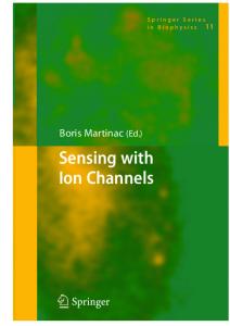
Fig. 1 Voltage- and Ca2+o-gated CALHM channels. a Transmembrane topology of CALHM channel. Asn72, Pro86, Asp121, and Asn140 are indicated for human CALHM1. ECL1 and ECL2 are extracellular loops 1 and 2, respectively. N amino terminus, C carboxyl terminus, TM transmembrane domain. b Normalized conductance-voltage (G-V) relations for human CALHM1 in 5 mM Ca2+ and 0 Ca2+. Solid lines are
Boltzmann function fits to the experimental data. c CALHM1 channel contains a voltage sensor and a Ca2+ sensor that detects extracellular Ca2+ concentration. The CALHM channel gate is closed by physiological [Ca2+]o and hyperpolarized voltages (left). Depolarization and/or lowering [Ca2+]o can open the gate, enabling Na+ and Ca2+ influx and ATP efflux (right)
VRAC also share other secondary structural features. All three families possess an amino terminal α-helix, a beta-sheet in extracellular loop 2 (ECL2), cysteines in both ECLs, and, except for connexins, α-helical regions in the carboxyl termini that align well [36]. Nevertheless, CALHM1 lacks homology with any of these channel families, and it does not have any sequence similarity to other known ion channels, suggesting that it belongs to a novel ion channel family [36].
observed in oocytes expressing CALHM channels, CALHM1 could either be an ion channel or activate an endogenous conductance. To distinguish between these possibilities, potential pore-lining residues were mutated and ion selectivity was determined. Site-directed mutagenesis of the asparagine at position 72 (Fig. 1a), within the sequence reminiscent of that of the NMDA receptor ion selectivity filter, was without effect on the conductance or ion selectivity [20], suggesting that if CALHM1 was indeed an ion channel, it was unlikely to have structural similarity with the NMDA receptor pore region. CALHM1 is predicted to have four TM helices, similar to Ca2+-selective Orai1 channels, which have acidic residues near the end of TM domains that play key roles in ion permeation [24, 48]. Mutation of aspartic acid at position 121, predicted to reside at the extracellular end of putative TM3 (Fig. 1a), to either alanine, cysteine or arginine changed CALHM1 ion selectivity, whereas mutation to glutamic acid to preserve the negative charge was without effect [20]. These results suggest that CALHM1 is likely a pore-forming subunit of a novel ion channel [20]. To determine whether CALHM1 is the founding member of an ion channel family, the C. elegans CALHM homolog CLHM-1 was expressed in Xenopus oocytes and electrophysiological studies were performed. Although CLHM-1 and hCALHM1 have only ∼16 % sequence identity, CLHM-1 localized to the plasma membrane and exhibited channel properties very similar to CALHM1 [37]. Mutation of aspartic
CALHM1 is the pore-forming subunit of an ion channel To explore the possible ion channel function of CALHM1, recombinant human CALHM1 (hCALHM1) was expressed in Xenopus oocytes. In solutions containing 2 mM Ca2+ and 1 mM Mg2+, depolarizing the membrane voltage generated large outward currents that activated with slow kinetics (τ∼ 3 s at +60 mV) and deactivated at hyperpolarized voltages (τ= 0.2 s at −80 mV) specifically in CALHM1-expressing oocytes [20]. Similar currents were observed in oocytes co-injected with Xenopus connexin-38 antisense oligonucleotide to inhibit endogenous Cx38 currents and with 1,2-bis(oaminophenoxy)ethane-N,N,N′,N′-tetraacetic acid (BAPTA) to inhibit Ca2+-activated Cl− currents [5]. An EGFP-tagged hCALHM1 had similar gating properties and localized strongly to the plasma membrane [20]. Although currents were
Pflugers Arch - Eur J Physiol
acid at position 125 at the extracellular end of putative TM3 altered ion selectivity as observed for the homologous mutation in CALHM1 [37]. It remains to be determined whether TM3 lines the ion permeation pathway in CALHM channels, and how this conserved aspartic acid (Asp121/Asp125) contributes to CALHM channel ion selectivity and conductance. Nevertheless, these results indicate that CALHM proteins are an evolutionarily conserved family of ion channels. Whether other vertebrate CALHM homologs function as ion channels remains unknown.
CALHM channels have weak ion selectivity Analyses of the ion permeability properties of hCALHM1 and CLHM-1 revealed that they have weak ion selectivities [20, 36, 37]. The relative permeabilities were estimated as PNa/PCa/ PK/PCl = 1:11:1.2:0.6 for hCALHM1 and 1:3.6:1.1:0.5 for CLHM-1, respectively. Similar results were obtained with bath Na+ replaced by K+ in either 0 or 2 mM Ca2+o. Thus, the permeability properties of both channels are quite similar, with a notable anion permeability. It is possible that the relative permeability of Ca2+ may be overestimated, because the changes of the reversal potentials in response to [Ca2+]o were measured at a low NaCl concentration due to difficulty in measuring changes in Ca2+ reversal potentials in normal extracellular NaCl concentration using two-electrode voltage clamp in Xenopus oocytes. The weak relative ion selectivity of CALHM channels suggests that either the ion selectivity filter is nondiscriminatory or the ion conduction pore is wide. To distinguish these possibilities, reversal potentials were measured with various tetraalkylammonium monovalent cations with different sizes, including tetramethylammonium (TMA + , ionic radius 3.47 Å), tetraethylammonium (TEA + , 4.00 Å), and tetrabutylammonium (TBA+, 4.94 Å) as permeant ions. Relative to Na+, PTMA/PTEA/PTBA is equal to 0.31:0.21:0.07 [36]. A plot of the molecular masses of each amine as well as small monovalent cations against their permeabilities was well fitted with an exponential relationship, suggesting that the size of the cation, rather than binding within the pore, is the major determinant of its permeation [36]. Incorporating viscous drag of each ion in an excluded volume model was used to estimate the functional diameter of the CALHM1 pore to be 14.2 Å. This estimated pore diameter was independently confirmed by optical analyses of the permeation of fluorescent dyes of different sizes [36]. Thus, a wide pore of CALHM channels likely accounts for its weak ion selectivity. The relative ion permeabilities and pore size of CALHM channels are similar to those of connexin hemichannels [1, 21, 46]. However, CALHM1 does not form gap junction channels [36].
CALHM channels are regulated by extracellular [Ca2+] Removal and subsequent add-back of Ca2+o strongly elevated [Ca2+]i in CALHM1-transfected HT-22 [8] and N2A cells [20]. The relationship between the currents recorded from CALHM1-expressing cells and the Ca2+ add-back response was explored by exposing CALHM1-expressing oocytes to solutions with different [Ca2+]o [20]. Reductions of [Ca2+]o induced large inward currents at −80 mV in oocytes expressing CALHM1 in a reversible and concentration- and timedependent fashion with half-maximal inhibitory [Ca2+]o of ∼220 μM with a Hill coefficient ∼2. Importantly, simple surface charge effects could not account for this [Ca2+]o regulation [20]. Similar Ca2+o-dependent CALHM1 gating was also observed in mammalian cells, demonstrating that CALHM1 channel gating is activated by reduced [Ca2+]o as well as depolarization [20]. This Ca2+o-dependent gating of CALHM1 is the biophysical basis for the Ca2+ add-back responses in CALHM1-expressing cells. The dose-response relation for Mg2+ is similar in shape to that of Ca2+ at the same holding potential, but with an apparent affinity over tenfold lower than that for Ca2+ (IC50 of Mg2+ =3.3 mM) [20]. CLHM-1 is similarly regulated by [Ca2+]o and [Mg2+]o [37]. CALHM1 currents exhibit outward rectification in the presence of 2 mM Ca2+o. The rectification could be caused either by voltage-dependent pore block by extracellular divalent cations or by modulation of an intrinsic voltagedependent gating mechanism. Ca2+o did not alter the linear instantaneous current-voltage (I-V) relation or the singlechannel current amplitude and conductance [20, 36, 37], indicating that Ca2+o is unlikely a voltage-dependent pore blocker. Like CALHM channels, connexin hemichannels are activated by a reduction of [Ca2+o] [19, 32]. Neutralization of an aspartic acid residue at position 50 (D50N) in the first ECL of Cx26 strongly altered Ca2+o regulation [19, 35]; however, the mechanism remains poorly defined [12]. Furthermore, as CALHM channels have a much shorter ECL1 (