473
Restorative Neurology and Neuroscience 27 (2009) 473–491 DOI 10.3233/RNN-2009-0494 IOS Press
Can we change brain functioning with cognition-focused interventions in Alzheimer’s disease? The role of functional neuroimaging Jorien van Paasschen, Linda Clare ∗ , Robert T. Woods and David E.J. Linden School of Psychology, Bangor University, UK
Abstract. Purpose: This review considers the application of functional magnetic resonance imaging (fMRI) to identify treatment effects and brain plasticity in cognition-focused interventions aimed at people with Alzheimer’s disease (AD). At present there is little evidence available that bears directly on this question. Associative memory function is affected in the early stages of AD and also deteriorates disproportionately in comparison to other types of memory in healthy ageing. Methods: We review paradigms from the literature on face-name learning in fMRI in three groups (AD, mild cognitive impairment, and healthy ageing) with the aim of developing a paradigm to measure treatment effects and functional plasticity following cognitive intervention. Results: Previous studies have commonly selected participants with high levels of education, and have generally used challenging tasks, with considerable variations in level of task performance across studies. The findings of the review indicate that there is a need for a simple face-name learning paradigm that can be used with people with AD, and which can be applied either as a single assessment tool to compare various subject groups or as an outcome tool to assess functional changes following a period of cognitive intervention. Conclusions: We make recommendations for such a paradigm and discuss pilot data showing the successful application of our paradigm in an individual with MCI. Keywords: Neural plasticity, mild cognitive impairment, dementia, associative memory, fMRI, treatment outcome
1. Introduction Associative memory declines in healthy ageing, and shows a significant impairment in early-stage Alzheimer’s disease (AD) (Fowler et al., 2002; Grady and Craik, 2000). A number of studies have investigated changes in associative memory in healthy ageing, mild cognitive impairment (MCI), and AD, using a variety of stimulus sets such as face-name associations, word pairs, words and fonts, pictures and spatial ∗ Corresponding author: Linda Clare, PhD, School of Psychology, Bangor University, Bangor, Gwynedd, LL57 2AS, UK. Tel.: +44 1248 388178; Fax: +44 1248 382599; E-mail:
[email protected].
positions, or words and background colour (Chalfonte and Johnson, 1996; Naveh-Benjamin, 2000; NavehBenjamin et al., 2004; Sperling et al., 2003a). Many healthy older people as well as people with MCI and people with AD complain of difficulties in associative memory, such as being unable to recall the names of people who are familiar to them. As little is known about functional brain activity and mechanisms of plasticity during face-name learning in healthy ageing, MCI, and AD, the current review focuses on memory for face-name associations. We will first discuss why it is important to use neuroimaging as a tool to assess treatment efficacy. The review will then focus on methodological issues aris-
0922-6028/09/$17.00 2009 – IOS Press and the authors. All rights reserved
474
J. van Paasschen et al. / Can we change brain functioning with cognition-focused interventions
ing from previous studies using face-name learning paradigms with fMRI in healthy older adults, people with MCI, and people with AD. Next, we introduce a novel face-name association task based on the issues emerging from the reviewed papers. Finally, we discuss pilot data from a participant with amnestic mild cognitive impairment (aMCI) who performed our facename task in the scanner prior to and following eight weeks of cognitive rehabilitation.
2. Using neuroimaging to assess the efficacy of cognition-focused intervention fMRI has been applied to demonstrate interventionrelated plasticity in a range of areas. For example, children with developmental dyslexia showed improved reading performance coupled with increased brain activity in areas that were associated with phonologic processing in control subjects after a reading training programme (e.g. Aylward et al., 2003; Temple et al., 2003). In healthy young adults, working memory capacity, traditionally considered to be constant, was increased following a five-week training programme (Olesen et al., 2004; Westerberg and Klingberg, 2007). Importantly, behavioural changes were associated with increased activation in frontal and parietal areas related to working memory function (Linden, 2007). People with acquired brain injury who received individual cognitive rehabilitation sessions each improved on different neuropsychological measures, and showed varying patterns of change in brain activity (Laatsch and Krisky, 2006; Laatsch et al., 2004). Whereas some participants demonstrated overall increased brain activation, others showed a redistribution of activated areas, or a general decrease in brain activity. Recent studies have used functional magnetic resonance imaging (fMRI) to examine age differences and changes related to AD during associative learning of faces and names. These methods are useful in determining changes in function of brain areas involved in associative memory, and can also identify involvement of additional, perhaps compensatory, brain regions in AD compared to healthy ageing. However, functional imaging methods have rarely been applied to assess the outcome of cognitive interventions in ageing and dementia. This is remarkable given that numerous behavioural studies have been dedicated to identifying effective memory-enhancing strategies for both healthy older adults and people with AD. Identifying biological
markers of such interventions is important for a number of reasons. Firstly, it would aid our understanding of neural mechanisms of treatment effects. It has been suggested that neural systems that are mildly damaged behave differently to systems that are severely damaged (Prvulovic et al., 2005). This implies that the way in which a neural system responds to cognitive intervention may be different depending on the degree of neuronal loss. Mild dysfunction in a neural system may be caused by decreased processing efficiency combined with almost intact processing capacity. In such a scenario, it may be possible to (partially) restore neural function. For example, it has been demonstrated that healthy older adults who learned word lists showed similar brain activation patterns to young adults if they were instructed to encode the words semantically (e.g. Logan et al., 2002). Wexler et al., (2000) found that participants with a diagnosis of schizophrenia improved on a working memory task following working memory training, and showed higher activation in the left inferior frontal cortex, an area that is activated in a verbal memory task in healthy subjects and shows reduced activation in people with schizophrenia. Similarly, people with schizophrenia who successfully completed a cognitive remediation therapy programme showed increased brain activation in areas associated with verbal working memory following the intervention (Wykes et al., 2002). Alternatively, cognitive intervention might operate through promoting the use of additional, compensatory brain areas. In a recent review, Grady (2008) pointed out that altered patterns of activation in older participants may indicate that, to aid task performance, older adults recruit additional areas compared to younger adults. Some cross-sectional studies comparing memory-related brain activation in healthy older adults and people with AD have also suggested that people with AD recruit additional brain regions compared to their healthy counterparts, and that these regions compensate for loss of function in typical memory areas (e.g. Grady et al., 2003; Pariente et al., 2005). A third mechanism through which cognition-based intervention may be effective is the reduction of aberrant brain activity. In healthy ageing, brain activation changes may reflect nonselective activation of areas in the older group that are not recruited by the younger group, and which play no significant role in task performance (e.g. Logan et al., 2002). In AD, it has been proposed that brain regions associated with a resting state network (Raichle et al., 2001) are disrupted, and
J. van Paasschen et al. / Can we change brain functioning with cognition-focused interventions
that these may not be efficiently inhibited during engagement in a cognitive task (e.g. Buckner et al., 2005; Lustig et al., 2003). Identifying biomarkers for effects of cognitive treatment can provide an additional outcome measure alongside task performance and measures of everyday functioning. In the field of pharmacological intervention aimed at improving cognitive function in people with MCI and AD, identification of neural mechanisms by which a drug operates is a key question, and there appears to be a rich tradition of assessing treatment effects using neuroimaging methods in such studies (e.g. Goekoop et al., 2006; Kircher et al., 2005; Rombouts et al., 2002; Saykin et al., 2004). When conducting cognition-focused interventions, there are good reasons to base the evaluation of outcome on an integration of neural and cognitive outcome measures. Finally, if it were possible to identify brain activation patterns predictive of treatment success prior to the actual intervention, this knowledge could be used to decide on allocation to treatment group. In cases where different treatments options are available, for example a pharmacological and a psychological intervention, classifying patients based on their likely response to either of these would surely be beneficial. By obtaining baseline neuroimaging data before treatment and later comparing responders in the different treatment groups on these measures, we may ultimately be able to develop biomarkers of treatment response that can serve as a basis for individual therapy design (Linden, 2006). Currently, it is not possible to draw firm conclusions regarding the identification of brain regions that correspond to the efficacy of a particular cognitive intervention. To further our knowledge in this area, there are some general issues that future research needs to address. For example, in order to establish biomarkers for cognitive intervention it is important to determine the reliability of particular findings following the use of a given paradigm. From a methodological perspective, studies are needed that re-test a paradigm once it appears to yield a specific pattern of brain activity in a large number of participants. Additionally, the effects of simply re-testing participants who have not received cognitive treatment need to be examined to ensure that any activation differences do not merely reflect practice effects, or the result of testing at two different time points. Alternatively, studies could make use of carefully designed control conditions, such as the inclusion of a placebo therapy group, to ensure specificity of the findings with regard to the efficacy of the experimental treatment. In addition, to avoid practice ef-
475
fects and present a strong argument in favour of therapy efficacy, researchers must choose a paradigm that addresses cognitive processes similar to those tackled in the intervention, while ensuring that the specific task used in the scanner has not previously been used as part of the treatment. Next, studies examining neural mechanisms of treatment need to ensure the validity of their paradigm. Ideally, any alterations on a neural level should be supported by a change in task performance on the paradigm (Donders, 1969), although some have proposed that the relation between neural activation and behaviour is not straightforward and that under some circumstances one can interpret imaging data in the absence of behavioural evidence (Wilkinson and Halligan, 2004). Nevertheless, although ‘absence of evidence is not evidence of absence’, as these authors point out, the efficacy of an intervention is generally demonstrated through behavioural changes in the first instance. It is important that a paradigm taps into the type of cognitive task in which the participants experience difficulties. This allows for a demonstration of possible generalisability of the intervention effects to other tasks within the same cognitive domain. As the focus of the current review is specifically on identifying suitable paradigms to study associative memory in healthy ageing and AD in conjunction with cognitive intervention, in the following section we explore whether existing studies on face-name learning in healthy ageing, MCI and AD can inform us as to an appropriate choice of paradigm for investigating this question.
3. Using neuroimaging to study associative memory in healthy ageing and AD The use of fMRI in studying associative memory in healthy older adults and clinical populations brings into play a number of important methodological issues, for example task difficulty and the number of stimuli used. We will consider the paradigms that previous studies have employed, the groups that were compared, the success rate on the chosen task, and the findings in relation to their objectives. Our main questions are: 1. Based on evidence from the included studies, what would be the optimal parameters of a facename learning paradigm suitable for older people with MCI or AD? 2. What evidence is available about using such a paradigm to evaluate the effects of cognitionfocused interventions?
476
J. van Paasschen et al. / Can we change brain functioning with cognition-focused interventions
3.1. Studies included in the review A search of the PubMed and PsycInfo databases was conducted on 28 March 2008. We combined the search terms ‘aging OR ageing’, ‘age difference ∗’, ‘Alzheimer∗’, and ‘mild cognitive impairment’ with ‘associative OR relational learning’, ‘associative encoding’, ‘associative memory’, ‘associative retrieval’, ‘associative recognition’, and ‘fMRI OR imaging OR neuroimaging’. Studies were included if they used fMRI, if they included healthy older people, people with MCI, or people diagnosed with probable AD according to NINCDS-ADRDA criteria, or a combination of these groups, and if they focused on associative memory using a face-name learning paradigm. The search detected 46 published papers, of which eight studies fulfilled all inclusion criteria. A description of these studies, including their objectives, participant samples, experimental tasks, success rates, and outcomes, is provided in Table 1. Several methodological issues arise from these studies. In the next section we will consider matters relating to study design, experimental paradigm, and participant characteristics. 3.2. Using an event-related or a blocked design The studies included in this review all used either a blocked or an event-related design. A blocked design offers the simplest way to compare the amount and the location of brain activity between, for example, a healthy control group and a patient sample in a given task. In an alternating blocked design, trials in a particular condition are grouped together and contrasted with a block of trials from another condition to identify brain regions specific to a particular task (Huettel et al., 2004). The majority of studies reviewed here used more or less the same alternating blocked design to study memory function in ageing and AD (Celone et al., 2006; Dickerson et al., 2005; Petrella et al., 2006; Petrella et al., 2007; Sandstrom et al., 2006; Sperling et al., 2003a), comparing brain activation during blocks of novel face-name pairs to that in blocks of familiar face-name pairs and blocks of fixation. The length of encoding blocks in these studies varied across studies, and lasted between 35 and 50 seconds. The blocked design in these studies is justified, as their main aim was to compare activation patterns between different groups. Although a blocked design allows for the detection of active voxels, it cannot give a good estimate of the time course or the shape of the BOLD signal
because it groups together all trials from one condition. It is also not possible to separate brain activation during successful and unsuccessful trials. A general limitation of the comparison of a condition of interest, for example novel face-name pairs, with a reference condition, for example familiar face-name pairs, is that it relies on the theory of pure insertion (e.g. Donders, 1969). This approach assumes that there are no interactions between different cognitive processes involved in a task. That is, a reference condition is supposed to control for any processes that are not of specific interest, in this case anything but associative memory encoding, but does not induce separate psychological processes. Although designs of cognitive subtraction have been well established in imaging studies (e.g. Petersen et al., 1989), this assumption will never hold completely (e.g. Friston et al., 1996), since the brain cannot be viewed as a linear system. Two studies opted for a mixed event-related design (Pariente et al., 2005; Rand-Giovannetti et al., 2006). In both cases, the design was a mix between a blocked and an event-related design because,as it was a memory experiment, it was necessary to group trials into encoding and recognition blocks. An event-related design allows for post-hoc trial sorting, and gives a good estimation of the time course of the hemodynamic response (Huettel et al., 2004). The disadvantage of this type of design is that it may result in reduced detection power if the hypothesised hemodynamic response that is used to define the statistical model for data analysis differs from the actual response. Moreover, in the memory studies discussed here, very similar events (face-name pairs) are presented at a rate of between 4 and 7 seconds. This requires that the inter-trial intervals (ITI) are of different lengths and that these different ITIs are interspersed with the target trials in a random fashion (Dale and Buckner, 1997). This so-called ‘jittering’ of fast event-related trials allows for estimation of single hemodynamic responses by deconvolution. In addition, to be effective the jitter must be of sufficient length in relation to the BOLD response. Rand-Giovannetti and colleagues, (2006) aimed to compare functional brain activation in response to repeated presentation of the face-name pairs, and therefore needed to be able to distinguish which trials were first and which were repeated presentations. Their paradigm was based on an earlier study in which ITI varied between 0.25 and 10 seconds with a mean of 2.84 seconds (Sperling et al., 2003b). However, in this study no specific details were given as to interval length between trials. Pariente et al., (2005) opted for an event-related design because
Objective To investigate patterns of encodingrelated brain activity across a continuum from healthy ageing to AD.
To investigate whether hippocampal and entorhinal activation during learning is altered in the earliest phase of MCI, using fMRI
Study Celone et al., 2006
Dickerson et al., 2005
Participants A total of 52 participants were included in the study: – 15 healthy older controls (OC) (8 females; mean age 75.5 ± 6 years; 16.5 ± 2.1 years of education; MMSE 29.5 ± 0.5) – 15 people with MCI with a CDR sum-of-box score between 0.5–1.5 [low-SB group] (7 females; mean age 75.1 ± 7.1 years; 17.1 ± 2.6 years of education; MMSE 29.3 ± 0.9) – 12 people with MCI with a CDR sum-of-box score between 2.0–3.5[high-SB group] (6 females; mean age 80 ± 4.5 years; 15.3 ± 3.7 years of education; MMSE 28.6 ± 1.2) – 10 people with probable AD in accordance with NINCDSADRDA criteria (7 females; mean age 77.6 ± 8 years; 12.5 ± 2.8 years of education; MMSE 21.1 ± 3.2) The AD group had a significantly lower education and MMSE score than the other three groups. The High-SB MCI group was significantly older than the other three groups. – 10 people with probable AD (NINCDS-ADRDA criteria); 6 females; mean age 77.6 ± 8.0 years; mean years of education 13.0 ± 3.1; mean MMSE score 21.1 ± 3.1; none were on cholinesterase inhibitors.
Findings A specific set of large-scale distributed brain networks is thought to mediate the process of associative encoding. There was a strong reciprocal relationship between the memory related activation in the hippocampus and deactivation of medial and lateral parietal regions. Also, the results provided support for a nonlinear trajectory of memory-related activation and deactivation over the course of prodromal AD. Participants with very mild MCI demonstrated increased memory-related activation, as well as increased deactivation in a default network compared to older controls. Subjects with more impaired MCI showed decreased hippocampal activation compared to normal controls. These findings suggest that a widely distributed memory-network is altered in preclinical AD, and that there is an interaction between MTL and neocortical pathology.
Results show that MCI subjects with relatively mild cognitive difficulties show more hippocampal activation during a memory task compared to healthy older adults, but people with AD show less hippocampal activation compared to healthy ageing. The MCI group performed similarly to the OC group. The hyperactivation of the hippocampal
Success rate AD – face recognition 64.6%, FCAR 65.7% high-SB MCI – face recognition 75.3%, FCAR 87.0% lowSB MCI – face recognition 78.8%; FCAR 83.0% OC – face recognition 74.8%; FCAR 87.7% On both tasks the AD group scored significantly lower than the other three groups.
AD – 66.0% MCI – 85.0% OC – 87.0% There was no significant difference between the OC and the MCI groups, but both these groups performed significantly
Paradigm The paradigm was very similar to that used by Sperling et al. in 2003 and also consisted of 84 novel and 2 repeated face-name pairs. There were three blocks of fixation between novel and repeated blocks, each lasting 25 seconds. Participants were instructed to try to remember the name associated with each face. For each face-name combination, they also were instructed to make a decision on whether the name ‘fit’ the face. Approximately 5 minutes after scanning participants performed two postscan recognition tasks: a face recognition task and a forced choice associative recognition test. In the face recognition test, participants made ‘old – new’ judgments on 12 ‘old’ faces, the two repeated faces, and eight novel faces. In the forced choice associative recognition test (FCAR), another set of 12 ‘old’ faces was shown, each paired with two names: the original name and another ‘old’ but incorrect name for that face. Participants indicated the correct name by pointing to it on the computer monitor.
For the encoding task, see Sperling et al., (2003). Approximately 5 minutes after the scanning session, subjects underwent a brief forced-choice recognition task for a subset of face-name pairs (14) presented in the scanner. Each face was shown with two names underneath: one that was previously paired with that face, and one
Table 1 Studies exploring face-name learning with fMRI in healthy older adults, and people with Alzheimer’s disease and/or mild cognitive impairment
J. van Paasschen et al. / Can we change brain functioning with cognition-focused interventions 477
Objective
To gain a better understanding of potentially compensatory networks during a memory task in AD compared to healthy older controls
Study
Pariente et al., 2005
Success rate better on the task than the AD group.
AD – 39.5%; range 15–22 out of 48 OC: 61.5%, range 18–38 out of 48 Although performance in the AD group was significantly lower than that of the OC group, patient accuracy was signifiantly above chance (25%).
Paradigm that was previously paired with another face. Subjects indicated the correct name by pointing to it on the screen.
During event-related fMRI participants studied a total of 48 face-name pairs. Images were acquired for both encoding and recognition. There were 12 face-name pairs in each study phase; each pair was presented for 6.4 seconds with an inter-trial interval of 0.1 second. Participants were instructed to associate the faces and names, and were asked to press a key as soon as a new association appeared on the screen. Each study phase was followed by a test phase in which a face from the study phase was presented with four different names which had all been seen in the study phase. Participants were asked to select the corresponding name using the keys on the response box. A distraction task was presented between study and retrieval phase to prevent rehearsal. The task was explained outside the scanner and there was a 20 minute practise session with different faces than those used in the actual task. There was another 10-minute practise session inside the scanner to ensure that all participants understood the task well. Task duration was 20 min 24 sec.
Participants – 9 people with MCI (CDR sum of boxes between 0.5 and 1.5); 4 females; mean age 73.9 ± 7.3; mean years of education 18.4 ± 2.4; mean MMSE score 29.6 ± 0.5. – 10 healthy OC; 7 females; mean age 71.5 ± 2.9 years; mean years of education 14.9 ± 3.1; mean MMSE score 29.7 ± 0.5. Educational level in the MCI group was significantly higher than in the other two groups. The MMSE score in the AD group was significantly lower than that of the MCI or the OC group.
– 12 people with probable AD (NINCDS-ADRDA criteria); 8 females; mean age 70.9 ± 6.4 years; mean years of education 12.9 ± 2.3; mean MMSE score 25.1 ± 1.8; none were on cholinesterase inhibitors. AD patients were selected by their ability to perform above chance on the paradigm. – 17 healthy OC (OC); 13 females; mean age 70.6 ± 5.6 years; mean years of education 13.2 ± 3.8; mean MMSE score 29.0 ± 1.0. The AD group scored significantly lower on the MMSE than the OC group.
Table 1, continued Findings formation in MCI could imply the recruitment of additional neural resources in response to AD pathology; a difference in processing strategy; a change in recruitment of other neocortical regions; or the increasing abnormality of mechanisms involved in plasticity. Even after correcting for volume, hippocampal activation was higher in MCI and diminished in AD, compared to the OC group. This finding is similar to results showing that individuals who are cognitively intact but are genetically at risk for AD show increased MTL activation. The results of the present study show that there is a phase of increased medial temporal activation early in the course of prodromal AD, prior to clinical dementia. This increase exists in the absence of MTL atrophy, suggesting that physiological changes may precede significant structural abnormalities in very early AD. The control group demonstrated activation in right hippocampus for correctly versus incorrectly encoded and recognised pairs. The right hippocampus was hypoactivated in the AD group for both encoding and recognition. The AD group, on the other hand, showed hyperactivation in parts of the parietal and frontal lobes. Even in young participants, there is greater activation as the stimuli and the task become more complex. This effect has also been observed in people with neurodegenerative disease. It is believed that the hyperactivation in the AD group reflects successful compensation, and could indicate recruitment of additional cognitive resources or greater cognitive effort. However, the hyperactivation is thought not to be restricted to memory processes, but rather is linked with the involvement of a general attentional system.
478 J. van Paasschen et al. / Can we change brain functioning with cognition-focused interventions
Objective To assess abnormalities in brain activation patterns during encoding and retrieval in people with MCI, using 4 T fMRI
To identify brain regions in which memory-related changes in activation correlate with actual impairment in AD, MCI, and healthy older adults.
Study Petrella et al., 2006
Petrella et al., 2007
– 13 AD (6 females); mean age 71.2 ± 6.8 years; mean education 12.7 ± 2.3 years; mean MMSE 24.6 ± 2.4. – 34 MCI (18 females); mean age 74.5 ± 8.6 years; mean education 15.1 ± 2.5 years; mean MMSE 26.7 ± 1.9. – 28 OC (14 females); mean age 72.0 ± 5.0 years; mean education 16.3 ± 2.8 years; mean MMSE 28.3 ± 1.4. There were group differences in education and MMSE scores.
Participants – 20 people with MCI; 8 females; mean age 75.0 ± 7.6 years; mean years of education 15.0 ± 2.2; mean MMSE 26.7 ± 1.5. – 20 healthy older controls; 11 females; mean age 71.2 ± 4.5 years; mean years of education 15.9 ± 2.9; mean MMSE 28.4 ± 1.4. The MCI group obtained a significantly lower score on the MMSE than the OC group.
Table 1, continued Paradigm The task was adapted from Sperling et al., (2003) and consisted of encoding and retrieval of 60 novel and two familiar face-name pairs, presented for 5 seconds each. Ten pairs were shown in one encoding block, followed immediately by a retrieval block. In the retrieval block two faces were presented with one name underneath them, and subjects indicated via a button press which face the name belonged to. However, it is not cl;ear whether the faces in the retrieval phase were novel or previously seen during the encoding phase. There were four encoding blocks (two novel, two familiar) and four retrieval blocks in one run. There were three runs in total. Each run lasted 6.8 minutes, bringing the total scan time to 20 min 24 sec. See Petrella et al., (2006).
Findings Both prefrontal and medial temporal lobe regions were activated by associative memory tasks in healthy older controls. Compared to healthy older adults, people with MCI showed reduced brain activity during associative encoding in bilateral superior and inferior frontal gyri and in the cerebellum. During recognition of face-name associations the MCI group showed reduced activity in bilateral frontal areas, and left hippocampus, compared to the OC group. Participants with MCI showed higher activation compared to the OC participants in the left insula and the right precentral gyrus. This increase in MCI may represent a compensatory response to neural loss. fMRI signal decreased significantly across groups (OC > MCI > AD) in the left anterior cingulate and MTL regions. A significant increase across groups (OC < MCI < AD) occurred in posterior medial cortices as a result of progressive lack of deactivation in MCI and AD. Delayed recall scores obtained on a word learning list correlated positively with activity in left fusiform gyrus and negatively with activation in posterior medial cortices (PMCs). Because the extent and magnitude of activations were greater in PMCs than in MTL areas, and because neuropsychological test scores correlated with activity levels in the PMCs, it is suggested that functional changes in this area may be a better marker for functional changes as a result of disease than MTL areas.
Success rate The MCI group scored significantly lower (60.0% correct) on the recognition task than the OC group (74.0% correct). There was no significant difference between the groups on the number of nonrecorded (missed) responses.
Recognition scores for novel face-name pairs: AD: 46% correct MCI: 59% correct OC: 71% correct There was a significant difference in performance between the three groups.
J. van Paasschen et al. / Can we change brain functioning with cognition-focused interventions 479
Objective To investigate the effects of repetition (a memory enhancing technique) during encoding on brain activation, especially hippocampal and neocortical regions, using event-related fMRI
Participants 12 healthy older adults (7 females; mean age 72.6 years (range 65–82); mean years of education unknown; mean MMSE score 29.5 ± 0.7).
Table 1, continued
Paradigm Subjects viewed a total of 40 face-name pairs, presented over 10 runs in a mixed event-related and blocked design. During encoding, each face-name pair was presented for 4.75 seconds and shown three times. All pairs were intermixed with a fixation cross. The presentation of the faces occurred in random order. Upon each presentation, the name was shown three times, to correspond with visual stimulus complexity during retrieval, where three names were presented. Subjects were instructed to view and remember the face name pairs. Recognition occurred immediately after presentation, and at a delay of 5 minutes. In the recognition blocks subjects were presented with a face paired with three names: the target name, a name previously paired with one of the other faces shown in that encoding block, and a distracter name not previously seen. The total scan time was 31 min 15 sec. Sandstrom et To assess whether hip- – 20 MCI (8 females); 75 ± 7.6 See Petrella et al., (2006). al., 2006 pocampal atrophy conyrs; 15 ± 2.2 yrs of education; founds measurements mean MMSE 26.7 ± 1.5 of hippocampal activa- – 20 OC (11 females; 71.2 ± 4.5 tion in subjects with yrs; 15.9 ± 2.9 yrs of educaMCI, using fMRI durtion; mean MMSE 28.4 ± 1.4) ing encoding and retrieval. Activations in template-based regions of interest (ROIs) were compared to manually drawn ROIs.
Study RandGiovannetti et al., 2006
Findings The findings in this study suggest that the MTL memory system is largely preserved in healthy ageing. There was a striking difference in response between the hippocampus and neocortical areas to subsequent encoding trials. The neocortical regions were continually activated during repeated stimulus presentation, particularly in prefrontal and superior parietal cortices. Activation in these areas has previously been associated with maintaining complex attention. It was concluded that techniques that improve performance through repeated stimulus presentation may do so by modulating activation in neocortical attentional networks.
Template-based analysis is very useful for whole-brain exploratory analyses when the aim is to detect patterns of cortical (dys)function across subjects. However, when comparing different clinical groups and healthy participants, template-based analyses might report significant results which could reflect morphological instead of functional changes. The authors argue that increased activation in the hippocampus in very mild MCI is considered likely to be confounded by activation in the surrounding tissue. It is suggested that template-based analyses are useful when comparing across subjects with MCI. However, when comparing MCI subjects with healthy controls, template-based analysis may lead to confounds in activation patterns because activity in tissue in the MTL but outside the hippocampus may be included in the template.
Success rate Immediate recognition – 94.3% Delayed recognition – 93.2% There was no significant loss of information over time.
Not mentioned in this paper. Since it appears to use the same data set as Petrella et al., (2006), it can be assumed that success rates are identical to the rates reported in that paper.
480 J. van Paasschen et al. / Can we change brain functioning with cognition-focused interventions
Objective Participants et Using fMRI to com- – 7 people with probable AD (NINCDS-ADRDA criteria); pare activation patterns 6 females; mean age 80.6 ± during associative en6.9 years; mean years of educoding in normal agecation unknown; mean MMSE ing and mild AD score 22.6 ± 2.2; none were on cholinesterase inhibitors – 10 healthy OC; 8 females; mean age 74.1 ± 7.3 years; years of education unknown; mean MMSE score unknown – 10 young controls; 6 females; mean age 24.9 ± 3.5 years
Paradigm A total of 84 novel face-name pairs was presented in blocks of 7 pairs. Two familiar items were repeatedly presented and formed the control task. All stimuli were shown for 5 seconds. Participants were explicitly instructed to learn the faces and names for a later memory test. There is no mention of subjects practicing on the task prior to being scanned. There were two novel blocks, two repeated blocks and two fixation blocks per run. In total there were six runs; each lasted 4 min 15 seconds. Post-scan memory testing took place immediately after scanning. Twelve faces were presented: six from the novel block, four distractor faces, and the faces from the repeated block. Subjects indicated whether they had seen the face before with a yes/no answer. If yes, they were asked to recall the name associated with that face.
Success rate AD – novel face recognition 60%; name recall 12%; OC – novel face recognition 78%; name recall 40%; young – novel face recognition 94%; name recall 58% Young adults performed better on all tasks than the older adults apart from the name recall for the familiar faces. There was no significant difference in face recognition between the AD group and the older adults, but the AD group recalled significantly fewer names than the older group
Findings The AD group showed greater activation in precuneus and posterior cingulate, compared to the OC group. These regions have been found to show disruptions in resting state metabolism in AD and may demonstrate exaggerated activation during encoding following substantial neuronal loss in the hippocampus. Similar activation patterns for all three groups were observed in striate and extrastriate cortices, suggesting that people with AD can mount to a significant BOLD signal in response to stimuli. It is argued that age differences in memory performance may be related to changes in frontoparietal regions involved in complex attentional processes, whereas changes related to AD seem to reflect changes in hippocampus and medial temporal lobe.
CDR = Clinical Dementia Rating scale; FCAR = Forced choice associative recognition; MMSE = Mini Mental State Examination; NINCDS-ADRDA criteria = National Institute of Neurological and Communicative Disorders and Stroke-Alzheimer’s Disease and Related Disorder Association.
Study Sperling al., 2003
Table 1, continued
J. van Paasschen et al. / Can we change brain functioning with cognition-focused interventions 481
482
J. van Paasschen et al. / Can we change brain functioning with cognition-focused interventions
one of their aims was to compare correctly and incorrectly recognised trials. However, it is unclear whether the design actually allowed for trial separation. Each stimulus was presented for 6.4 seconds with an intertrial interval of 0.1 seconds. This is extremely short for an event-related design, nor does it meet the criteria of jittered ITIs (Dale and Buckner, 1997). Thus, it is unclear whether activation in each separate trial truly reflected activity for that trial, or whether there was added activation from the hemodynamic response during the previous trial. 3.3. Studying encoding, retrieval, or both? Many imaging studies of memory have focused on the encoding process, as did four of the studies reviewed here (Celone et al., 2006; Dickerson et al., 2005; Petrella et al., 2007; Sperling et al., 2003a). However, the remaining four studies explored activation patterns during both encoding and recognition (Pariente et al., 2005; Petrella et al., 2006; Rand-Giovannetti et al., 2006; Sandstrom et al., 2006). A significant advantage of this approach is that information is obtained for both memory processes, which can be especially useful in the field of AD where it remains unclear whether the impairments are more encoding-related or more retrievalrelated (e.g. Greene et al., 1996). Studying recognition processes in an event-related paradigm may also add to our understanding of successful retrieval or recognition in comparison to failure to recognise a set of items. As will be discussed later in more detail, activation patterns may be quite similar between groups if comparisons are made using only successful trials (e.g. Daselaar et al., 2003). 3.4. Choosing an appropriate reference condition In order to identify patterns of functional brain activation related to associative memory, an appropriate reference condition is needed to allow for a contrast with the experimental task. The reference condition must provide similar visual input to the experimental task, but must elicit no, or much less, memory processing. Following the procedures used by Sperling and colleagues, (2003a), the studies in this review have all opted for a control task in which two familiar face-name associations are shown repeatedly. The familiarity is achieved by showing these face-name pairs to participants several times before the start of the experiment, for example in a practice session. Only two studies reported actually having tested recall and recognition
memory for the repeated face-name pairs (Petrella et al., 2007; Sperling et al., 2003a). Petrella and colleagues found ceiling effects for recognition of familiar face-name pairs in their groups of healthy older adults and people with MCI (96% and 91% correct, respectively). The AD group correctly recognised the familiar face-name pairs on 80% of trials. However, Sperling et al. obtained a different result. Two out of ten healthy older adults in that study were unable to recall one of the names of the two repeated face-name pairs, which were shown 49 times each over the course of the experiment. Two out of seven people with AD failed to recognise one of the repeated faces, and their recall of the names of the repeated pairs was significantly lower than that of the healthy older adults. Such poor recognition despite the extensive repetition raises doubts about the engagement of participants during this reference task. If participants are not engaged, then this type of reference task may not be optimal. A useful example of a reference condition that may be more engaging for participants was proposed in a study that was not included in this review because of the absence of distinct age groups and the use of positron emission tomography (PET) (Herholz et al., 2001). In their study, the reference condition required identification of gender. Participants viewed unfamiliar male and female faces labelled either ‘male’ or ‘female’, and decided for each pair whether or not the label was valid. Because a decision needs to be made upon viewing each stimulus, this reference condition may ensure that participants are actively engaging in and directing their attention to the task. 3.5. Testing memory for associations A wide variety of retrieval tasks was used in the selected studies, ranging from free recall of names – in which people with AD only scored 12% correct (Sperling et al., 2003a) – to forced-choice of one out of a number of names to match a particular face (e.g. Celone et al., 2006; Pariente et al., 2005). The difficulty levels of these retrieval tasks varied considerably, and they tapped into very different memory processes. It has been suggested that, when studying associative memory, care must be taken at the retrieval stage to ensure that recollection of the association and not of a single item is promoted (Naveh-Benjamin, 2000). For example, free recall of a name associated with a particular face requires the initiation of an unstructured memory search, and while the face may serve as a cue, the memory tested in this case is arguably that for a single
J. van Paasschen et al. / Can we change brain functioning with cognition-focused interventions
item. Naveh-Benjamin has proposed that to study associative retrieval, one must present participants with associations at encoding as well as retrieval. With respect to the studies included in this review, recognition paradigms have been used on a subset of the target stimuli (Celone et al., 2006; Dickerson et al., 2005; Sperling et al., 2003a) as well as on the full set of target items (Pariente et al., 2005; Petrella et al., 2006; Petrella et al., 2007; Rand-Giovannetti et al., 2006; Sandstrom et al., 2006). 3.6. The role of instructions All of the studies reviewed here have made use of intentional learning instructions which indicate that the associations are to be learned for a later memory test. Importantly, in most of the studies, participants were explicitly encouraged to study both the face and the name. The way in which participants are instructed to learn is known to affect their memory for the stimuli at the retrieval stage (Naveh-Benjamin, 2000; Troyer et al., 2006). In a study conducted with healthy older people, Troyer and colleagues, (2006) showed that matching of faces and names was best when participants were aware that their memory for the faces and names would later be tested (intentional learning), compared to when participants were unaware of any later memory task (incidental learning). Besides intentional learning, recognition of the face-name combinations was best when participants had learned these through linking the semantic meaning of the name to a prominent feature in the face. Troyer et al. give the example of a person named ‘Ms. Rowe’, for whom the link might be ‘A row is a line of things. This person’s prominent feature is their teeth: they are in a very straight row’. However, the advantage was only observed when the semantic association was generated by the participant, and not when it was provided by the experimenter. NavehBenjamin, (2000) asked young and older participants to memorise either the font (perceptual), the meaning of the word (contextual), or both (associative), in a verbal learning task. Both groups performed better on a subsequent recognition test if they had been instructed during encoding to pay attention to the feature tested. In a study comparing various learning conditions in people with AD, Parkinson’s disease (PD), and healthy older adults, participants were asked to generate words (Barrett et al., 2000). In one condition, they were also asked to remember the words they had generated for a later memory test. Although the PD group and the healthy older adults benefited from the intention to remember,
483
the AD group remembered fewer self-generated words if participants knew they would be tested later. The study concluded that perhaps people with AD should be encouraged to generate links to things they need to remember under incidental and not intentional learning conditions. However, the study did not test whether or not intentional learning was beneficial to people with AD if the information to be learned was externally generated (by the experimenter). A lot of the information we need to remember in daily life is externally generated, for example appointments, or events one would like to attend. There is evidence that people with AD are very impaired on tests of incidental learning, and it has been suggested that this might serve to discriminate between AD and other types of dementia (Lindeboom et al., 2002). Moreover, clinical studies aimed at improving memory performance in people with early-stage AD have also yielded positive results using a variety of techniques under intentional learning conditions (Clare et al., 2000; Clare et al., 2002). For example, in individualised interventions based on errorless learning, Clare et al., (2000) successfully taught four participants with AD strategies to remember names of people in their social environment, names of famous people, or personal information. In a second study, twelve people with early-stage AD participated in a cognitive rehabilitation programme in which they learned face-name associations (Clare et al., 2002). The group improved significantly on free recall of names, and this treatment gain was maintained for up to six months. This suggests that there may be beneficial effects of intentional instructions at encoding. 3.7. Task difficulty A classical conundrum regarding the comparison between healthy and clinical participant groups is whether the experimental task needs to be adapted in order to achieve similar difficulty levels in each group. It has been suggested that in a memory task, brain activation differences in healthy older adults and people with AD may occur due to differences in task performance and effort (Gould et al., 2005). Gould and colleagues adapted difficulty levels to individual participants’ performance in an fMRI study of associative memory and found that during successful associative learning similar networks of frontal, parietal and occipital regions were activated in healthy older controls and people with AD. What such an approach does not tell us, however, is which neural processes account for the difficulties that people with AD have with tasks that pose no
484
J. van Paasschen et al. / Can we change brain functioning with cognition-focused interventions
problem for a healthy control group. The studies reviewed here have all opted for a design in which all participants engage in the same task. Clinical groups performed well above chance level in all studies that included a clinical sample (Celone et al., 2006; Dickerson et al., 2005; Pariente et al., 2005; Sperling et al., 2003a). Pariente and colleagues used an event-related design, which allowed them to compare performance for only the successfully encoded and retrieved items in the AD group and the healthy older control group. Given that all participants performed significantly better than chance, this method offers a good alternative to correcting for task difficulty. In some studies a memory test was administered that included only a small subset of the learned associations(Celone et al., 2006; Dickerson et al., 2005; Sperling et al., 2003a). This makes it harder to draw inferences about how well participants performed on the task as a whole. 3.8. Number of stimuli As noted earlier, some fMRI studies investigating face-name learning in AD have used relatively large stimulus sets of, for example, 60 (Petrella et al., 2006; Petrella et al., 2007; Sandstrom et al., 2006) or even 84 face-name associations (Celone et al., 2006; Dickerson et al., 2005; Sperling et al., 2003a). In contrast, neuropsychological tests aimed at assessing memory for single faces, or face-name associations, have used smaller numbers of stimuli. The Rivermead Behavioural Memory Test (RBMT-II; Wilson et al., 1985) asks people to encode one face-name association and recall it after a delay. Similarly, the Doors and People Test (Baddeley et al., 1994) presents four face-nameoccupation associations to be learned for immediate and delayed recall. The Faces subtest in the Wechsler Memory Scale (WMS-III; Wechsler, 1997) uses 24 stimuli to assess face recognition, while participants are required to study 50 faces in the Face Recognition subtest of the Recognition Memory Test (Warrington, 1984). Although this latter number may seem similar to the 60 stimuli as described in some of the fMRI studies, note that the test requires people to encode single faces as opposed to face-name associations, which is an easier task to perform (e.g. Achim and Lepage, 2005; Naveh-Benjamin, 2000). The striking differences in numbers of stimuli used raises the question as to what sort of task is feasible in an fMRI environment for people with AD. Surprisingly, participants in most of the included studies performed well above chance level in the case of associative recognition, even with a high
number of stimuli (Celone et al., 2006; Dickerson et al., 2005; Pariente et al., 2005; Petrella et al., 2006; Petrella et al., 2007; Rand-Giovannetti et al., 2006). One of the reasons for the overall high performance may be related to the generally high educational level of the participants in these studies. Mean number of years of education averaged across the included studies was 15.5 years for healthy older people, 16.0 years for people with MCI, and 12.8 years for people with AD (for details of the individual studies, see Table 1). This is somewhat higher than that of the average older population in the US and the UK. According to a survey assessing educational attainment in the United States, 73% of people aged 65 and over were high school graduates or equivalent (US Census Bureau, 2006), translating to 12 years of education. In England, a government survey showed that 64% of people aged 55–64 had 12 years or less of formal education; more than half of this group obtained no qualifications and thus may have left school after 10 or even 8 years of education (Department for Children, Schools, and Families, 2007). In Wales, where the research described here was conducted, 79% of people aged 60–64 had 12 years or less of formal education, and half of this group obtained no qualifications (National Assembly for Wales, 2006). While it can be desirable in terms of efficacy and costliness to include very able participants, it remains difficult to imagine that the challenging paradigms used by most of the reviewed studies could be performed by the average older person or by the full range of people typically seen by clinicians in routine clinical practice.
4. Developing an evidence-based face-name learning paradigm for people with MCI and AD In summary, based on the studies discussed above, we can conclude that it is useful to study both encoding and retrieval processes. There appears to be only one study so far that has done this. Some of the papers discussed used only a subset of stimuli to assess subsequent recall of the associations, and did so by using the face as a cue and asking participants to recall the matching name. As discussed, testing participants’ memory for all trials is desirable as it gives a more complete insight into their performance. The studies that have used repeating face-name associations as a reference condition yielded rather poor results for these associations, suggesting that perhaps participants were not paying much attention to them. Therefore an engaging task should be used as a reference condition to ensure
J. van Paasschen et al. / Can we change brain functioning with cognition-focused interventions Table 2 Characteristics and success rates of the pilot sample (n = 16) Mean age in years ± SD female/male Mean education in years ± SD Mean MMSE score ± SD Mean success rate two-item version Mean success rate three-item version Mean success rate six-item version
76.9 ± 7.7 10/6 9.8 ± 2.4 23.9 ± 4.2 64.8%∗ 48.9% 52.0 %
MMSE = Mini Mental State Examination (score out of a possible 30 points). SD = standard deviation. ∗ significantly different to the three- and six-item version at p < 0.05.
participants’ interest in and attention to the task. We should test recognition rather than free recall to assess memory for associations so as to ensure recollection of the association instead of a single item. When participants are aware that their memory is being tested their performance tends to be better than when learning occurs incidentally; thus instructions need to make clear that the information to be learned needs to be recalled later on. Task performance needs to be above chance level to ensure that participants understand the task and are engaging in memory processing. We have pointed out earlier that stimulus sets in some fMRI studies are very large and that a small number of face-name pairs may be more feasible for people with AD. The existing paradigms to study associative memory in AD in an fMRI environment do not seem easily reconcilable with the cognitive abilities of the average population. To address these issues, we set out to design a facename learning task that could be used to evaluate treatment effects in groups that are representative of people with AD seen in routine clinical practice. First of all, we established what number of stimuli can be presented to this group of people in order to yield a reasonable, above chance level of performance. A pilot task was carried out with 16 people with various types of dementia (AD, vascular dementia, and mixed AD/vascular dementia) who were attending a Memory Clinic or a Day Hospital in a rural area of the UK. Characteristics of this pilot sample are shown in Table 2. Initially, three versions of a face-name learning task were generated, each version varying with regard to the number of stimuli presented (two, three, or six face-name pairs). Participants were instructed to try to learn which name and face went together. They were aware that they would later be tested on what they had learned since the literature reviewed earlier showed that performance was better under intentional learning conditions. Mean scores are presented in Table 2.
485
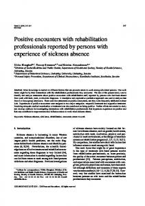
Performance was best when two face-name pairs were presented. Consequently, we have adapted the two-item face-name association task for use in an fMRI environment. Although event-related and blocked designs both have advantages and disadvantages, here we opted for a blocked design as it allows for detection of active voxels with far fewer trials than an eventrelated design, and thus minimises the time spent in the scanner – an important consideration for research involving patient groups. This design enables us to compare different groups as well as to assess functional change over time. Below we present a description of the stimuli, procedure, and image acquisition used in our proposed paradigm, as well as pilot data from a participant with aMCI who performed the task in the scanner both before and after an eight-week period of cognitive rehabilitation. 4.1. A face-name association paradigm for use in MCI and AD 4.1.1. Stimuli Figure 1 shows an example of the stimuli and presents a schematic overview of the different conditions. Twelve grey-scale pictures of Caucasian faces (6 males, 6 females, aged between 20 and 70 years) were obtained from the AR Face Database (Martinez and Benavente, 1998). All were adjusted to match a size of 7*6 cm. Each face was paired with a common two-syllable first name obtained from public lists on the internet of the most popular English names from each decade of the past century. Each face-name association was formed by a face centred on a black background with the first name printed in white below. On each learning trial, the face-name association is accompanied by a box with a tick (?) to stress that the face and name should be associated. All recognition trials are accompanied by a box with a question mark to elicit a response. The words ‘yes’ and ‘no’, together with an arrow pointing either to the left or right, are presented on the top right and left of the screen to aid participants in using the correct, previously identified button for responding. The paradigm consists of six runs with three blocks each. Each block starts and ends with a fixation dot in the centre of the screen. In each run, participants perform three tasks: associating faces with names (Encoding), deciding whether a particular association is correct or not (Recognition), and making decisions on whether a face and a gender are correctly matched or not (Control). All faces together with either a name or a gender are shown for 5800 ms with an
486
J. van Paasschen et al. / Can we change brain functioning with cognition-focused interventions
Fig. 1. Schematic representation of the face-name association task. This figure shows stimuli used during encoding (A), recognition (B), and the gender control task (C). D represents one run which includes encoding (diagonal stripes), recognition (dotted), and a gender control task (horizontal stripes). The grey bars represent the instructions given prior to the start of each condition. The black bars represent the blocks in which a fixation cross was shown.
inter-stimulus-interval of 200 ms. During the learning block, two face-name combinations are presented three times in alternating order, bringing the total viewing time to 17400 ms per association. In the recognition block, six face-name combinations are presented (three correct, three incorrect). 4.1.2. Procedure A 10-minute training session is conducted outside the scanner prior to the start of the experiment. During Encoding, an instruction is given to try to learn which faces and names belong together. In the Recognition phase, an instruction is given to answer ‘yes’ if the face and name were paired during the encoding phase, and ‘no’ if they were not, via a button press. The associations remain on the screen for the allocated time, regardless of whether a response is given. To avoid the introduction of novel stimuli, only the existing faces and names from the preceding learning block are used in a recognition block. The order of presentation is pseudo-randomised in that a face-name combination (either correct or incorrect) can never be directly fol-
lowed by the same combination, and an equal number of correct and incorrect combinations is presented. The control task block consists of one male and one female face with either ‘male’ or ‘female’ written below the picture (three correct and three incorrect combinations). Participants are asked to indicate whether the combination of face and gender is correct by giving ‘yes’ and ‘no’ answers via a button press. The stimuli are randomised as in the recall blocks. The control task is always administered last in order to reduce task switching. Two different versions are available so that a different version can be used pre and post treatment. The order of the blocks and the hand with which participants are asked to answer ‘yes’ or ‘no’ is counterbalanced. Functional images are collected for six runs. Following the functional image acquisition, once outside the scanner participants are asked to view 48 pictures (12 from the FN task, 12 from the control task, and 24 novel pictures), and indicate by means of a button press on a laptop keyboard whether they have seen the face anywhere in the experiment or not. The order of presentation is randomised. The experiment,
J. van Paasschen et al. / Can we change brain functioning with cognition-focused interventions
including functional image acquisition and post-scan recognition, has a duration of about 45 minutes. 4.2. Pilot data Here we discuss briefly some preliminary data from a participant with MCI in order to illustrate the application of the paradigm (Clare et al., in press). The participant was a 77-year-old female with eight years of education. She met criteria for amnestic MCI (Petersen, 2004), and was taking acetylcholinesterase inhibitors (Aricept, 10 mg). She scored 27/30 on the Mini Mental State Examination (MMSE; Folstein et al., 1975). The participant received eight weekly sessions of cognitive rehabilitation (Clare, 2007). She was scanned using the two-item face-name association task prior to (T1) and following (T2) the treatment period. She performed above chance level on immediate recognition of the face-name pairs both at T1 and at T2 and scored at ceiling level on the control task. She showed improvement in face-name recognition at T2 (mean score 97% correct) compared to T1 (mean score 72% correct). Functional MRI data were pre-processed and analysed using BrainVoyager QX (Brain Innovation, Maastricht, The Netherlands). The first two volumes of each run were discarded to avoid differences in T1 saturation. All images were motion-corrected and low frequency drifts were removed using a temporal high pass filter (0.0044 Hz). All data were spatially smoothed using a 4 mm FWHM Gaussian kernel. Temporal smoothing with a Gaussian kernel of 2.8 seconds FWHM was also applied to remove high frequency fluctuation. The functional data were manually coregistered with the three-dimensional anatomical scans and then resampled to isometric 3 × 3 × 3 mm voxels with trilinear interpolation. The 3D scans were transformed into Talairach space (Talairach and Tournoux, 1988). Subsequently, the co-ordinates of this transformation were applied to the co-registered functional data, which were resampled to 1 × 1 × 1 mm voxels. All blocks in the experimental task were convolved with a two gamma hemodynamic response function in order to obtain predictors for Encoding, Recognition, and Control task prior to and following the treatment period. These were entered into a fixed effects general linear model. We compared brain activation pre and post treatment for each condition separately using a t-test of this contrast. Differences in activation were considered significant at a threshold of p < 0.001 (Bonferroni corrected for multiple comparisons).
487
We detected several mainly frontally located brain regions that showed activation differences prior to and following the treatment period during encoding and recognition. No areas were detected that showed a change over time during the control task. Figure 2 shows two areas in the left inferior and middle frontal gyrus that demonstrated increased activation during encoding following treatment. During recognition we found involvement of the right inferior frontal gyrus (IFG) and right inferior parietal lobule (IPL) following the treatment period, while these areas appeared not to be involved in task performance prior to the intervention. This pattern of results supports the potential use of the task to evaluate treatment effects.
5. Discussion fMRI can be valuable for identifying biological markers of cognitive intervention. Such information can aid our understanding of the neural mechanisms through which cognitive treatment might operate. These may include restoration of neural function; promotion of the use of additional, compensatory brain regions; or reduction of aberrant brain activity. In addition, identification of brain activity predictive of treatment success could potentially be used to allocate patients to treatment groups. Currently, as far as the authors are aware, there are no fMRI studies that explore functional brain activation differences following cognitive intervention in people with MCI and AD. fMRI has been applied as an outcome measure for cognitive intervention in studies including other clinical groups such as people with schizophrenia (Wexler et al.; Wykes et al., 2002) and people with traumatic brain injury (TBI) (Laatsch and Krisky, 2006; Laatsch et al., 2004). Wexler and colleagues suggested that verbal memory could be ameliorated in some people with schizophrenia following training, in which case a behavioural improvement was accompanied by an increase in brain regions associated with verbal memory performance in healthy individuals. Similarly, Wykes et al. reported increased brain activation in people with schizophrenia following cognitive remediation therapy. Those participants for whom the intervention had been successful showed increased brain activation in areas associated with verbal working memory. Thus, although these studies are exploratory and it is not possible to draw firm conclusions from their results, their findings point in the direction of restoration of function. The participants
488
J. van Paasschen et al. / Can we change brain functioning with cognition-focused interventions Left inferior frontal gyrus
2
2
1.5
1.5
Beta value
Beta value
Left middle frontal gyrus
1 0.5
1 0.5
0
0
-0.5
-0.5 Pre-treatment
A.
Post-treatment
Pre-treatment
B.
Right te mporo-parie tal junction
2
2
1.5
1.5
Beta value
Beta value
Right inferior frontal gyrus
1 0.5
1 0.5
0
0
-0.5
-0.5 Pre-treatment
Post-treatment
Post-treatment
Pre-treatment
Post-treatment
Fig. 2. Brain areas showing a change following eight weeks of cognitive rehabilitation therapy during encoding (A) and recognition processes (B). The bar graphs represent the beta values in the brain structures showing the largest activation differences prior to and following treatment.
with TBI each showed varying patterns of change in brain activity following cognitive rehabilitation of reading ability (Laatsch and Krisky, 2006; Laatsch et al., 2004). Whereas some participants demonstrated overall increased brain activation, others showed a redistribution of activated areas, or an overall decrease in brain activity. Understandably, the participants varied in the location and extent of their lesions and in the length of time that had passed since the injury. The current paper explored the optimal parameters for a face-name learning paradigm to capture treatment effects of cognitive rehabilitation on associative memory in people with MCI and AD by reviewing existing studies that used face-name learning in the scanner with healthy older adults and people with MCI and AD. Previous studies have mainly focused on the encoding process, but we suggest it is worthwhile to collect data from both encoding and retrieval processes as it is still
unclear which of these processes is more affected in AD. Although an event-related design allows separation of successful and unsuccessful trials, a blocked design may be more realistic for frail older participants as this reduces the number of trials needed and, therefore, the time spent in the scanner. Because studies using repeated stimulus sets as a reference condition yielded poor results, we question whether this type of reference condition actually engages participants, and suggest a more engaging task in which participants make gender judgments. Some of the reviewed studies tested memory for face-name associations through free recall, but testing associative memory may be more effectively undertaken by presenting an association at retrieval as well as encoding. One striking characteristic of the reviewed studies was the high number of face-name associations participants were asked to learn. Participants in most of the studies also had high levels of educa-
J. van Paasschen et al. / Can we change brain functioning with cognition-focused interventions
tion. We suggest that these paradigms may be too challenging for the average older person, or for the range of people seen in routine clinical practice. Taking the above information into account, we designed a simpler face-name task with the aim of using it as a treatment outcome measure for people with MCI and AD. Our paradigm was tested in a pilot study with a participant with aMCI who received eight sessions of cognitive rehabilitation. Although these data are preliminary and represent only a single participant, it is nevertheless worthwhile to discuss the results in the light of the neural mechanisms that might underlie cognitive intervention. As mentioned earlier, such mechanisms could operate through restoration of function, promotion of the use of additional, compensatory brain areas, or reduction of aberrant brain activation. The pilot data discussed here show increased activation following treatment in inferior frontal brain areas during encoding, in conjunction with improved performance on the face-name association task. Inferior frontal regions are thought to be activated when participants are engaged in semantic memory strategies (Logan et al., 2002). The cognitive rehabilitation intervention may have encouraged the spontaneous use of such strategies, and reactivated an area that showed decreased processing efficiency coupled with (nearly) intact processing capacity. This explanation is compatible with the theory of restoration of function. On the other hand, during recognition there was activation in the right IFG and IPL following treatment, two areas that showed no involvement in the task prior to the intervention. It is possible that these activations represent the use of compensatory brain areas. However, it is difficult to interpret the nature of these findings based on a single participant, and these data are presented here mainly to illustrate that our paradigm can capture changes in functional brain activation following cognitive intervention. To the best of our knowledge, fMRI has not previously been used as a tool to assess the outcome of cognition-focused interventions in healthy ageing, MCI, or AD. This is important because we need to understand more about the neural mechanisms that underlie the potential success of cognitive interventions in order to begin to identify what factors predict positive outcomes in intervention studies. This information may eventually be used to identify a priori which participants may benefit from a given cognitive intervention. The current evidence on the use of fMRI as a treatment outcome measure is inconsistent, and more attention should be directed towards ensuring reliabil-
489
ity and validity of a paradigm used to assess neural change over time. Careful scrutiny of cross-sectional studies employing face-name paradigms has revealed a number of important issues that such a paradigm might need to take into account. Based on this information, we have presented a simpler face-name association task that may be used with people who have AD or MCI as a single measurement in time, for example to compare different populations, as well as to evaluate the effects of intervention through repeated measurement. Acknowledgements JVP was supported by a PhD studentship from the School of Psychology, Bangor University. The research described here was supported by a grant from the Alzheimer’s Society (UK) to L. Clare (PI), D.E.J. Linden, R.T. Woods and M.D. Rugg. We thank Sue Evans and Caroline Parkinson for their contribution to obtaining pilot data. References Achim, A. M., & Lepage, M. (2005). Neural correlates of memory for items and for associations: an event-related functional magnetic resonance imaging study. Journal of Cognitive Neuroscience, 17, 652-667. Aylward, E. H., Richards, T. L., Berninger, V. W., Nagy, W. E., Field, K. M., Grimme, A. C., et al. (2003). Instructional treatment associated with changes in brain activation in children with dyslexia. Neurology, 61, 212-219. Baddeley, A. D., Emslie, H., & Nimmo-Smith, I. (1994). Doors and People: A Test of Visual and Verbal Recall and Recognition. Bury St. Edmunds: Thames Valley Test Company. Barrett, A. M., Crucian, G. P., Schwartz, R. L., & Heilman, K. M. (2000). Testing memory for self-generated items in dementia: method makes a difference. Neurology, 54, 1258-1264. Buckner, R. L., Snyder, A. Z., Shannon, B. J., LaRossa, G., Sachs, R., Fotenos, A. F., et al. (2005). Molecular, structural, and functional characterization of Alzheimer’s disease: evidence for a relationship between default activity, amyloid, and memory. J Neurosci, 25, 7709-7717. Celone, K. A., Calhoun, V. D., Dickerson, B. C., Atri, A., Chua, E. F., Miller, S. L., et al. (2006). Alterations in memory networks in mild cognitive impairment and Alzheimer’s disease: an independent component analysis. J Neurosci, 26, 10222-10231. Chalfonte, B. L., & Johnson, M. K. (1996). Feature memory and binding in young and older adults. Mem Cognit, 24, 403-416. Clare, L. (2007). Neuropsychological rehabilitation and people with dementia. Hove: Psychology Press. Clare, L., Van Paasschen, J., Evans, S., Parkinson, C., Woods, R. T., & Linden, D. E. (in press). Goal-oriented cognitive rehabilitation for an individual with Mild Cognitive Impairment: behavioural and neuroimaging outcomes. Neurcase.
490
J. van Paasschen et al. / Can we change brain functioning with cognition-focused interventions
Clare, L., Wilson, B. A., Carter, G., Breen, K., Gosses, A., & Hodges, J. R. (2000). Intervening with everyday memory problems in dementia of Alzheimer type: an errorless learning approach. J Clin Exp Neuropsychol, 22, 132-146.
Huettel, S. A., Song, A. W., & McCarthy, G. (2004). Functional Magnetic Resonance Imaging. Sunderland, MA: Sinauer Associates.
Clare, L., Wilson, B. A., Carter, G., Roth, I., & Hodges, J. R. (2002). Relearning face-name associations in early Alzheimer’s disease. Neuropsychology, 16, 538-547.
Kircher, T. T., Erb, M., Grodd, W., & Leube, D. T. (2005). Cortical activation during cholinesterase-inhibitor treatment in Alzheimer disease: preliminary findings from a pharmacofMRI study. Am J Geriatr Psychiatry, 13, 1006-1013.
Dale, A. M., & Buckner, R. L. (1997). Selective averaging of rapidly presented individual trials using fMRI. Human Brain Mapping, 5, 329-340.
Laatsch, L., & Krisky, C. (2006). Changes in fMRI activation following rehabilitation of reading and visual processing deficits in subjects with traumatic brain injury. Brain Inj, 20, 1367-1375.
Daselaar, S. M., Veltman, D. J., Rombouts, S. A., Raaijmakers, J. G., & Jonker, C. (2003). Neuroanatomical correlates of episodic encoding and retrieval in young and elderly subjects. Brain, 126, 43-56.
Laatsch, L. K., Thulborn, K. R., Krisky, C. M., Shobat, D. M., & Sweeney, J. A. (2004). Investigating the neurobiological basis of cognitive rehabilitation therapy with fMRI. Brain Inj, 18, 957-974.
Department for Children, Schools, and Families (2007). Retrieved 15 August, 2008, from http://www.dcsf.gov.uk/rsgateway/DB/ SFR/s000777/sfrdius01-2008.pdf.
Lindeboom, J., Schmand, B., Tulner, L., Walstra, G., & Jonker, C. (2002). Visual association test to detect early dementia of the Alzheimer type. J Neurol Neurosurg Psychiatry, 73, 126-133.
Dickerson, B. C., Salat, D. H., Greve, D. N., Chua, E. F., RandGiovannetti, E., Rentz, D. M., et al. (2005). Increased hippocampal activation in mild cognitive impairment compared to normal aging and AD. Neurology, 65, 404-411.
Linden, D. E. (2006). How psychotherapy changes the brain–the contribution of functional neuroimaging. Mol Psychiatry, 11, 528-538.
Donders, F. C. (1969). On the speed of mental processes. Acta Psychol (Amst), 30, 412-431. Folstein, M. F., Folstein, S. E., & McHugh, P. R. (1975). Mini-mental state. A practical method for grading the cognitive state of patients for the clinician. J Psychiatr Res, 12, 189-198.
Linden, D. E. (2007). The working memory networks of the human brain. Neuroscientist, 13, 257-267. Logan, J. M., Sanders, A. L., Snyder, A. Z., Morris, J. C., & Buckner, R. L. (2002). Under-recruitment and nonselective recruitment: dissociable neural mechanisms associated with aging. Neuron, 33, 827-840.
Fowler, K. S., Saling, M. M., Conway, E. L., Semple, J. M., & Louis, W. J. (2002). Paired associate performance in the early detection of DAT. J Int Neuropsychol Soc, 8, 58-71.
Lustig, C., Snyder, A. Z., Bhakta, M., O’Brien, K. C., McAvoy, M., Raichle, M. E., et al. (2003). Functional deactivations: change with age and dementia of the Alzheimer type. Proc Natl Acad Sci USA, 100, 14504-14509.
Friston, K. J., Price, C. J., Fletcher, P., Moore, C., Frackowiak, R. S., & Dolan, R. J. (1996). The trouble with cognitive subtraction. Neuroimage, 4, 97-104.
Martinez, A. M., & Benavente, R. (1998). The AR Face database. CVC technical report # 24, June.
Goekoop, R., Scheltens, P., Barkhof, F., & Rombouts, S. A. (2006). Cholinergic challenge in Alzheimer patients and mild cognitive impairment differentially affects hippocampal activation–a pharmacological fMRI study. Brain, 129, 141-157. Gould, R. L., Brown, R. G., Owen, A. M., Bullmore, E. T., Williams, S. C., & Howard, R. J. (2005). Functional neuroanatomy of successful paired associate learning in Alzheimer’s disease. Am J Psychiatry, 162, 2049-2060. Grady, C. L. (2008). Cognitive neuroscience of aging. Ann N Y Acad Sci, 1124, 127-144. Grady, C. L., & Craik, F. I. (2000). Changes in memory processing with age. Curr Opin Neurobiol, 10, 224-231. Grady, C. L., McIntosh, A. R., Beig, S., Keightley, M. L., Burian, H., & Black, S. E. (2003). Evidence from functional neuroimaging of a compensatory prefrontal network in Alzheimer’s disease. J Neurosci, 23, 986-993.
National Assembly for Wales (2006). Retrieved 15 August, 2008, from http://new.wales.gov.uk/docrepos/40382/40382313/ statistics/post16/post16-2006/sb79-2006r.pdf?lang=en. Naveh-Benjamin, M. (2000). Adult age differences in memory performance: tests of an associative deficit hypothesis. J Exp Psychol Learn Mem Cogn, 26, 1170-1187. Naveh-Benjamin, M., Guez, J., Kilb, A., & Reedy, S. (2004). The associative memory deficit of older adults: further support using face-name associations. Psychol Aging, 19, 541-546. Olesen, P. J., Westerberg, H., & Klingberg, T. (2004). Increased prefrontal and parietal activity after training of working memory. Nat Neurosci, 7, 75-79. Pariente, J., Cole, S., Henson, R., Clare, L., Kennedy, A., Rossor, M., et al. (2005). Alzheimer’s patients engage an alternative network during a memory task. Ann Neurol, 58, 870-879. Petersen, R. C. (2004). Mild cognitive impairment as a diagnostic entity. Journal of Internal Medicine, 256, 183-194.
Greene, J. D., Baddeley, A. D., & Hodges, J. R. (1996). Analysis of the episodic memory deficit in early Alzheimer’s disease: evidence from the doors and people test. Neuropsychologia, 34, 537-551.
Petersen, S. E., Fox, P. T., Posner, M. I., Mintun, M. A., & Raichle, M. E. (1989). Positron emission tomography of the processing of single words. Journal of Cognitive Neuroscience, 1, 153-170.
Herholz, K., Ehlen, P., Kessler, J., Strotmann, T., Kalbe, E., & Markowitsch, H. J. (2001). Learning face-name associations and the effect of age and performance: a PET activation study. Neuropsychologia, 39, 643-650.
Petrella, J. R., Krishnan, S., Slavin, M. J., Tran, T. T., Murty, L., & Doraiswamy, P. M. (2006). Mild cognitive impairment: evaluation with 4-T functional MR imaging. Radiology, 240, 177-186.
J. van Paasschen et al. / Can we change brain functioning with cognition-focused interventions Petrella, J. R., Wang, L., Krishnan, S., Slavin, M. J., Prince, S. E., Tran, T. T., et al. (2007). Cortical deactivation in mild cognitive impairment: high-field-strength functional MR imaging. Radiology, 245, 224-235. Prvulovic, D., Van de Ven, V., Sack, A. T., Maurer, K., & Linden, D. E. (2005). Functional activation imaging in aging and dementia. Psychiatry Research, 140, 97-113. Raichle, M. E., MacLeod, A. M., Snyder, A. Z., Powers, W. J., Gusnard, D. A., & Shulman, G. L. (2001). A default mode of brain function. Proc Natl Acad Sci USA, 98, 676-682. Rand-Giovannetti, E., Chua, E. F., Driscoll, A. E., Schacter, D. L., Albert, M. S., & Sperling, R. A. (2006). Hippocampal and neocortical activation during repetitive encoding in older persons. Neurobiol Aging, 27, 173-182. Rombouts, S. A., Barkhof, F., Van Meel, C. S., & Scheltens, P. (2002). Alterations in brain activation during cholinergic enhancement with rivastigmine in Alzheimer’s disease. J Neurol Neurosurg Psychiatry, 73, 665-671. Sandstrom, C. K., Krishnan, S., Slavin, M. J., Tran, T. T., Doraiswamy, P. M., & Petrella, J. R. (2006). Hippocampal atrophy confounds template-based functional MR imaging measures of hippocampal activation in patients with mild cognitive impairment. AJNR Am J Neuroradiol, 27, 1622-1627. Saykin, A. J., Wishart, H. A., Rabin, L. A., Flashman, L. A., McHugh, T. L., Mamourian, A. C., et al. (2004). Cholinergic enhancement of frontal lobe activity in mild cognitive impairment. Brain, 127, 1574-1583. Sperling, R. A., Bates, J. F., Chua, E. F., Cocchiarella, A. J., Rentz, D. M., Rosen, B. R., et al. (2003a). fMRI studies of associative encoding in young and elderly controls and mild Alzheimer’s disease. J Neurol Neurosurg Psychiatry, 74, 44-50. Sperling, R. A., Chua, E., Cocchiarella, A., Rand-Giovannetti, E., Poldrack, R., Schacter, D. L., et al. (2003b). Putting names to faces: successful encoding of associative memories activates the anterior hippocampal formation. Neuroimage, 20, 14001410. Talairach, J., & Tournoux, P. (1988). Co-Planar Stereotaxic Atlas of the Human Brain: 3-Dimensional Proportional System: An Approach to Cerebral Imaging. Stuttgart: Thieme Medical
491
Publishers. Temple, E., Deutsch, G. K., Poldrack, R. A., Miller, S. L., Tallal, P., Merzenich, M. M., et al. (2003). Neural deficits in children with dyslexia ameliorated by behavioral remediation: evidence from functional MRI. Proc Natl Acad Sci USA, 100, 28602865. Troyer, A. K., H¨afliger, A., Cadieux, M. J., & Craik, F. I. (2006). Name and face learning in older adults: effects of level of processing, self-generation, and intention to learn. J Gerontol B Psychol Sci Soc Sci, 61, P67-74. US Census Bureau (2006). Retrieved 15 August, 2008, from http:// factfinder.census.gov/servlet/STTable? bm=y&-geo id= 01000US&-qr name=ACS 2006 EST G00 S1501&-ds name =ACS 2006 EST G00 . Warrington, E. K. (1984). Recognition Memory Test manual. Windsor, UK: Nfer-Nelson. Wechsler, D. (1997). Wechsler Memory Scale - Third edition. San Antonio, TX: Psychological Corporation. Westerberg, H., & Klingberg, T. (2007). Changes in cortical activity after training of working memory–a single-subject analysis. Physiol Behav, 92, 186-192. Wexler, B. E., Anderson, M., Fulbright, R. K., & Gore, J. C. (2000). Preliminary evidence of improved verbal working memory performance and normalization of task-related frontal lobe activation in schizophrenia following cognitive exercises. Am J Psychiatry, 157, 1694-1697. Wilkinson, D., & Halligan, P. (2004). The relevance of behavioural measures for functional-imaging studies of cognition. Nat Rev Neurosci, 5, 67-73. Wilson, B., Cockburn, J., & Baddeley, A. D. (1985). The Rivermead Behavioural Memory Test. Titchfield: Thames Valley Test Company. Wykes, T., Brammer, M., Mellers, J., Bray, P., Reeder, C., Williams, C., et al. (2002). Effects on the brain of a psychological treatment: cognitive remediation therapy: functional magnetic resonance imaging in schizophrenia. Br J Psychiatry, 181, 144-152.


![[Read] ['Creative] [Change;] [Why] [We] [Resist] [It...How] [We] [Can ...](https://m.moam.info/img/260x300/read-creative-change-why-we-resist-ithow-we-can-_6479a77e097c476c028b579e.jpg)