Sep 3, 1998 - scopic examination of the patient's urine demonstrated ú1,000 pyelonephritis and ... developed increasing testicular swelling and pain, and findings manipulation, may be .... Urinary retention, intermittent self-. Urine culture ...
942
Candidal Epididymo-Orchitis: Case Report and Review Grant A. Jenkin, Marcus Choo, Patrick Hosking, and Paul D. R. Johnson
From the Infectious Diseases Service and Department of Anatomical Pathology, Austin & Repatriation Medical Centre, Heidelberg, Victoria, Australia
We describe a case of epididymo-orchitis with candiduria and histologically proven epididymal abscesses due to Candida albicans and review six previously reported cases. Candidal epididymoorchitis occurs in patients with recognized risk factors for candidal infection, often after instrumentation of the urinary tract. Cases caused by both C. albicans and Candida glabatra have been described. Drainage or orchidectomy may be required for definitive diagnosis and treatment. Treatment with oral antifungals alone has been effective in two cases.
Candida species — predominantly C. albicans, but including C. glabatra — are commonly isolated from urine, particularly among patients with indwelling catheters. This circumstance most often represents colonization, but the spectrum of candidal urinary tract disease includes cystitis, fungus ball formation, pyelonephritis and perinephric abscess, emphysematous cystitis and pyelonephritis, and prostatitis [1]. Identified risk factors include the presence of an indwelling catheter or other bladder instrumentation, diabetes mellitus, and prior antibiotic therapy. The increase in numbers of elderly and immunocompromised patients, combined with the frequency of genitourinary tract manipulation, may be expected to lead to an increase in the incidence of urinary candidiasis. We present a case of C. albicans epididymo-orchitis with epididymal abscesses and review six previously reported cases.
Case Report A 72-year-old man with type 2 diabetes mellitus and a history of completely resected lung carcinoma without evidence of recurrence underwent elective transurethral resection of the prostate (TURP) for benign prostatic hyperplasia. After the surgery, he developed a group B streptococcal urinary tract infection and received intravenous cefotaxime for 2 days, followed by oral cephalexin for 10 days. One month later he presented with bilateral epididymo-orchitis. Microscopic examination of a urine specimen showed ú1,000 1 103 WBCs/mL, 60 1 103 RBCs/mL, and 1/ yeast cells; culture yielded C. albicans (10,000 colonies/mL). Treatment with intravenous amoxicillin and cefotaxime was begun for presumed bacterial epididymo-orchitis despite the fact that no bacteria had been isolated from the urine at this time. A scrotal ultrasonogram
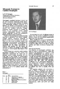
showed marked epididymal swelling and moderate bilateral hydroceles but no abscess. Blood cultures were negative. Oral fluconazole (200 mg daily) was added to the regimen 7 days after admission because of slow clinical response. After nine days of treatment with oral fluconazole, repeated microscopic examination of the patient’s urine demonstrated ú1,000 1 103 WBCs/mL, 120 1 103 RBCs/mL but no yeasts, and culture was negative. Fluconazole therapy was ceased at this point because it was considered that the pathogen was most likely bacterial, and the patient was discharged with a prescription for oral amoxicillin/clavulanic acid. Twelve days later he developed increasing testicular swelling and pain, and findings on an ultrasonogram suggested an abscess in the upper pole of the right testis. Bilateral orchidectomy was performed. Macroscopically, both specimens showed inflammation and thickening of the tunica vaginalis, which contained a moderate amount of purulent fluid. Bilateral epididymal abscesses were found that measured 10 1 10 mm on the right and 15 1 15 mm on the left. Microscopic examination with periodic acid – Schiff and Grocott-Gomori methenamine – silver nitrate stains revealed fungal hyphae and spores, consistent with a Candida species (figure 1). Ziehl-Neelsen and gram stains were negative. Swabs of the operative specimens showed 3/ polymorphonuclear leukocytes, but cultures were negative, and surgical tissue specimens were not submitted for culture. It seems likely that the patient’s infection was due to the C. albicans previously isolated from urine despite the negative intraoperative swab culture. A review of the histopathological findings after TURP showed adenomatous hyperplasia of prostatic glands and stroma but no evidence of active prostatitis or fungal infection, and prostatic fluid was not submitted for culture. The patient received a further 6-week course of oral fluconazole (200 mg daily) and recovered satisfactorily. Discussion
Received 21 July 1997; revised 14 November 1997. Reprints or correspondence: Dr. Grant Jenkin, Infectious Diseases Registrar, Victorian Infectious Diseases Service, Royal Melbourne Hospital, Grattan St. Parkville, Victoria 3050, Australia. Clinical Infectious Diseases 1998;26:942–5 q 1998 by The University of Chicago. All rights reserved. 1058–4838/98/2604–0022$03.00
/ 9c4a$$ap25
03-09-98 18:11:45
Candidal epididymo-orchitis is an uncommon manifestation of candidal urinary tract disease. Our case is the seventh reported in the English-language literature and the fifth in which the diagnosis was confirmed by either histology or culture of an aspirate (table 1).
cida
UC: CID
CID 1998;26 (April)
Candidal Epididymo-Orchitis
943
Figure 1. A, Epididymal abscess in the upper left of the microscopic field, separated from adjacent uninvolved testicular parenchyma by a fibrous tissue capsule, from a patient with candidal epididymo-orchitis. (Original magnification, 140; stain, hematoxylin-eosin.) B, High-power view showing thin septate hyphae (large arrow) with oval budding bodies (short arrow), typical of Candida species, within a background of degenerate inflammatory cells in the center of the abscess. (Original magnification, 1400; stain, Grocott-Gomori methenamine – silver.)
All but one of the patients were elderly (mean age, 69.1 years) and had identifiable risk factors for candidal urinary tract infection — i.e., diabetes mellitus, bladder instrumentation or urinary outflow obstruction, previous broad-spectrum antibiotic therapy, or HIV infection. It therefore seems likely that the etiology of candidal epididymo-orchitis in these patients was retrograde spread of organisms from urine into the epididymis. This is the usual mechanism for the development of bacte-
rial epididymo-orchitis in men ú35 years of age, particularly after instrumentation of the urinary tract. Hematogenous dissemination seems less likely, and in both cases where candidemia was diagnosed, the authors considered the epididymoorchitis to be the primary infection. Three patients, including ours, had documented negative blood cultures. All patients presented with scrotal swelling and pain typical of epididymo-orchitis, which was bilateral in three; the duration
Table 1. Cases of candidal epididymo-orchitis described in the English-language literature. Patient no. [reference]
Age (y)
Candida species
Treatment
Urine culture (ú100,000 colonies/mL) Urine culture Urine culture (ú100,000 colonies/mL), orchidectomy culture, orchidectomy histology
C. albicans
Ketoconazole, 200 mg/d, for 6 w
C. albicans C. albicans
Fluconazole, 200 mg/d, for 6 w Orchidectomy (unilateral), amphotericin B bladder irrigation
Blood culture, urine culture, culture of abscess, testicular biopsy histology Urine culture, blood culture, abscess aspirate culture Urine culture (ú100,000 colonies/mL), aspirate culture, histology Urine culture (10,000 colonies/mL), histology
C. glabatra
Open drainage, amphotericin B
C. glabatra
Open drainage, amphotericin B, 0.7 g, / flucytosine, 1.5 g b.i.d., for 2 w Fluconazole, 200 mg/d, open drainage, orchidectomy (unilateral) Orchidectomy (bilateral), fluconazole, 200 mg/d for 9 d and 6 w
Underlying condition(s)
1 [2]
45
HIV infection
2 [3] 3 [4]
65 66
4 [5]
84
Diabetes mellitus, TURP Diabetes mellitus, recurrent bacterial urinary tract infections, vesicoureteric reflux, urethral false passage from catheterization, broad-spectrum antibiotic therapy Diabetes mellitus
5 [6]
83
6 [7]
69
7 [PR]
72
Diabetes mellitus, TURP, squamous cell carcinoma of bladder, urinary outflow obstruction Urinary retention, intermittent selfcatheterization, broad-spectrum antibiotic therapy Diabetes mellitus, TURP, broadspectrum antibiotic therapy
Diagnosis
C. glabatra
C. albicans
NOTE. PR Å present report; TURP Å transurethral resection of prostate.
/ 9c4a$$ap25
03-09-98 18:11:45
cida
UC: CID
944
Jenkin et al.
of symptoms ranged from 5 days to 5 months. In contrast, acute bacterial epididymo-orchitis is usually unilateral, with onset over 1 – 2 days. Cultures of urine yielded Candida species in pure growth for all patients, although three patients were receiving antibiotic treatment at the time that the first urine specimen was obtained. On urine microscopy, budding yeast forms were seen in five patients, and pyuria was reported in four cases but not commented on in three. A Candida colony count of ú10,000/mL in a midstream catch urine specimen has been used to differentiate infection from colonization, although the validity of this criterion is uncertain [8]. Colony counts were reported for four of the patients described in table 1, and three had counts of ú100,000/mL; however, one of the specimens was from a catheter, which invalidates colony counts. Nevertheless, our patient’s count was 10,000/mL; using this figure as a cutoff could lead to misdiagnosis of true infections. Although persistence of candiduria in at least two separate urine specimens does not imply disease, and indeed asymptomatic candiduria may persist for many months [9], we would expect that patients with candidal epididymo-orchitis would have persistent candiduria. Unfortunately, results of multiple urine examinations were reported for only two of the patients before antifungal therapy was initiated, although both had persistent candiduria. Assays for detection of Candida antigen have variable sensitivity but may assist in the diagnosis of disseminated candidiasis in conjunction with other laboratory and clinical features [10]. These assays may be of less value for patients with focal invasive candidiasis [11], and the only patient (patient 3) for whom a serum Candida antigen test was reported had an equivocal result and negative blood cultures. Although no deaths were reported, the associated morbidity, particularly local abscess formation, was significant: orchidectomy was necessary in three cases. In addition, two patients developed candidemia. The age and/or underlying medical conditions of these patients may have contributed to abscess formation, but the delay in diagnosis could also have been important. Where it could be determined, the initiation of antifungal therapy occurred from 5 to 10 days after the first documentation of candiduria and from 7 days to 5 months after the first symptoms of epididymo-orchitis. The delay in commencing appropriate antifungal therapy despite the presence of documented candiduria probably reflects the difficulty in differentiating candidal colonization from true infection. For four patients, antifungal treatment was begun presumptively because they did not respond to antibiotic therapy. For three other patients, treatment was started after blood cultures or culture of an aspirate was reported to be positive. Relying on lack of response to antibacterials to diagnose candidal epididymo-orchitis can contribute significantly to therapeutic delays. Failure to isolate a causative bacterium, in association with the presence of candiduria and risk factors for candidal infection, should prompt early consideration of a diagnosis of candidal epididymo-orchitis. For patients with
/ 9c4a$$ap25
03-09-98 18:11:45
CID 1998;26 (April)
complicated epididymo-orchitis, percutaneous or open drainage should be considered to obtain specimens for diagnostic microbiology and histology as well as for therapeutic reasons. This approach is particularly important in the setting of immunosuppression, where other unusual organisms have been reported and progression to abscess formation may be more common [12]. Treatment with oral antifungals alone for 6 weeks was successful in patients 1 and 2, both of whom had C. albicans isolated from urine. The success of antifungals alone in these two patients was likely not related to early diagnosis because patient 1 had symptoms for 6 weeks before receiving therapy with oral ketoconazole. Neither of these patients had evidence of local abscess formation. In contrast, patient 6 required surgical drainage of an epididymal abscess despite the fact that he had undergone prior percutaneous drainage and had received fluconazole. Susceptibility results were not reported for his C. glabatra isolate, although subsequent treatment with fluconazole was successful. Although there may be a role for antifungal therapy alone with close monitoring of such patients, open drainage or orchidectomy will generally be required if an abscess, pyocele, or other local complication develops, as indicated by clinical evidence of treatment failure and findings on ultrasonography. Oral fluconazole, in a dose of 200 – 400 mg daily for £6 weeks, is an attractive treatment option for this infection. Fluconazole has proven efficacy for candidal infections, including candidemia in nonneutropenic hosts, at a dose of 400 mg/d [13]. Oral administration results in high concentrations of the drug in urine and tissues, although specific data on its penetration into epididymal and testicular tissue are not available [14], and fluconazole is generally well tolerated. C. glabatra accounted for three of the seven isolates recovered. Isolation of this organism has therapeutic implications because of its relative intrinsic resistance to fluconazole [15, 16], as seen in the case of patient 5, and because of the possibility of resistance developing during treatment [17]. Treatment with systemic amphotericin B, in addition to surgical drainage, was successful in two patients (in one of these patients, it was successful in combination with flucytosine for a fluconazole-resistant C. glabatra isolate). Susceptibility testing may be of value in guiding therapy, particularly with non-albicans Candida species [18], and amphotericin B, with or without flucytosine, would generally be an effective alternative agent in this situation. In conclusion, we recommend that the diagnosis of candidal epididymo-orchitis be considered in patients with risk factors for candiduria and histories of current or recent urinary tract instrumentation who present with epidiymo-orchitis, particularly bilateral or complicated, and candiduria. Repeating a urine culture is likely to be of benefit because if a patient’s candiduria resolves, the development of candidal epidiymo-orchitis would be unlikely. Urine cultures are usually accurate in predicting the etiology of epididymo-orchitis in this age group, and clinicians
cida
UC: CID
CID 1998;26 (April)
Candidal Epididymo-Orchitis
should probably trust the results. We recommend that such patients be seen by a urologist and that early scrotal ultrasonography be performed to detect local complications, since surgical intervention is likely to be required if an abscess forms. Antibiotic therapy should be ceased if not required for other reasons; diabetes mellitus, if present, should be controlled; and urinary catheters should be removed, if possible. We recommend starting therapy with oral fluconazole, 200 – 400 mg/d, choosing the higher dose particularly if the Candida species is likely to be relatively resistant to fluconazole or if an abscess or candidemia is present. If resistance to fluconazole is likely (e.g., the isolate is C. krusei) or confirmed on testing, then intravenous amphotericin B (0.5 mg/[kgrd]), with or without flucytosine, would be preferred. The duration of treatment should, of course, be guided by clinical and microbiological response. If drainage is not performed, we would favor a 6-week course of systemic antifungal therapy. If complete surgical drainage or resection is required, then 2 weeks of postoperative therapy would probably suffice.
References 1. Crislip MA, Edwards JE. Candidiasis. In: Systemic fungal infections — diagnosis and treatment part II. Infect Dis Clin North Am 1989; 3: 103 – 33. 2. Swartz DA, Harrington P, Wilcox R. Candidal epididymitis treated with ketoconazole [letter]. N Engl J Med 1988; 319:1485. 3. Gordon DL, Maddern J. Treatment of candida epididymo-orchitis with oral fluconazole [letter]. Med J Aust 1992; 156:744. 4. Docimo SG, Rukstalis DB, Rukstalis MR, Kang J, Cotton D, DeWolf WC. Candida epididymitis: newly recognized opportunistic epididymal infection. Urology 1993; 41:280 – 2.
/ 9c4a$$ap25
03-09-98 18:11:45
945
5. Sheaff M, Ahsan Z, Badenoch D, Baithun S. A rare cause of epididymoorchitis. Br J Urol 1995; 75:250 – 1. 6. Jenks P, Brown J, Warnock D, Barnes N. Candida glabatra epididymoorchitis: an unusual infection rapidly cured with surgical and antifungal treatment. J Infect 1995; 31:71 – 2. 7. Lyne JC, Flood HD. Bilateral fungal epididymo-orchitis with abscess. Urology 1995; 46:412 – 4. 8. Edwards JE Jr. Candida species. In: Mandell GL, Bennett JE, Dolin R, eds. Mandell, Douglas and Bennett’s principles and practice of infectious diseases. 4th ed. New York: Churchill Livingstone, 1995:2289 – 306. 9. Wong-Beringer A, Jacobs RA, Guglielmo J. Treatment of funguria. JAMA 1992; 267:2780 – 5. 10. Hopfer RL. Contemporary techniques for molecular diagnoses of mycoses. Clinical Microbiology Newsletter 1997; 19:169 – 73. 11. Phillips P, Dowd A, Jewesson P, et al. Nonvalue of antigen detection immunoassays for diagnosis of candidaemia. J Clin Microbiol 1990; 28: 2320 – 6. 12. Parr NJ, Prasad BRP, Hayhurst V, McMillan A, Leen CS, Fowler JW. Suppurative epididymo-orchitis in young ‘‘high risk’’ patients — a new problem? Br J Urol 1993; 72:949 – 51. 13. Rex JH, Bennett JE, Sugar AM, et al. A randomized trial comparing fluconazole with amphotericin B for the treatment of candidaemia in patients without neutropenia. N Engl J Med 1994; 331:1325 – 30. 14. Debruyne D, Ryckelynck JP. Clinical pharmacokinetics of fluconazole. Clin Pharmacokinet 1993; 24:10 – 27. 15. Como JA, Dismukes WE. Oral azole drugs as systemic antifungal therapy. N Engl J Med 1994; 330:263 – 72. 16. Price MF, LaRocco MT, Gentry LO. Fluconazole susceptibilities of Candida species and distribution of species recovered from blood cultures over a 5-year period. Antimicrob Agents Chemother 1994; 38:1422 – 4. 17. Hitchcock CA, Pye GW, Troke PF, Johnson EM, Warnock DW. Fluconazole resistance in Candida glabatra. Antimicrob Agents Chemother 1993; 37:1962 – 5. 18. Rex JH, Pfaller MA, Galgiani JN, et al. Development of interpretive breakpoints for antifungal susceptibility testing: conceptual framework and analysis of in vitro – in vivo correlation data for fluconazole, itraconazole, and candida infections. Clin Infect Dis 1997; 24:235 – 47.
cida
UC: CID