Cannabidiol, unlike synthetic cannabinoids, triggers activation of RBL-2H3 mast cells Elda Del Giudice,* Luciano Rinaldi,* Marzia Passarotto,* Fabrizio Facchinetti,* Antonello D’Arrigo,* Adriano Guiotto,† Maurizio Dalle Carbonare,* Leontino Battistin,‡ and Alberta Leon*,‡,1 *Research and Innovation (R&I) Company, Padova, Italy; †Department of Pharmaceutical Sciences, University of Padova, Padova, Italy; and ‡San Camillo Hospital, IRCCS, Venezia-Lido, Italy
Abstract: Cannabidiol (CBD), a prominent psychoinactive component of cannabis with negligible affinity for known cannabinoid receptors, exerts numerous pharmacological actions, including antiinflammatory and immunosuppressive effects, the underlying mechanisms of which remain unclear. In the current study, we questioned whether CBD modulates activation of mast cells, key players in inflammation. By using the rat basophilic leukemia mast cell line (RBL-2H3), we demonstrate that CBD (3–10 M) augments -hexosaminidase release, a marker of cell activation, from antigenstimulated and unstimulated cells via a mechanism, which is not mediated by Gi/Go protein-coupled receptors but rather is associated with a robust rise in intracellular calcium ([Ca2ⴙ]i) levels sensitive to clotrimazole and nitrendipine (10 –30 M). This action, although mimicked by ⌬9-tetrahydrocannabinol (THC), is opposite to that inhibitory, exerted by the synthetic cannabinoids WIN 55,212-2 and CP 55,940. Moreover, the vanilloid capsaicin, a full agonist of transient receptor potential channel VR1, did not affect [Ca2ⴙ]i levels in the RBL2H3 cells, thus excluding the involvement of this receptor in the CBD-mediated effects. Together, these results support existence of yet-to-be identified sites of interaction, i.e., receptors and/or ion channels associated with Ca2ⴙ influx of natural cannabinoids such as CBD and THC, the identification of which has the potential to provide for novel strategies and agents of therapeutic interest. J. Leukoc. Biol. 81: 1512–1522; 2007. Key Words: -hexosaminidase 䡠 calcium 䡠 ion channels 䡠 ⌬9tetrahydrocannabinol 䡠 CB receptors 䡠 VR1 receptors 䡠 TRP channels
INTRODUCTION Plant-derived cannabinoids, such as ⌬9-tetrahydrocannabinol (THC) and cannabidiol (CBD), the main psychoactive and nonpsychoactive components of cannabis, respectively, possess myriad pharmacological properties, many of which are mimicked by their synthetic (e.g., CP 55,940) as well as endogenous counterparts, the endocannabinoids (e.g., arachi1512
Journal of Leukocyte Biology Volume 81, June 2007
donylethanolamide, also known as anandamide). However, although much evidence points to a role of known cannabinoid receptors, namely CB1 and CB2 [1, 2], in determining some of the effects produced by most cannabinoids/endocannabinoids, CBD binds with low affinity to these receptors [3, 4]. Yet, CBD exerts, in analogy to THC and other CB receptor agonists, immunosuppressive and anti-inflammatory properties, the combination of which has, for example, been shown to culminate in potent, antiarthritic effects in experimental models of chronic joint inflammation [5]. CBD also reduces edema and hyperalgesia in a rat paw model of carrageenan-induced inflammation as well as serum TNF-␣ levels in LPS-treated mice [6, 7]. Studies directly investigating the effects of CBD on specific immune cell populations include suppression of NO production and cytokine release in macrophages, modulation of cytokine release in PBMC and suppression of chemokine production in a human B cell line [8 –11]. Notwithstanding this, the mode(s) of action of CBD are still largely obscure. Although mechanisms related with the endocannabinoid system have been proposed in, at least, the antihyperalgesic actions of CBD, other evidence provides for alternative mechanisms by which CBD can induce immunomodulatory and anti-inflammatory effects [3, 6, 7, 12, 13], the further elucidation of which has the potential to open new avenues in the pharmacology of cannabinoids. Mast cells, traditionally associated with immediate hypersensitivity and allergic reactions, play significant roles in the defense against bacteria and parasites as well as in the pathogenesis of a variety of disorders (e.g., autoimmune diseases such as rheumatoid arthritis and multiple sclerosis, congestive heart failure, and pain, among others) [14 –16]. These cells, widely distributed throughout connective and mucosal tissues, often adjacent to blood and lymphatic vessels and in close contact to nerves, can in fact be activated by a variety of stimuli to secrete and produce a battery of mediators with proinflammatory and immunoregulatory potential. However, although activated mast cells may, by virtue of their location and mediator expression, be pivotal in the genesis, magnitude, or
1
Correspondence: Research and Innovation (R&I) Company, Via Svizzera 16, 35127 Padova, Italy. E-mail:
[email protected] Received December 20, 2006; revised January 26, 2007; accepted February 5, 2007. doi: 10.1189/jlb.1206738
0741-5400/07/0081-1512 © Society for Leukocyte Biology
duration of an inflammatory response in many tissues, the balance of engaging inhibitory and activatory cell-surface receptors on mast cells determines whether the cell becomes active upon a challenge. Moreover, complex molecular, intracellular networks control the extent, persistence, or both of the secretory response, and genetic and environmental factors control their mediator content as well as other aspects of their phenotype. Therefore, elucidating how mast cell function may be modulated has the potential to provide insight into how mast cells might be manipulated to achieve therapeutic ends. Given the anti-inflammatory properties of CBD together with the potential, although controversial, of cannabinoids/endocannabinoids to modulate mast cell activation [17–22], we questioned here, for the first time, whether CBD affects mast cell function. To this end, we used RBL-2H3 cell cultures, a rat cell line of basophilic-leukemia origin, displaying characteristics of mucosal mast cells, used extensively to study signaling pathways, leading to degranulation and release of inflammatory mediators upon antigen-induced aggregation of the IgE-bound, high-affinity receptors (FcεRIs) expressed on their surface [23–25]. It is surprising that our results show that exposure of the RBL-2H3 cells to CBD results in their activation. This effect, similar to that exerted by THC, is opposite to that inhibitory of the synthetic CB receptor agonists WIN 55,212-2 and CP 55,940.
MATERIALS AND METHODS Materials CP 55,940 [(–)-cis-3-[2-hydroxy4-(1,1-dimethylheptyl)phenyl]-trans-4-(3-hydroxypropyl) cyclohexanol]; WIN 55,212-2 [R-(⫹)-[2,3-dihydro-5-methyl-3[(morpholinyl)methyl]pyrrolo[1,2,3-de]-1,4-benzoxazin-yl]-(1-naphthalenyl)methanone mesylate]; JWH-133 [(6aR,10aR)-3-(1,1-dimethylbutyl)6a,7,10,10a-tetrahydro-6,6,9-trimethyl-6H-dibenzo[b,d]pyran]; AM630 [6-iodo-2-methyl-1-[2-(4-morpholinyl)ethyl]-1H-[indol-3-yl](4-methoxyphenyl)methanone]; and AM281 [1-(2,4-dichlorophenyl)-5-(4-iodophenyl)-4methyl-N-4-morpholinyl-1H-pyrazole-3-carboxamide] were purchased from Tocris (UK). Pertussis toxin (PTX) was obtained from List (Campbell, CA, USA). DMEM, FCS, L-glutamine, and penicillin/streptomycin were purchased from Biochrom (Berlin, Germany). Culture dishes and plates were purchased from Iwaki (Japan). Fluo-3/AM [1-[2-amino-5-(2,7-dichloro-6-hydroxy-3-oxo9-xanthenyl)phenoxy]-2-(2-amino-5-methylphenoxy)ethane-N,N,N⬘,N⬘tetraacetic acid], pentaacetoxymethyl ester, was purchased from Molecular Probes (Oregon, WA, USA). All the other reagents were obtained from Sigma Chemical Co. (St. Louis, MO, USA).
RBL-2H3 cells and immunologic activation RBL-2H3 cells were purchased from American Type Culture Collection (Manassas, VA, USA). The cells were cultured in DMEM supplemented with 10% FBS, 2 mM L-glutamine, 100 U/ml penicillin, and 100 g/ml streptomycin. Cells were grown in 75 cm2 culturing flasks at 37°C in a humidified atmosphere of 5% CO2, and the medium was refreshed three times a week. RBL-2H3 were activated by the IgE receptor cross-linking; in brief, RBL2H3 cells were seeded into 96-well plates at a density of 105 cells per well in 100 l culture medium in the presence of 0.5 g/ml antidinitrophenol (antiDNP) IgE (Sigma Chemical Co., Clone SPE-7) and incubated overnight at 37°C in a humidified atmosphere of 5% CO2 in air. The adherent cells were washed in PBS and incubated with DNP-human serum albumin (HSA) antigen (HSA conjugated to DNP at 0.1 g/ml final concentration) or vehicle (PBS) in DMEM phenol red-free containing 1 mg/ml BSA and 2 mg/ml glucose. Cannabinoid receptor agonists, with the exception of THC, and antagonists were dissolved in DMSO at a final concentration of 0.1% and added to culture cells 5 min
before antigen (DNP-HSA) stimulation. Controls were at all times exposed to the same solvent concentration. After 1 h, activation was measured by assessing the release of -hexosaminidase in the culture medium (see below).
Measurement of -hexosaminidase release -Hexosaminidase activity was assayed according to the method described by Smith et al. [26]. Briefly, 50 l cell supernatant was added to 50 l substrate (1.4 mM p-nitrophenyl N-acetyl-d-glucosaminide in 0.2 M citrate buffer, pH 4.5) in a 96-well plate and incubated for 3 h at 37°C. The reaction was stopped by addition of 100 l Tris buffer, pH 9, and the absorbance was read at 405 nm on a Spectra Count plate reader (Packard, Downers Grove, IL, USA). Release of -hexosaminidase is presented as optical absorbance of the -hexosaminidase-converted product per 50 l cell culture medium.
RT-PCR Total RNA was isolated from RBL-2H3 cell cultures by using Trizol (Life Technologies, Gaithersburg, MD, USA), according to the manufacturer’s instruction. First-strand cDNA was prepared by RT of 1 g total RNA by using oligo(dT) to prime Moloney murine leukemia virus RT (Promega, Madison, WI, USA). The RT-PCR was performed on identical amounts of cDNA for each sample. Primers used were designed from sequences in the GeneBank database using Primer Express software (Perkin-Elmer, Boston, MA, USA). Primer sequences were the following: rat CB1 sense primer: 5⬘-GGC ATC TCT TTC TCA GTC AC-3⬘, and antisense primer: 5⬘-ATC AGG TAG GTC TCG TCA AT-3⬘ (890 bp product); and rat CB2 sense primer: 5⬘-CAA CGA CTA CCT CCT GGG C-3⬘, and antisense: 5⬘-TCA GCA GTT GGA GCA GCC-3⬘ (523 bp product). For PCR amplification of specific cDNAs, the 50-l reactions contained the following reagents: 0.1 mM each deoxy (d)CTP, dGTP, dATP, and dTTP, 2.5 mM MgCl2, 50 mM KCl, 10 mM Tris (pH 9.0), 0.1% Triton X-100, 500 nM each primer, 1 U Taq polymerase, and 40 ng of the cDNA synthesized in the RT reaction. The number of cycles and reaction temperature conditions were optimized to provide a linear relationship between the amount of input template and the amount of PCR product. Amplification programs were the following: 35 cycles of 94°C for 1 min, 60°C for 1 min, and 72°C for 1 min for CB2 and 35 cycles of 94°C for 1 min, 55°C for 2 min, and 74°C for 1 min for CB1. PCR products were separated by electrophoresis through a 1.4% agarose gel and stained with ethidium bromide. An image of the gel was captured digitally by using PhotoCapt 99.01 software (Bioprofil, Germany). The identities of the amplicones were confirmed with sequencing.
Measurements of intracellular calcium ([Ca2⫹]i) by flow cytometry RBL-2H3 cells (106 cells/ml) were loaded with 5 M Fluo-3/AM in DMEM phenol red-free containing 1.8 mM Ca2⫹ plus 1 mg/ml BSA and 2 mg/ml glucose for 1 h at room temperature. Subsequently, cells were washed twice in PBS and resuspended (106 cells/ml) in DMEM phenol red-free containing 1.8 mM Ca2⫹ plus 1 mg/ml BSA and 2 mg/ml glucose, and 500 l was used for the analysis. Fluo-3 fluorescence was monitored using a FACScan (Becton Dickinson, San Jose, CA, USA) flow cytometer and CellQuest software. A baseline value was obtained for each sample by fluorescence measurement for 10 s before addition of the compounds. Cannabinoids were added to cells 5 min before antigen (DNP-HSA) stimulation. Fluo-3 fluorescence was expressed in arbitrary fluorescence intensity units and plotted as fluorescence-1 versus time. Mean fluorescence intensity (MFI) was expressed as the percent of increase in fluorescence with respect to baseline fluorescence.
Statistical analysis Results were represented as mean ⫾ SEM. Significance of difference between groups was assessed by nonparametric ANOVA (Kruskal-Wallis test) followed by post-hoc, multiple comparisons test (Dunn).
RESULTS Effects of CBD on -hexosaminidase release from the RBL-2H3 cells To assess the modulatory effects of CBD on activation of RBL-2H3 cells, CBD was added to IgE-sensitized RBL-2H3 Del Giudice et al. Cannabidiol activates RBL-2H3 cells
1513
cells in the presence or absence of antigen, i.e., DNP (100 ng/ml), conjugated with HSA, for 60 min. Activation was evaluated as release of -hexosaminidase in the culture medium. As shown in Figure 1A, CBD stimulated basal and IgE-DNP-induced -hexosaminidase release in a concentrationdependent manner (1–10 M). In contrast, the synthetic CB1/ CB2 receptor agonists (⫹)WIN 55,212-2 and CP 55,940, but not the inactive stereoisomer (–)WIN 55,212-3 and the selective CB2 receptor agonist JWH-133, reduced (0.1–10 M) the IgE-DNPinduced -hexosaminidase release from the RBL-2H3 cells in a concentration-dependent manner (Fig. 1, B and C). Moreover, unlike CBD, none of the cannabinoid receptor agonists used affected basal -hexosaminidase release in unstimulated cells. Also, to further confirm the activating effects of CBD in the RBL-2H3 cells, we evaluated whether CBD could oppose the inhibitory actions of (⫹)WIN 55,212-2 and CP 55,940 on antigeninduced -hexosaminidase release. As shown in Figure 2, CBD (1, 3, and 10 M) was able to fully counteract the inhibitory effects of WIN 55,212-2 (Fig. 2A) and CP 55,940 (Fig. 2B).
Effects of CB receptor antagonists on CBDinduced -hexosaminidase release In line with previous reports [19], we found that RBL-2H3 cells express CB1 and CB2 receptor mRNA (Fig. 3A), the identity
of which was confirmed by sequencing. To examine whether CBD-evoked -hexosaminidase release was mediated by CB1 or CB2 receptors, we exposed RBL-2H3 cells to CBD (10 M) in the presence or absence of selective CB1, AM281, or CB2, AM630, receptor antagonists/inverse agonists [27, 28]. Results in Figure 3B show that AM281, when used at concentrations of 0.1 M, was without effect in basal and antigen-stimulated conditions, and AM630 (1 M) modestly reduced -hexosaminidase release from the antigen-stimulated cells in the absence or presence of CBD. At higher concentrations, both antagonists/inverse agonists displayed, particularly in conditions of antigen stimulation, inhibitory effects per se (data not shown), thus limiting their use. Notwithstanding this, at the concentrations used, the CB1 but not CB2 receptor antagonist significantly, although partially, counteracted the inhibitory effect of (⫹)WIN 55,212-2 (10 M, Fig. 3C) on IgE-DNP-induced -hexosaminidase release. These latter results are in accord with previous findings showing that ligation of CB1, but not CB2, receptors suppresses FcεRI-induced serotonin release in RBL-2H3 cells [19, 20]. Rather, it appears that CB2 ligation may, given the ability of JWH-133 (10 M) to facilitate mediator release from the IgE-DNP-stimulated RBL-2H3 cells (Fig. 1C), result in opposite effects from those induced by CB1 ligation.
Fig. 1. Effect of cannabinoids on -hexosaminidase release in RBL-2H3 cells, which were sensitized with anti-DNP-IgE (0.5 g/ml) for 18 –20 h and then incubated with vehicle (DMSO, 0.1%) or increasing concentrations of CBD (A) or (⫹)WIN 55,212-2 [WIN(⫹)] and (–)WIN 55,212-3 [WIN(–); B] or CP 55,940 (CP) and JWH-133 (JWH; C), with (IgE-DNP) or without (basal) antigen (DNP-HSA, 0.1 g/ml). Results are expressed as mean ⫾ SEM of values from five to 10 independent experiments, each one conducted in triplicate. °, P ⬍ 0.05; °°, P ⬍ 0.01; and °°°, P ⬍ 0.001, with respect to basal release, and *, P ⬍ 0.05; **, P ⬍ 0.01; and ***, P ⬍ 0.001, with respect to IgE-DNP stimulation.
1514
Journal of Leukocyte Biology Volume 81, June 2007
http://www.jleukbio.org
Fig. 4B) and CP 55,940 (1–10 M, Fig. 4C), did not prevent the CBD (1–10 M)-activating effect in the presence or in the absence of antigenic stimulation, thus indicating no functional requirement of Gi3 protein-linked receptors [30]. Rather, in conditions of antigen stimulation, PTX pretreatment augmented -hexosaminidase release in the presence but not in the absence of CBD, raising the hypothetical possibility of an involvement of the PTX-insensitive Gq protein [31]. These results exclude participation of the G␣i/o-coupled CB1 and CB2 receptor isoforms found in the RBL-2H3 cells [20], thus raising speculation of a contribution of an endogenous ligand in the ability of AM630 to affect -hexosaminidase release from the antigen-stimulated cells in the presence or absence of CBD (Fig. 3B).
Effects of CBD on intracellular Ca2⫹ levels Degranulation of mast cells is known to be triggered by an increase of [Ca2⫹]i [32]. As monitored by flow cytometry in a time-resolved mode, CBD alone evoked, in a concentrationdependent manner (1–10 M), a persistent rise of [Ca2⫹]i in RBL-2H3 cells (Fig. 5A). Moreover, upon IgE-DNP stimulation, CBD (10 M) caused a higher increase of intracellular Ca2⫹ level than did IgE-DNP stimulation alone (Fig. 5B). The effect of CBD (10 M) on [Ca2⫹]i was dependent on extracellular Ca2⫹, as it was abolished almost completely in the presence of the Ca2⫹ chelator, BAPTA (2 mM), in the extracellular medium (Fig. 5C). On the contrary, (⫹)WIN 55,212-2 (10 M, Fig. 6A) and CP 55,940 (10 M, Fig. 6C) counteracted the antigen-evoked rise of [Ca2⫹]i, also in the presence of the Ca2⫹ chelator, BAPTA (2 mM), in the extracellular medium (Fig. 6, B and D). The inactive enantiomer (–)WIN 55,212-3, as well as the CB2 receptor agonist JWH-133, did not modify antigen-induced elevation of [Ca2⫹]i (data not shown). Unlike CBD, (⫹)WIN 55,212-2 (10 M, Fig. 6A) and CP 55,940 (10 M, Fig. 6C) did not affect, per se, basal [Ca2⫹]i in RBL-2H3 cells.
Involvement of cation channels in CBD-evoked [Ca2⫹]i rise Fig. 2. Effect of CBD on synthetic cannabinoid modulation of -hexosaminidase release. RBL-2H3 cells sensitized with anti-DNP-IgE (0.5 g/ml) for 18 –20 h were pretreated with increasing concentrations of CBD (1–10 M) or vehicle (DMSO, 0.1%) for 5 min, followed by increasing concentrations of (⫹)WIN 55,212-2 (A) or CP 55,940 (B) in the presence of antigen (DNP-HSA, 0.1 g/ml). Results, expressed as percent of IgE-DNP or IgE-DNP-CBDinduced release, are represented as mean ⫾ SEM of values from three independent experiments conducted in triplicate. *, P ⬍ 0.05; **, P ⬍ 0.01; and ***, P ⬍ 0.001, with respect to vehicle-treated.
Effects of PTX on CBD-induced -hexosaminidase release As CB receptors are known to be coupled with the Gi/Go GTP-binding protein [29], we evaluated whether PTX, which inactivates the Gi/Go GTP-binding protein, could prevent CBD-evoked -hexosaminidase release. As shown in Figure 4A, 18 –20 h pretreatment with PTX (100 ng/ml), capable of preventing the inhibitory effects of WIN 55,212-2 (1–10 M,
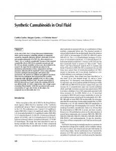
To better understand the nature of the prominent influx of extracellular Ca2⫹ evoked by CBD, we questioned, given evidence of the capability of CBD to interact with the vanilloid receptor type 1 (VR1) subgroup of cation channels within the transient receptor potential (TRP) family, whether such an influx was mimicked by capsacin, a full agonist of VR1 receptors [6, 33]. However, capsacin (0.1–10 M) did not affect [Ca2⫹]i levels (Fig. 7A), a result in accord with a previous report showing no sensitivity of RBL-2H3 cells to capsaicin (CPS) [34]. Next, we challenged RBL-2H3 cells with CBD (5 M) in the presence of the dihydropyridine, nitredipine (3–30 M), recently shown to affect Ca2⫹ entry in nonexcitable cells [35]. Results reported in Figure 7B showed that nitrendipine inhibited (3–30 M) the CBD-evoked [Ca2⫹]i rise in RBL-2H3 cells in a concentration-dependent manner. Analogous results were obtained with the imidazole derivative clotrimazole (3–30 M, Fig. 7C), another cationic channel blocker. Both channel blockers, at the highest concentration tested (30 M), deDel Giudice et al. Cannabidiol activates RBL-2H3 cells
1515
Fig. 3. RT-PCR analysis of CB1 and CB2 expression in RBL-2H3 cells. Total RNA extracted from RBL-2H3 cells was retro-transcribed, and cDNA was amplified with specific primers for CB1 and CB2 receptors. Gel image is representative of three independent experiments (A). Effect of cannabinoid receptor antagonists on cannabinoid modulation of -hexosaminidase release. RBL-2H3 cells sensitized with anti-DNP-IgE (0.5 g/ml) for 18 –20 h were pretreated with the CB1 receptor antagonist AM281 (0.1 M) or the CB2 receptor antagonist AM630 (1 M) or vehicle (vh; DMSO, 0.1%) for 5 min, followed by 10 M CBD or vehicle (DMSO, 0.1%) in the presence (IgE-DNP) or absence (basal) of antigen (DNP-HSA, 0.1 g/ml; B). Results are represented as mean ⫾ SEM of values from three to five independent experiments conducted in triplicate. °°, P ⬍ 0.01, with respect to CBD-treated; *, P ⬍ 0.05; **, P ⬍ 0.01; and ***, P ⬍ 0.001, with respect to vehicle-treated. RBL-2H3 cells sensitized with anti-DNP-IgE (0.5 g/ml) for 18 –20 h were pretreated with AM281 (0.1 M) or AM630 (1 M) for 5 min, followed by addition of 10 M (⫹)WIN 55,212-2 (WIN) or vehicle (DMSO, 0.1%) in the presence of antigen (DNP-HSA, 0.1 g/ml; C). Results are represented as mean ⫾ SEM of values from three to five independent experiments conducted in triplicate. **, P ⬍ 0.01, and ***, P ⬍ 0.001, with respect to vehicle-treated, and °, P ⬍ 0.05, with respect to (⫹)WIN 55,212-2-treated.
Fig. 4. Effect of PTX on cannabinoid modulation of -hexosaminidase release. RBL-2H3 cells were sensitized with anti-DNP-IgE (0.5 g/ml) in the presence or absence of PTX (100 ng/ml) for 18 –20 h, followed by addition of increasing concentrations of CBD (A), (⫹)WIN 55,212-2 (B), or CP 55,940 (C) or vehicle (DMSO, 0.1%) in the presence (IgE-DNP) or absence (basal) of antigen (DNP-HSA, 0.1 g/ml). Results are represented as mean ⫾ SEM of values from at least three independent experiments conducted in triplicate. °°, P ⬍ 0.01, and °°°, P ⬍ 0.001, with respect to basal release, and *, P ⬍ 0.05; **, P ⬍ 0.01; and ***, P ⬍ 0.001, with respect to the IgE-DNPstimulated cells. ns, Not significant.
1516
Journal of Leukocyte Biology Volume 81, June 2007
http://www.jleukbio.org
Fig. 5. Effect of CBD on [Ca2⫹]i. RBL-2H3 cells, loaded with Fluo-3/AM (5 M) and resuspended in phenol red-free DMEM, were incubated with increasing concentrations of CBD (1–10 M, A) or with CBD (10 M) in the presence or absence of 1,2-bis(2-aminophenoxy)ethane-N,N,N⬘,N⬘-tetraacetic acid (BAPTA; 2 mM, C). RBL-2H3 cells, sensitized with anti-DNP-IgE for 18 –20 h, followed by loading with Fluo-3/AM (5 M), were incubated with antigen (DNP-HSA, 0.1 g/ml) or vehicle (PBS) in the presence or absence of CBD (10 M, B). Fluo-3 fluorescence intensity was monitored in a time-resolved mode by flow cytometry. Results, expressed as percent of Fluo-3 MFI variation with respect to baseline fluorescence intensity of unstimulated cells, are representative of at least three independent experiments.
creased, per se, basal [Ca2⫹]i slightly in RBL-2H3 cells (Fig. 7, B and C).
Effects of THC on RBL-2H3 cell activation In a final series of experiments, we also evaluated, for comparative purposes, whether THC, the major natural psychoactive component of marijuana, affects [Ca2⫹]i in RBL2H3 cells in basal or antigen-stimulated conditions. THC has been reported to induce histamine secretion from rat peritoneal mast cells in vitro via CB receptor-independent interactions [21]. Moreover, THC elevates [Ca2⫹]i levels in resting T cells in a PTX-insensitive manner and exerts, unlike other cannabinoids, analgesic effects also via nonCB1/CB2 receptor-related mechanisms involving extracellular Ca2⫹ influx, independent of VR1 receptors [36, 37]. Our results, reported in Figure 8A, show that THC (5–20 M) induced, similarly to CBD, a concentration-dependent rise of [Ca2⫹]i in RBL-2H3 cells, an effect antagonized by clotrimazole (30 M, Fig. 8B). Moreover, THC (15 M) caused PTX-insensitive release of -hexosaminidase in the culture medium (Table 1).
DISCUSSION Our results show that CBD, in contrast to the synthetic CB1/ CB2 receptor agonists CP 55,940 and (⫹)WIN 55,212-2, induces concentration-dependent activation of RBL-2H3 cells alone and in concert with FcεRI immunoreceptor stimulation via a mechanism that is not mediated by Gi/Go protein-coupled receptors but rather, is associated with a robust rise in [Ca2⫹]i levels. Indeed, unlike CBD, CP 55,940 and (⫹)WIN 55,212-2, but not the inactive stereoisomer (–)WIN 55,212-3 and the selective CB2 agonist JWH-133 [38], inhibited -hexosaminidase release induced by the IgE receptor FcεRI cross-linking. Whereas the inhibitory effect of (⫹)WIN 55,212-2 was counteracted partially by the CB1 but not the CB2 receptor antagonists/inverse agonists (AM281 and AM630, respectively) as well as PTX-sensitive, the activating effect of CBD was neither sensitive to CB1/2 receptor antagonism nor prevented but rather, potentiated by pretreatment with PTX. In addition, unlike CBD, (⫹)WIN 55,212-2 and CP 55,940 did not affect basal [Ca2⫹]i but rather, inhibited antigen-induced [Ca2⫹]i rise, also in the absence of extracellular Ca2⫹. All this not only confirms previous results but also most importantly, excludes Del Giudice et al. Cannabidiol activates RBL-2H3 cells
1517
Fig. 6. Effect of synthetic cannabinoids on [Ca2⫹]i in the presence or absence of extracellular Ca2⫹. RBL-2H3 cells, loaded with 5 M Fluo-3/AM, were resuspended in phenol red-free DMEM, with (B, D) or without (A, C) BAPTA (2 mM). RBL-2H3 cells, sensitized with anti-DNP-IgE and loaded with Fluo-3/AM, were incubated with 10 M (⫹)WIN 55,212-2 (A, B) or 10 M CP 55,940 (C, D) and subsequently stimulated with (IgE-DNP) or without antigen (DNP-HSA, 0.1 g/ml). Fluo-3 fluorescence intensity was monitored, in a time-resolved mode, by flow cytometry. Results, expressed as percent of Fluo-3 MFI variation with respect to baseline fluorescence intensity of unstimulated cells, are representative of at least three independent experiments.
direct or indirect involvement of known CB and other receptors coupled to Gi/o proteins, herein including the abnormal CBD receptor, in the CBD-induced activation of the RBL-2H3 cells [13, 22, 25, 39]. Recently, by using resting human and murine T cells, it has been postulated that only cannabinoids, which possess a THClike, three-ring structure, are able to increase [Ca2⫹]i robustly in a CB receptor-independent manner [40]. Although our and other findings [41] with CBD, a bicyclic cannabinoid, do not support such a hypothesis, we found that THC induces, like CBD, PTX-insensitive -hexosamidase release from the RBL2H3 cells, an effect likely consequent to the elevation of [Ca2⫹]i. THC is, at difference with (⫹)WIN 55,212-2 and CP 1518
Journal of Leukocyte Biology Volume 81, June 2007
55,940, a low-affinity, partial agonist for CB1 receptors and unlike (⫹) WIN55,212-2, largely ineffective in coupling CB1 receptors in a conformation that enables Gq signaling [4, 42, 43]. Moreover, THC has, in analogy to our results, been reported to induce in vitro histamine secretion from rat peritoneal mast cells via mechanisms independent of CB1/CB2 receptors and importantly, solely, partly sensitive to PTX [21]. These and other findings, showing that CB1 and CB2 receptor isoforms are G␣i/o coupled in RBL-2H3 cells, exclude a mechanism of THC action, which involves CB receptors [20]. Rather, it may be postulated that the capability of THC, like CBD, to activate the RBL-2H3 cells may be mediated by specific non-CB receptors. Moreover, the structural difference between the two http://www.jleukbio.org
A 220
CPS 1µM
150
CPS10µM
140 MFI (% of basal)
200 MFI (%of basal)
B 160
CPS 0.1µM
180 160 140 120 0
30
120 110 100 90
60
90
120
150
0
100
70 60
CBD 5µM Clotr. 3µM+CBD Clotr. 10µM+CBD Clotr. 30µM+CBD Clotr. 30µM
160 150 140
MFI (%of basal)
130
80
100
C
CBD 5µM Nitr. 30µM+CBD Nitr. 10µM+CBD Nitr. 3µM+CBD Nitr. 30µM
130 120 110 100 90
100
0
Fig. 7. Effect of CPS on [Ca2⫹]i. RBL-2H3 cells loaded with Fluo-3/AM (5 M) and resuspended in phenol red-free DMEM were incubated with increasing concentrations of CPS (0.1–10 M, A). Effect of cation channel blockers on the CBD-evoked elevation of [Ca2⫹]i in RBL-2H3 cells, which when loaded with 5 M Fluo-3/AM, were treated with 5 M CBD alone or in the presence of increasing concentrations (3–30 M) of nitrendipine (Nitr.; B) or clotrimazole (Clotr.; C). Results, expressed as percent of Fluo-3 MFI variation with respect to baseline fluorescence intensity of unstimulated cells, are representative of at least three independent experiments.
80 70 60
time (sec.)
molecules raises the question as to whether the same or different sites are implicated. Ca2⫹ mobilization from intracellular stores and Ca2⫹ entry from the extracellular space are key features of antigen-induced degranulation of RBL-2H3 cells [44]. Here, although FcεRI clustering was, upon removal or chelation of extracellular Ca2⫹, found to induce a transient increase in [Ca2⫹]i, likely reflecting Ca2⫹ release from intracellular stores, the CBD-induced [Ca2⫹]i rise was abolished completely. Moreover, pretreatment with SKF96365, a cation channel blocker commonly used to block voltage-independent Ca2⫹ channels, abolished the CBD-induced [Ca2⫹]i rise completely (data not shown). These findings, suggesting that the Ca2⫹ source is mainly attributable to extracellular Ca2⫹ influx, raise the possibility that ligand-gated or receptor-operated Ca2⫹ influx channels are implicated, the most promising molecular candidates of which are represented by the TRP gene superfamily of Ca2⫹-permeable cation channels [45]. However, although CBD has been described to be capable of interacting with the TRPV1 channel (also termed VR1 receptors), the vanilloid
CPS, a full agonist of TRPV1 receptors, did not affect [Ca2⫹]i in the RBL-2H3 cells [3, 33]. Also THC, although able to increase [Ca2⫹]i in the RBL-2H3 cells, does not induce Ca2⫹ transients in cells expressing VR1 receptors [37]. Together, this excludes the possibility that TRPV1 channels mediate the activation of the RBL-2H3 cells induced by CBD or THC. Nevertheless, as TRP channels other than VR1, e.g., ANKTM1 (TRPA1) and TRPC1, have been implicated in CB1/CB2/VR1 receptor-independent effects of THC and cannabinol, another nonpsychoactive component of marijuana, it would not be surprising if a member of the TRP ion channel family other than TRPV1, directly or indirectly via receptor-operated mechanisms, is implicated [46 –52]. Conversely, irrespective of the Ca2⫹ influx channels implicated, flow of ions, such as K⫹, plays an important role in the activation responses of RBL-2H3 cells, as they regulate cell membrane potential and thus serve to sustain the Ca2⫹ influx necessary for degranulation [53–56]. Among these, RBL-2H3 cells display, upon antigen stimulation, an outwardly rectifying, Ca2⫹-dependent K⫹ efflux pathway, recently proposed to Del Giudice et al. Cannabidiol activates RBL-2H3 cells
1519
220 MFI (%of basal)
B
THC 5uM THC 10uM THC 15uM THC 20uM
240
200 180 160 140
THC 20µM Clotr.+THC Clotri. 30µM
200 180 160 140 120
120 100
240 220
MFI (%of basal)
A
100 0
20
40 60 time (sec.)
80
100
80 60
100
0 time (sec.)
Fig. 8. Effect of THC on [Ca2⫹]i. RBL-2H3 cells loaded with Fluo-3/AM (5 M) and resuspended in phenol red-free DMEM were incubated with increasing concentrations of THC (5–20 M, A). Effect of clotrimazole on THC-⌬9-evoked elevation of [Ca2⫹]i in RBL-2H3 cells, which when loaded with 5 M Fluo-3/AM, were treated with 20 M THC alone or in the presence of clotrimazole (30 M, B). Results, expressed as percent of Fluo-3 MFI variation with respect to baseline fluorescence intensity of unstimulated cells, are representative of at least three independent experiments.
involve conductances of the intermediate conductance Ca2⫹activated K⫹ channel (IKCa)-type, the opening of which is expected to increase the electrical driving force for Ca2⫹ entry [55, 56]. Here, we found that the CBD- and THC-evoked, intracellular Ca2⫹ rise was inhibited dose-dependently by the cation channel blockers clotrimazole (10 –30 M) and nitrendipine (10 –30 M). It is intriguing that clotrimazole is also a relatively selective blocker of IKCa channels, an effect mimicked by nitrendipine at the concentrations used [57–59]. Although this raises the possibility of an action of these receptors on IKCa channels, clotrimazole had, in spite of its ability to markedly reduce the [Ca2⫹]i rise induced by CBD, no effect on the CBD-induced -hexosaminidase release from the RBL2H3 cells (data not shown). Moreover, clotrimazole did not inhibit antigen-mediated secretion of the RBL-2H3 cells, a result in line with previous reports [55]. Also, 1-ethyl-2-benzimidazolinone, a specific IKCa opener, reportedly did not elicit histamine release from mast cells on its own, suggesting that IKCa opening alone is insufficient for degranulation [60, 61]. Thus, although it may be speculated that the concerted action between Ca2⫹ influx channels and IKCa channels on the cell TABLE 1.
Effect of PTX on ⌬9-THC-Mediated -hexosaminidase Release in RBL-2H3 Cells
Treatment Vehicle (ethanol 0.1%) THC (15 M) PTX (100 ng/ml) PTX ⫹ THC
Mean of -hexo release OD405
SEM ⫾
0.3997 0.639 0.387 0.603
0.022 0.037 0.035 0.061
Cells were incubated for 18 –20 h in the presence or absence of PTX (100 ng/ml) and then incubated with vehicle or ⌬9-THC (15 M). The results are presented as mean ⫾ SEM of three independent experiments.
1520
Journal of Leukocyte Biology Volume 81, June 2007
surface may allow the RBL-2H3 cells to sustain elevated [Ca2⫹]i in response to CBD, other signaling pathways are likely necessary for the effects of CBD on their mediator release. However, given the promiscuous capability of clotrimazole to block several types of channels, further studies are warranted to confirm and expand these speculations. Whereas activation of mast cells has, at least traditionally, been implicated in the genesis and/or amplification of inflammatory reactions, most studies show that CBD as well as THC exert, in contrast to our in vitro results, anti-inflammatory and immunosuppressive proprieties in vivo [5]. Although difficult to reconcile, it is noteworthy that mast cells are tissue-dwelling cells, heterogeneous in phenotype and function, the activation of which results from the transient displacement of a physiological balance between positive and negative signals delivered by activating and inhibitory receptors and as such, profoundly affected by the microenvironment. Moreover, CBD activates, likely via TRPV1-mediated mechanisms, CPS-sensitive nerve fibers in inflamed tissues, and THC causes CB1/CB2-independent release of a calcitonin gene-related peptide from CPSsensitive nerves in isolated rat and mouse mesenteric arterial rings, even from TRPV1 knockout animals [35]. Given the known role of these nociceptors in not only in hyperalgesia but also in development and progression of inflammation, it cannot be excluded that their activity-induced desensitization by CBD and THC may contribute to their anti-inflammatory action in vivo. Other, although not exclusive, possibilities include effects on endocannabinoid production or degradation, as well as effects on adenosine signaling in vivo [3, 7, 41]. In sum, results from our study lend support to existence of yet-to-be-identified sites, i.e., receptors and/or ion channels, of interaction of natural cannabinoids, such as CBD and THC. Such sites are, at least in RBL-2H3 mast cells, coupled, http://www.jleukbio.org
directly or indirectly, with Ca2⫹ influx channels, the opening of which triggers and/or participates in their activation. Although future studies are necessary to identify and address the site(s) implicated, our results not only have the potential of opening new avenues in the pharmacology of plant-derived cannabinoids but also raise the possibility that mast cell activation may contribute to the adverse bronchopulmonary consequences of chronic marijuana smoking.
ACKNOWLEDGMENTS This work was in part supported by the Italian Ministry for Education, University and Scientific Research (MIUR), grant #3933.
REFERENCES 1. Matsuda, L. A., Lolait, S. J., Brownstein, M. J., Young, A. C., Bonner, T. I. (1990) Structure of a cannabinoid receptor and functional expression of the cloned cDNA. Nature 346, 561–564. 2. Munro, S., Thomas, K. L., Abu-Shaar, M. (1993) Molecular characterization of a peripheral receptor for cannabinoids. Nature 365, 61– 65. 3. Bisogno, T., Hanus, L., De Petrocellis, L., Tchilibon, S., Ponde, D. E., Brandi, I., Moriello, A. S., Davis, J. B., Mechoulam, R., Di Marzo, V. (2001) Molecular targets for cannabidiol and its synthetic analogues: effect on vanilloid VR1 receptors and on the cellular uptake and enzymatic hydrolysis of anandamide. Br. J. Pharmacol. 134, 845– 852. 4. Howlett, A. C., Barth, F., Bonner, T. I., Cabral, G., Casellas, P., Devane, W. A., Felder, C. C., Herkenham, M., Mackie, K., Martin, B. R. et al. (2002) International Union of Pharmacology. XXVII. Classification of cannabinoid receptors. Pharmacol. Rev. 54, 161–202. 5. Malfait, A. M., Gallily, R., Sumariwalla, P. F., Malik, A. S., Andreakos, E., Mechoulam, R., Feldmann, M. (2000) The nonpsychoactive cannabis constituent cannabidiol is an oral anti-arthritic therapeutic in murine collagen-induced arthritis. Proc. Natl. Acad. Sci. USA 97, 9561–9566. 6. Costa, B., Giagnoni, G., Franke, C., Trovato, A. E., Colleoni, M. (2004) Vanilloid TRPV1 receptor mediates the antihyperalgesic effect of the nonpsychoactive cannabinoid, cannabidiol, in a rat model of acute inflammation. Br. J. Pharmacol. 143, 247–250. 7. Carrier, E. J., Auchampach, J. A., Hillard, C. J. (2006) Inhibition of an equilibrative nucleoside transporter by cannabidiol: a mechanism of cannabinoid immunosuppression. Proc. Natl. Acad. Sci. USA 103, 7895– 7900. 8. Coffey, R. G., Yamamoto, Y., Snella, E., Pross, S. (1996) Tetrahydrocannabinol inhibition of macrophage nitric oxide production. Biochem. Pharmacol. 52, 743–751. 9. Sacerdote, P., Martucci, C., Vaccani, A., Bariselli, F., Panerai, A. E., Colombo, A., Parolaro, D., Massi, P. (2005) The nonpsychoactive component of marijuana cannabidiol modulates chemotaxis and IL-10 and IL-12 production of murine macrophages both in vivo and in vitro. J. Neuroimmunol. 159, 97–105. 10. Watzl, B., Scuderi, P., Watson, R. R. (1991) Marijuana components stimulate human peripheral blood mononuclear cell secretion of interferon-␥ and suppress interleukin-1 ␣ in vitro. Int. J. Immunopharmacol. 13, 1091–1097. 11. Srivastava, M. D., Srivastava, B. I., Brouhard, B. (1998) ⌬9 Tetrahydrocannabinol and cannabidiol alter cytokine production by human immune cells. Immunopharmacology 40, 179 –185. 12. Begg, M., Mo, F. M., Offertaler, L., Batkai, S., Pacher, P., Razdan, R. K., Lovinger, D. M., Kunos, G. J. (2003) G protein-coupled endothelial receptor for atypical cannabinoid ligands modulates a Ca2⫹-dependent K⫹ current. Biol. Chem. 278, 46188 – 46194. 13. Offertaler, L., Mo, F. M., Batkai, S., Liu, J., Begg, M., Razdan, R. K., Martin, B. R., Bukoski, R. D., Kunos, G. (2003) Selective ligands and cellular effectors of a G protein-coupled endothelial cannabinoid receptor. Mol. Pharmacol. 63, 699 –705. 14. Williams, C. M., Galli, S. J. (2000) The diverse potential effector and immunoregulatory roles of mast cells in allergic disease. J. Allergy Clin. Immunol. 105, 847– 859.
15. Nigrovic, P. A., Lee, D. M. (2005) Mast cells in inflammatory arthritis. Arthritis Res. Ther. 7, 1–11. 16. Brown, M. A., Gregory, G. D. (2006) Mast cells in allergy and autoimmunity: implications for adaptive immunity. Methods Mol. Biol. 315, 35–50. 17. Lau, A. H., Chow, S. S. (2003) Effects of cannabinoid receptor agonists on immunologically induced histamine release from rat peritoneal mast cells. Eur. J. Pharmacol. 464, 229 –235. 18. Facci, L., Dal Toso, R., Romanello, S., Buriani, A., Skaper, S. D., Leon, A. (1995) Mast cells express a peripheral cannabinoid receptor with differential sensitivity to anandamide and palmitoylethanolamide. Proc. Natl. Acad. Sci. USA 92, 3376 –3380. 19. Samson, M. T., Small-Howard, A., Shimoda, L. M., Koblan-Huberson, M., Stokes, A. J., Turner, H. (2003) Differential roles of CB1 and CB2 cannabinoid receptors in mast cells. J. Immunol. 170, 4953– 4962. 20. Small-Howard, A. L., Shimoda, L. M., Adre, C. N., Turner, H. (2005) Anti-inflammatory potential of CB1-mediated cAMP elevation in mast cells. Biochem. J. 388, 465– 473. 21. Bueb, J. L., Lambert, D. M., Tschirhart, E. J. (2001) Receptor-independent effects of natural cannabinoids in rat peritoneal mast cells in vitro. Biochim. Biophys. Acta 1538, 252–259. 22. Jonsson, K. O., Persson, E., Fowler, C. J. (2006) The cannabinoid CB2 receptor selective agonist JWH133 reduces mast cell oedema in response to compound 48/80 in vivo but not the release of -hexosaminidase from skin slices in vitro. Life Sci. 78, 598 – 606. 23. Seldin, D. C., Adelman, S., Austen, K. F., Stevens, R. L., Hein, A., Caulfield, J. P., Woodbury, R. G. (1985) Homology of the rat basophilic leukemia cell and the rat mucosal mast cell. Proc. Natl. Acad. Sci. USA 82, 3871–3875. 24. Benhamou, M., Siraganian, R. P. (1992) Protein-tyrosine phosphorylation: an essential component of Fc ε RI signaling. Immunol. Today 13, 195–197. 25. Ozawa, K., Szallasi, Z., Kazanietz, M. G., Blumberg, P. M., Mischak, H., Mushinski, J. F., Beaven, M. A. (1993) Ca(2⫹)-dependent and Ca(2⫹)independent isozymes of protein kinase C mediate exocytosis in antigenstimulated rat basophilic RBL-2H3 cells. Reconstitution of secretory responses with Ca2⫹ and purified isozymes in washed permeabilized cells. J. Biol. Chem. 268, 1749 –1756. 26. Smith, J., Thompson, N., Thompson, J., Armstrong, J., Hayes, B., Crofts, A., Squire, J., Teahan, C., Upton, L., Solari, R. (1997) Rat basophilic leukaemia (RBL) cells overexpressing Rab3a have a reversible block in antigen-stimulated exocytosis. Biochem. J. 323, 321–328. 27. Gatley, S. J., Lan, R., Volkow, N. D., Pappas, N., King, P., Wong, C. T., Gifford, A. N., Pyatt, B., Dewey, S. L., Makriyannis, A. (1998) Imaging the brain marijuana receptor: development of a radioligand that binds to cannabinoid CB1 receptors in vivo. J. Neurochem. 70, 417– 423. 28. Hosohata, Y., Quock, R. M., Hosohata, K., Makriyannis, A., Consroe, P., Roeske, W. R., Yamamura, H. I. (1997) AM630 antagonism of cannabinoid-stimulated [35S]GTP ␥ S binding in the mouse brain. Eur. J. Pharmacol. 321, R1–R3. 29. Bayewitch, M., Avidor-Reiss, T., Levy, R., Barg, J., Mechoulam, R., Vogel, Z. (1995) The peripheral cannabinoid receptor: adenylate cyclase inhibition and G protein coupling. FEBS Lett. 375, 143–147. 30. Hoffman, H. M., Walker, L. L., Marquardt, D. L. (1997) Mast cell adenosine induced calcium mobilization via Gi3 and Gq proteins. Inflammation 21, 55– 68. 31. Mizota, K., Yoshida, A., Uccida, H., Fujita, R., Ueda, H. (2005) Novel type of Gq/11 protein-coupled neurosteroid receptor sensitive to endocrine disrupting chemicals in mast cell line (RBL-2H3). Br. J. Pharmacol. 145, 545–550. 32. Barker, S. A., Lujan, D., Wilson, B. S. (1999) Multiple roles for PI 3-kinase in the regulation of PLC ␥ activity and Ca2⫹ mobilization in antigen-stimulated mast cells. J. Leukoc. Biol. 65, 321–329. 33. Tominaga, M., Caterina, M. J., Malmberg, A. B., Rosen, T. A., Gilbert, H., Skinner, K., Raumann, B. E., Basbaum, A. I., Julius, D. (1998) The cloned capsaicin receptor integrates multiple pain-producing stimuli. Neuron 21, 531–543. 34. Biro, T., Maurer, M., Modarres, S., Lewin, N. E., Brodie, C., Petecs, G., Paus, R., Blumberg, P. M. (1998) Characterization of functional vanilloid receptors expressed by mast cells. Blood 91, 1332–1340. 35. Harper, J. L., Camerini-Otero, C. S., Kim, S. A., Jacobson, K. A., Daly, J. W. (2003) Dihydropyridines as inhibitors of capacitative calcium entry in leukemic HL-60 cells. Biochem. Pharmacol. 65, 329 –338. 36. Rao, G. K., Zhang, W., Kaminski, N. E. (2004) Cannabinoid receptormediated regulation of intracellular calcium by ⌬(9)-tetrahydrocannabinol in resting T cells. J. Leukoc. Biol. 75, 884 – 892. 37. Zygmunt, P. M., Andersson, D. A., Ho¨gesta¨tt, E. D. (2002) ⌬ 9-Tetrahydrocannabinol and cannabinol activate capsaicin-sensitive sensory nerves
Del Giudice et al. Cannabidiol activates RBL-2H3 cells
1521
38.
39.
40. 41. 42. 43. 44. 45. 46.
47. 48. 49.
via a CB1 and CB2 cannabinoid receptor-independent mechanism. J. Neurosci. 22, 4720 – 4727. Huffman, J. W., Liddle, J., Yu, S., Aung, M. M., Abood, M. E., Wiley, J. L., Martin, B. R. (1999) 3-(10,10-Dimethylbutyl)-1-deoxy-D8-THC and related compounds: synthesis of selective ligands for the CB2 receptor. Bioorg. Med. Chem. 7, 2905–2914. Jarai, Z., Wagner, J. A., Varga, K., Lak, K. D., Compton, D. R., Martin, B. R., Zimmer, A. M., Bonner, T. I., Buckley, N. E., Mezey, E. et al. (1999) Cannabinoid-induced mesenteric vasodilation through an endothelial site distinct from CB1 or CB2 receptors. Proc. Natl. Acad. Sci. USA 96, 14136 –14141. Rao, G. K., Kaminski, N. E. (2006) Cannabinoid-mediated elevation of intracellular calcium: a structure-activity relationship. J. Pharmacol. Exp. Ther. 317, 820 – 829. Drysdale, A. J., Ryan, D., Pertwee, R. G., Platt, B. (2006) Cannabidiolinduced intracellular Ca2⫹ elevations in hippocampal cells. Neuropharmacology 50, 621– 631. Wiley, J. L., Martin, B. R. (2002) Cannabinoid pharmacology: implications for additional cannabinoid receptor subtypes. Chem. Phys. Lipids 121, 57– 63. Lauckner, J. E., Hille, B., Mackie, K. (2005) The cannabinoid agonist WIN 55,212-2 increases intracellular calcium via CB1 receptor coupling to Gq/11 G proteins. Proc. Natl. Acad. Sci. USA 102, 19144 –19149. Narenjkar, J., Marsh, S. J., Assem, E. S. (1999) The characterization and quantification of antigen-induced Ca2⫹ oscillations in a rat basophilic leukaemia cell line (RBL-2H3). Cell Calcium 26, 261–269. Clapham, D. E., Runnels, L. W., Strubing, C. (2001) The TRP ion channel family. Nat. Rev. Neurosci. 2, 387–396. Jordt, S. E., Bautista, D. M., Chuang, H. H., McKemy, D. D., Zygmunt, P. M., Hogestat, E. D., Meng, I. D., Julius, D. (2004) Mustard oils and cannabinoids excite sensory nerve fibres through the TRP channel ANKTM1. Nature 427, 260 –265. Rao, G. K., Kaminski, N. E. (2006) Induction of intracellular calcium elevation by ⌬9 tetrahydrocannabinol in T cells involves TRPC1 channels. J. Leukoc. Biol. 79, 202–213. Pizzo, P., Burgo, A., Pozzan, T., Fasolato, C. (2001) Role of capacitative calcium entry on glutamate-induced calcium influx in type-I rat cortical astrocytes. J. Neurochem. 79, 98 –109. Ramsey, I. S., Delling, M., Clapham, D. E. (2006) An introduction to TRP channels. Annu. Rev. Physiol. 68, 619 – 647.
1522
Journal of Leukocyte Biology Volume 81, June 2007
50. Vriens, J., Watanabe, H., Janssens, A., Droogmans, G., Voets, T., Nilius, B. (2003) Cell swelling, heat, and chemical agonists use distinct pathways for the activation of the cation channel TRPV4. Proc. Natl. Acad. Sci. USA 101, 396 – 401. 51. Shimosato, G., Amaya, F., Ueda, M., Tanaka, Y., Decosterd, I., Tanaka, M. (2005) Peripheral inflammation induces up-regulation of TRPV2 expression in rat DRG. Pain 119, 225–232. 52. Stokes, A. J., Shimoda, L. M., Koblan-Huberson, M., Adra, C. N., Turner, H. (2004) A TRPV2-PKA signaling module for transduction of physical stimuli in mast cells. J. Exp. Med. 200, 137–147. 53. Labrecque, G. F., Holowka, D., Baird, B. (1991) Characterization of increased K⫹ permeability associated with the stimulation of receptors for immunoglobulin E on rat basophilic leukemia cells. J. Biol. Chem. 266, 14912–14917. 54. Bradding, P. (2005) Mast cell ion channels. Chem. Immunol. Allergy 87, 163–178. 55. Wischmeyer, E., Lentes, K. U., Karschin, A. (1995) Physiological and molecular characterization of an IRK-type inward rectifier K⫹ channel in a tumor mast cell line. Pflugers Arch. 429, 809 – 819. 56. Narenjkar, J., Marsh, S. J., Assem, el-S. K. (2004) Inhibition of the antigen-induced activation of RBL-2H3 cells by charybdotoxin and cetiedil. Eur. J. Pharmacol. 483, 95–106. 57. Alvarez, J., Montero, M., Garcia-Sancho, J. (1992) High affinity inhibition of Ca(2⫹)-dependent K⫹ channels by cytochrome P-450 inhibitors. J. Biol. Chem. 267, 11789 –11793. 58. Daly, J. W., Lueders, J., Padgett, W. L., Shin, Y., Gusovsky, F. (1995) Maitotoxin-elicited calcium influx in cultured cells. Effect of calciumchannel blockers. Biochem. Pharmacol. 50, 1187–1197. 59. Jensen, B. S., Odum, N., Jorgensen, N. K., Christophersen, P., Olesen, S. P. (1999) Inhibition of T cell proliferation by selective block of Ca(2⫹)-activated K(⫹) channels. Proc. Natl. Acad. Sci. USA 14, 10917– 10921. 60. Duffy, S. M., Lawley, W. J., Conley, E. C., Bradding, P. (2001) Resting and activation-dependent ion channels in human mast cells. J. Immunol. 167, 4261– 4270. 61. Mark Duffy, S., Berger, P. M. D., Cruse, G., Yang, W., Bolton, S. J., Bradding, P. D. M. (2004) The K⫹ channel iKCA1 potentiates Ca2⫹ influx and degranulation in human lung mast cells. J. Allergy Clin. Immunol. 114, 66 –72.
http://www.jleukbio.org