CELLULAR & MOLECULAR BIOLOGY LETTERS
Volume 8, (2003) pp 5 – 18 http://www.cmbl.org.pl Received 3 October 2002 Accepted 16 December 2002
CAPACITANCE AND RESISTANCE OF THE BILAYER LIPID MEMBRANE FORMED OF PHOSPHATIDYLCHOLINE AND CHOLESTEROL MONIKA NAUMOWICZ1, ANETA D. PETELSKA1 and ZBIGNIEW A. FIGASZEWSKI1,2 * 1 Institute of Chemistry, University of Białystok, al. J. Piłsudskiego 11/4, 15-443 Białystok, Poland, 2Laboratory of Interfacial Electrochemistry, Faculty of Chemistry, University of Warsaw, ul. Pasteura 1, 02-093 Warszawa, Poland Abstract: Capacity and electric resistance of lipid membranes composed of lecithin and cholesterol were determined. The components were chosen for the study because they were present in biological membranes. Capacitance of the lecithin and cholesterol membranes amounts to 0.38 and 0.61 µF/cm2, and resistance to 1.44×10⁴ and 2.12×10⁶ Ω cm2, respectively. A 1:1 complex appears as a result of lecithin-cholesterol membrane formation. Parameters of the membrane formed of the lecithin-cholesterol complex were determined: surface
( )
concentration (Γ3), capacitance (C3), and conductance R 3−1 , as well as the stability constant (K) of the complex. The mean values of those magnitudes are as follows: 4.265×10⁻⁶ mol/m2, 0.54 µF/cm2, 1.381×10⁻⁶ Ω-1 cm-2 and 3.748×10⁷, respectively. Key Words: Impedance Spectroscopy, Bilayer Lipid Membrane, Lecithin, Cholesterol INTRODUCTION Artificial membranes formed of components occurring in natural ones are often used in the studies of biological membranes. Many functions of biological membranes were reproduced and explained in the studies of artificial membranes. It has been demonstrated in numerous experiments that properties of the lipid membranes formed of artificial components are similar to those of natural cell membranes. Thus, properties of biological membranes can be studied using two well-defined model systems: liposomes [1,2] or not closed lipid bilayers [3].
* Corresponding author, Fax: +4885 745 75 81, E-mail:
[email protected]
6
CELL. MOL. BIOL. LETT.
Vol. 8. No. 1. 2003
Electric capacity of lipid bilayers is high, 0.3 - 1.3 µF/cm2 [4]. It depends on many factors, for example on the kind of component molecules, temperature, membrane potential, penetration of alien molecules or ions into the membrane. Capacity of the membrane also depends on the formation procedure. Capacities of the membranes formed by the Mueller-Rudin technique [3] are usually lower than of those formed by combining monolayers (the Montal-Mueller method [5,6]). For example, the specific capacity of a bilayer formed of monoglycerides with 18-carbon atom chains is 0.380 µF/cm2 when prepared by the MuellerRudin technique, or 0.745 µF/cm2 when prepared by the Montal-Mueller technique [7]. An amount of forming solvent is retained in the hydrophobic layer, provoking an increase in membrane thickness. The membranes formed by the Montal-Mueller method are solvent-free and their thickness is the same as that of biological membranes. The thickness and capacity of the membranes formed by the Mueller-Rudin method depend on the solvent used to dissolve the lipid. Short-chain hydrocarbons used as solvents increase membrane thickness and lower its capacity, while long-chain hydrocarbons (e.g. octadecane, squalene) allow to obtain membranes of the thickness and capacity values similar to those of membranes formed of monolayers (e.g. membranes formed of 1-glyceryl monooleate had a specific capacity of 0.380 µF/cm2 when prepared by Mueller-Rudin technique and if n-hexane was used as the solvent, and 0.750 µF/cm2 in the case of squalene [7]). Lipid bilayers formed of phospholipids and cholesterol were the subject of many studies because those compounds were present in biological membranes [8,9]. Various techniques were used to investigate interaction of phospholipids and cholesterol, for example monolayer surface chemistry [10], Raman spectroscopy [11], nuclear magnetic resonance [12], electron spin resonance [13], impedance spectroscopy [14]. Finean [15] demonstrated by his X-ray diffraction studies that in a monolayer containing phospholipid and cholesterol only one complex is formed, of 1:1 composition. The stability constant of the 1:1 lecithin-cholesterol complex in monolayers was given in [16]. Existence of the same complex was demonstrated in bilayers by interfacial tension measurements [17]. In our work, we would like to present the evidence for the formation of a 1:1 phosphatidylcholine-cholesterol complex in a lipid bilayer. Using equations derived in this paper the stability constant of the 1:1 complex was calculated. CAPACITANCE AND CONDUCTANCE OF TWO-COMPONENT LIPID MEMBRANES A two-component forming solution can be used to obtain a lipid bilayer. The components may or may not form another compound, a chemical complex. In the case where the membrane components do not form chemical compounds, we can describe their interaction by the equation expressing additivity of the capacitance or the conductance:
CELLULAR & MOLECULAR BIOLOGY LETTERS
C = C1S1 + C 2S 2
7
(1)
here:
S1 + S 2 = 1 m1 Γ1 m S2 = 2 Γ2
S1 =
m1 m1 + m 2 x1 + x 2 = 1 where: C [µF/cm2] C1, C2 [µF/cm2] x1 =
- capacitance of the membrane, - capacitance of the membrane built of component 1 and 2, respectively, S1 , S 2 - surface fractions of the membrane covered by component 1 and 2, respectively, m1, m2 [mol/m2] - quantities of component 1 and 2, respectively, per unit area of the membrane, 2 Γ1, Γ2 [mol/m ] - surface concentrations of the membrane built of component 1 and 2, respectively, x1, x 2 - molar fractions of component 1 and 2 in the solution forming the membrane, respectively. Elimination of S1, S2, m1 and m2 yields the equation: (2a) (C − C1 )Γ 2 x 1 + (C − C 2 )Γ1 x 2 = 0 The equation of conductance can be obtained in the same way from Eq. (1): (2b) R −1 − R1−1 Γ 2 x1 + R −1 − R −2 1 Γ1x 2 = 0 where: R −1 [Ω−1 cm-2] - conductance of the membrane,
(
)
(
R 1−1 , R −2 1 [Ω−1 cm-2]
)
- conductance of the membrane built of component 1 and 2, respectively.
If a 1:1 complex is formed in a two-component lipid bilayer then the capacity of the created membrane consist of, in an additive way, the capacities of three chemical compounds (component 1, component 2 and this complex): (3) C = C1S1 + C 2 S 2 + C 3S3 in which: S1 + S 2 + S3 = 1
8
CELL. MOL. BIOL. LETT.
Vol. 8. No. 1. 2003
m1 Γ1 m S2 = 2 Γ2 m S3 = 3 Γ3 S1 =
K=
m3 m1 ⋅ m 2
m1 + m 3 m1 + m 2 + 2m 3 x1 + x 2 = 1 where: C [µF/cm2] - capacitance of the membrane, 2 C3 [µF/cm ] - capacitance of the membrane compounds of the complex, S3 - surface fraction of the membrane covered by the complex, m3 [mol/m2] - quantity of complex per unit area of the membrane, Γ3 [mol/m2] - surface concentration of the membrane compounds of the complex, K - stability constant of the complex, x1, x 2 - molar fractions of element 1 and 2 in the solution forming the membrane, respectively. Elimination of S1, S2, S3, m1, m2 and m3 yields the basic equation: x1 =
[(C − C 1 )Α 2 x 1 + (C − C 2 )Α 1 x 2 ] [(C 3 − C 1 )Α 2 x 1 + (C 3 − C 2 )Α 1 x 2 + (C 1 − C 2 )(x 1 − x 2 )] (4a ) = KΓ 3 Α 1 Α 2 [(C − C 1 )(x 2 − x 1 ) + (C 3 − C )Α 1 x 2 ] [(C − C 2 )(x 1 − x 2 )Α 2 x 1 ]
where: Γ Α1 = 1 Γ3 Γ Α2 = 2 Γ3 The equation of conductance can be obtained in the same way from Eq. (3): [(R −1 − R 1−1 )Α 2 x 1 + (R −1 − R −21 )Α 1 x 2 ] [(R 3−1 − R 1−1 )Α 2 x 1 + (R 3−1 − R 2−1 )Α 1 x 2 + (R 1−1 − R 2−1 )(x 1 − x 2 )]
[(
)
(
)
= KΓ 3 Α 1 Α 2 R −1 − R 1−1 (x 2 − x 1 ) + R 3−1 − R −1 Α 1 x 2
] [(R
−1
)
− R 2−1 (x 1 − x 2 )Α 2 x 1
]
(4b )
where: R −1 [Ω−1 cm-2] - conductance of the membrane, R 3−1 [Ω−1 cm-2] - conductance of the membrane compounds of the complex.
CELLULAR & MOLECULAR BIOLOGY LETTERS
9
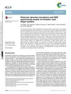
MATERIALS AND METHODS Materials The lipid bilayer was formed from 99% egg lecithin (3-sn-phosphatidylcholine, molar weight 752.08 g/mol), produced by Fluka, and from 98% cholesterol (molar weight 386.67 g/mol), produced by Sigma. Lecithin was dissolved in chloroform and the solvent was evaporated in argon atmosphere. Solutions containing 20 mg phosphatidylcholine, 20 mg cholesterol or 20 mg of their mixture in various proportions were prepared in 1 cm3 of an n-decane - nbutanol solvent system (10:1 volume ratio). The solvents were distilled and their purity was checked by measurements of refraction coefficient. Potassium chloride solution of 0.1 mol dm-3 was used as the electrolyte. The solution was prepared from mili-Q water and KCl (POCh). KCl was calcinated to remove organic impurities. Measurements Processes occurring in artificial lipid membranes were examined. Electric properties of bilayer lipid membranes can be examined thanks to a measuring vessel constructed in the Laboratory of Electrochemistry at the Institute of Chemistry in Białystok. The measuring vessel is presented in Figure 1. A syringe (1) with an external thread screw (2) and with a handwheel (3) was located in its upper part. An acid-resistant steel tube (4) with a tight teflon piston (5) was at the other end of the screw. A connector made of organic glass (polymethyl metacrylate) (6) with a platinum current electrode (7) and a silversilver chloride measuring electrode (8) was fixed to the syringe cone. The connector ended with a tight teflon attachment (9). The lower part of the vessel (10) made of organic glass contained a second current electrode (11) and a second silver-silver chloride measuring electrode (12). The lateral side of the vessel was a flat glass plate allowing to observe formed membranes. The syringe, the connector, and the teflon attachment formed a tight setup which could be filled with electrolyte solution. The forming solution was placed at the teflon cap tip and the setup was placed in the lower part of the measuring vessel, also filled with electrolyte solution. As a result, the teflon attachment was immersed in the electrolyte solution, the approximate level of which (13) is marked in Figure 1. A droplet of electrolyte could be squeezed out from the cap by rotating the handwheel (3) and a forming sphere (14) could be simultaneously observed under a microscope. N-butanol contained in the forming solution dissolved in aqueous solution, and n-decane able to wet teflon shifted into the cap. As a result, a bilayer in the form of a bubble built of lipids was produced. Electrochemical impedance spectroscopy (EIS) was applied to the study of variations in capacity and conductance. Impedance was measured with an AC impedance system (EG&G, Princeton Applied Research, Model 388) including a
10
CELL. MOL. BIOL. LETT.
Vol. 8. No. 1. 2003
potentiostat/galvanostat (Model 273), a two-phase lock-in analyzer (Model 5208) and a personal computer. For EIS measurements, a 4 mV amplitude sine wave was applied to the electrode at open circuit potential in the 0.01-10000 Hz frequency range. The silver-silver chloride electrodes used in the study showed a resistance of about 200 Ω, while the resistance of the measured membranes ranged from 105 to 108 Ω. As the electrodes were connected in a series, the double layer impedance of the electrode-solution interface could be neglected.
Fig. 1. The measuring vessel.
RESULTS AND DISCUSSION The phosphatidylcholine-cholesterol ratio in the forming solution was assumed to be similar to that of the membrane; it was impossible to determine the accurate composition of a lecithin-cholesterol bilayer [18]. Measurements were carried out at room temperature in the whole concentration range; the experimental capacitance and resistance values are related to surface area unit. Typical plots of impedance of chosen membranes are presented in Figure 2. Dependences of capacitance and resistance of the lecithin-cholesterol membrane on the mole fraction of cholesterol are presented in Figures 3 and 4, respectively. The measured capacitance values of the lecithin membrane and the cholesterol membrane amounted to 0.38 and 0.61µF/cm2, respectively, and are similar to the literature data [18,19]. Their measured resistance values were 1.44×10⁴ and 2.12×10⁶ Ω cm2. The Randles model was successfully applied to describe properties of bilayer lipid membranes. Its equivalent circuit presented in Figure 5 is the simplest, and it is characteristic for an artificial lipid bilayer only when ionophore systems, specific channels - pores and adsorption are absent [20].
CELLULAR & MOLECULAR BIOLOGY LETTERS
11
Fig. 2. Dependence of an imaginary part (-Z") on the real part (Z') for a membrane made of lecithin (a), lecithin and cholesterol (1:1 molar ratio) (b), and cholesterol (c).
Fig. 3. Dependence of capacitance of the lecithin-cholesterol membrane (C) on the mole fraction of cholesterol (x2).
Fig. 4. Dependence of resistance of the lecithin-cholesterol membrane (R) on the mole fraction of cholesterol (x2).
12
CELL. MOL. BIOL. LETT.
Vol. 8. No. 1. 2003
Cm R0 Rm Fig. 5. Equivalent circuit describing the membrane impedance: R0 - electrolyte resistance, Cm - capacitance of the membrane, Rm - resistance of the membrane.
The dependence resulting from Eq. (2a) expressed in the coordinate system in which the plot should be a straight line is presented in Figure 6. Its actual shape proves that it does not correspond to Eq. (2a) and it displays lecithin-cholesterol complex formation in the membrane. The dependence is also non-linear if the capacitance in Eq. (2a) is replaced by resistance yielding Eq. (2b).
Fig. 6. Plot illustrating Eq. (2a): C - capacitance of the lecithin-cholesterol membrane, C1 - capacitance of the lecithin membrane, C2 - capacitance of the cholesterol membrane, x1 - molar fraction of lecithin, x2 - molar fraction of cholesterol.
We suppose that phosphatidylcholine in bilayers formed by us is surrounded by a number of n-decane molecules. N-decane is weakly bound by cholesterol (or it is not bound at all); an amount of cholesterol reduces the amount of solvent in the bilayer [14]. Earlier studies [15-17] showed that a complex of 1:1 composition was formed in monolayers and bilayers containing lecithin and cholesterol. Therefore, it may be accepted that the dependences of capacity and conductance on solution composition are presented in Eqs. (4a) and (4b). Eqs. (4a) and (4b) are equations of second degree with respect to C and R, to lecithin-cholesterol complex composition, as well as with respect to the constants: C1, C2, C3, R1, R2, R3, A1, A2. Opening of parentheses results in a great complexity of the equation, and is troublesome when directly applied to the determination of constants. The constants of Eqs. (4) can be determined in individual cases using simplified forms of these equations.
CELLULAR & MOLECULAR BIOLOGY LETTERS
13
Eqs. (4) may be simplified to determine the parameters of the lecithincholesterol complex taking into account the high stability constant of the complex. Its literature value is 2.56×10⁶ from the measurements of monolayers [16], and 2.661×10⁷ in the case of bilayers [17]. Therefore, we presented Eqs. (4a) and (4b) as straight lines for low x 2 values. x1 − x 2 = −A 1C 3 + A 1C x2 x − x2 R 1−1 − R −1 1 = −A 1 R 3−1 + A 1 R −1 x2
(C1 − C)
(
)
(5a) (5b)
We can also describe them as straight lines for x2 values close to 1: x 2 − x1 = −A 2 C 3 + A 2 C x1 x − x1 R 2−1 − R −1 2 = − A 2 R 3−1 + A 2 R −1 x1
(C 2 − C)
(
)
(6a) (6b)
The plots of functions (5) and (6) are presented in Figures 7 and 8.
Fig. 7. Plot illustrating Eqs. (5a) and (6a), from which parameters A 1, A2 and Γ3 can be determined.
Fig. 8. Plot illustrating Eqs. (5b) and (6b), from which parameters A 1, A2 and Γ3 can be determined.
14
CELL. MOL. BIOL. LETT.
Vol. 8. No. 1. 2003
The A1 and A2 values determined from the slopes of straight lines are equal to 0.867 and 0.888 (Fig. 7), and 21.780 and 0.407 (Fig. 8). The straight lines mark the -A1C3 and -A2C3, as well as − A 1 R 3−1 and − A 2 R 3−1 on the abscissa. The C3 and R 3−1 values were determined from them; they are equal to 0.56 and 0.52 µF/cm2, and 1.351×10⁻⁶ and 1.411×10⁻⁶ Ω-1 cm-2. Surface concentrations of the membranes built of pure components can be determined from the equation: 1 Γ= s ⋅ NA here: NA = 6.02252×10²³ 1/mol - Avogadro constant, s - surface area occupied by a lecithin molecule (54 A2 [10]) or by a cholesterol molecule (38 A2 [21]). The calculated surface concentrations of lecithin (Γ1) and of cholesterol (Γ2) in the membranes built of pure components are 3.074×10⁻⁶ mol/m2 and 4.370×10⁻⁶ mol/m2, respectively. As the Γ1, Γ2, A1 and A2 values were known, it was possible to calculate the surface concentrations in the membrane built of the lecithin-cholesterol complex only. The resulting Γ3 values were 3.547×10⁻⁶, 4.921×10⁻⁶, 7.897×10⁻⁸ and 4.328×10⁻⁶ mol/m2. The stability constant of the lecithin-cholesterol complex was calculated by substituting C or R-1 equal to arithmetic mean values of capacitance or resistance reciprocal of the pure component into Eqs. (4). The resulting stability constant is 2.219×10⁷ and 5.276×10⁷, respectively (the Γ3 = 7.897⋅10-8 mol/m2 value was rejected because it was burdened by a too great error, which is probably due to the resistance values obtained at low mole fractions of cholesterol). The high stability constant obtained by us is close to the value obtained from interfacial tension measurements [17]. Correctness of simplification of Eqs. (4) is supported by a high K value. Other simplifications of Eqs. (4a) and (4b) can also be made [17]. Hydrogen bonds between lipid heads and between lipid chains should be taken in consideration in the interpretation of lipid-lipid interactions in the membrane. At the membrane/aqueous solution interface, water molecules take part in a network of hydrogen bonds which stretch between the lipids in the bilayer [22]. Lipid-lipid type interactions are stronger in the case of lipids whose amphiphilic phosphocholine heads are hydrated [23]. “Polar pores”, sometimes called defects, can be formed in a synthetic lipid bilayer due to even small differences in osmotic pressure [24]. Such defects can reduce electric resistance of the membrane to 106 Ω cm2, that is to values similar to those of natural membranes [20], even if they occupy less than 1% of its surface. With a lecithin membrane, hydrophilic heads of the lipid are oriented almost parallelly to the bilayer plane [25]. The lecithin head diameter is big compared with the diameter of the thin hydrophobic chain. This is the reason why their distances are mainly determined
CELLULAR & MOLECULAR BIOLOGY LETTERS
15
by the distances between the heads. Some of them can be directed to the interior of the membrane together with the water molecules hydrating them. Long hydrocarbon chains have freedom to move and there is a lot of place between them to accommodate molecules of water and the solvent used in forming the solution (Fig. 9a). The presence of water and solvent molecules in the lecithin bilayer (or in one where lecithin predominates) increases electric conductance of the bilayer. Probably, arrangement of water and solvent molecules in the hydrophobic part is random and irreproducible. The presence of cholesterol in the bilayer does not modify orientation of the lipid heads because the cholesterol molecules are located parallelly to the lecithin chains and their hydroxyl groups are oriented at the level of carbonyl groups of the lipid [26]. Mobility of the lipid chains is markedly reduced by cholesterol (the so-called condensation effect), but it has no effect on the terminal methyl group. Phosphatidylcholine chains fold back with their methyl ends to fill the space appearing in the presence of a shorter but voluminous cholesterol molecule [27]. Total membrane thickness is hereby reduced, as well as the amount of the solvent in the bilayer. The amount of water in the membrane is reduced in the presence of cholesterol because its molecule behaves as an “anti-pore former” owing to its small hydrophilic head and its voluminous hydrophobic tail. The presence of water is eliminated if the amount of cholesterol reaches about 50% (Fig. 9b). In a bilayer in which cholesterol predominates, or which is formed of cholesterol only, intermolecular distances are determined by the hydrophobic part diameter of its molecule. Therefore, the conditions are unfavourable for a marked number of water molecules being present in the hydrophobic part (Fig. 9c).
Fig. 9. Schematic representation of the membrane made of: a) lecithin, b) lecithin and cholesterol (1:1 molar ratio), c) cholesterol.
16
CELL. MOL. BIOL. LETT.
Vol. 8. No. 1. 2003
The above phenomenon can be observed in the dependences of capacitance and resistance on the mole fraction of cholesterol (Figures 3 and 4). A small scatter of experimental values is observed on the curve of capacitance, because the capacitance is little sensitive to the presence of a small amount of water molecules in a lipid layer (a similar shape of this dependence was also given by Ohki [19]). On the other hand, a marked scattering appears on the curve representing the dependence of electric resistance on the mole fraction of cholesterol in the range where lecithin predominates. The scatter is much smaller for the cholesterol content above 50%. Low resistance values of the membranes formed by us using a method similar to the Mueller-Rudin technique indicate that the bilayers are able to transport ions present on both membrane sides. It is intelligible that an amount of water molecules is assumed to be present in the membrane (which is probably small). The presence of cholesterol provokes changes in the lecithin bilayer, for example the mobility of the alkyl chains is reduced, the membrane is more ordered and more stiff and its permeability is lower. Many other compounds impose such properties to the membrane, among them cholesteryl esters (e.g. cholesteryl linoleate, cholesteryl oleate [28]), polar carotenoids (e.g. zeaxanthin, violaxanthin, lutein [29]), and some phosphate derivatives of polyisoprenols (e.g. hexadecaprenyl diphosphate, dolichyl phosphate [30]). These compounds are either membrane-spanning molecules able to reinforce the lipid bilayer by bracing together the two leaflets of the bilayers as rivets, or they make stiffer one leaflet by itself. Some of them are lipid bilayer membrane stabilizers in eukarya, bacteria and archaea [31]. The above-cited compounds can be useful as natural, non-toxic stabilizers in many applications, for example in drug delivery systems. REFERENCES 1. Jahnson, S.M., Bangham, A.D., Hill, M.W. and Korn, E.D. Single bilayer liposomes. Biochim. Biophys. Acta 233 (1971) 820-826. 2. Lasic, D.D. In: Liposomes: From Physics to Application (Elsevier Science B.V. Ed.) Netherlands, Amsterdam, 1995. 3. Mueller, P., Rudin, D.O., Tien, H.T. and Wescott, W.C. In: Recent Progress in Surface Science Vol. 1 (Academic Press, Inc. Ed.). New York, 1964, 379-393. 4. Tien, H.T. In: Bilayer Lipid Membrane: Theory and Practice (Marcel Dekker, Inc. Ed.) New York, 1974. 5. Montal, M. and Mueller, P. Formation of bimolecular membranes from lipid monolayers and a study of their electrical properties. Proc. Natl. Acad. Sci. U.S.A. 69 (1972) 3561-3566. 6. Montal, M. Formation of bimolecular membranes from lipid monolayers. Methods Enzymol. 32 (1974) 545-554.
CELLULAR & MOLECULAR BIOLOGY LETTERS
17
7. Benz, R., Frohlich, O., Lauger, O. and Montal, M. Electrical capacity of black films and of lipid bilayers made from monolayers. Biochim. Biophys. Acta 374 (1975) 323-334. 8. Singer, S.J. In: Structure and Function of Biological Membranes (Academic Press, Inc. Ed.). New York, 1971. 9. Chapman, D., Kramers, M.T.C. and Restall C.J. Cholesterol and biomembrane structures. In: Sterols and Bile Acids (Danielsson, H and Sjovall, J., Eds.), Elsevier Science B.V. Publ., Netherlands, Amsterdam, 1985, 151-174. 10. Joos, P. and Demel, R.A. The interaction energies of cholesterol and lecithin in spread mixed monolayers at the air-water interface. Biochim. Biophys. Acta 183 (1969) 447-457. 11. Lippert, J.L. and Peticolas, W.L. Laser Raman investigation of the effect of cholesterol on conformational changes in dipalmitoyl lecithin multilayers. Proc. Natl. Acad. Sci. U.S.A. 68 (1971) 1572-1576. 12. Taylor, R.P., Huang, C.H., Broccoli, A.V. and Leake, L. Nuclear magnetic resonance studies of amphiphile hydration. Effects of cholesterol on phosphatidylcholine hydration. Arch. Biochem. Biophys. 183 (1977) 83-89. 13. Hubbell, W.L. and McConnell, H.M. Molecular motion in spin-labeled phospholipids and membranes. J. Amer. Chem. Soc. 93 (1971) 314-326. 14. Fettiplace, R., Andrews, D.M. and Haydon, D.A. The thickness, composition and structure of some lipid bilayers and natural membranes. J. Membrane Biol. 5 (1971) 277-296. 15. Finean, J.B. Phospholipid-cholesterol complex in the structure of myelin. Experientia 9 (1953) 17-19. 16. Brzozowska, I and Figaszewski, Z.A. The equilibrium of phosphatidylcholine-cholesterol in monolayers at the air/water interface. Colloids Surf. B 23 (2002) 51-58. 17. Petelska, A.D. and Figaszewski, Z.A. Interfacial tension of the twocomponent bilayer lipid membrane modelling of cell membrane. Bioelectrochem. Bioenerg. 46 (1998) 199-204. 18. Hanai, T., Haydon, D.A. and Taylor, J. The influence of lipid composition and of some adsorbed proteins on the capacitance of black hydrocarbon membranes. J. Theoret. Biol. 9 (1965) 422-432. 19. Ohki, S. The electrical capacitance of phospholipid membranes. Biophys. J. 9 (1969) 1195-1205. 20. Krysiński, P. Zastosowanie impulsowych technik pomiarowych w badaniach sztucznych błon lipidowych. Post. Biochem. 28 (1982) 227-249. 21. Jain, M.K., Ramirez, F., McCaffrey, T.M., Ioannou, P.V., Marecek, J.F. and Leunissen-Bijvelt, J. Phosphatidylcholesterol bilayers a model for phospholipid-cholesterol interaction. Biochim. Biophys. Acta 600 (1980) 678-688.
18
CELL. MOL. BIOL. LETT.
Vol. 8. No. 1. 2003
22. Boggs, J.M. Lipid intermolecular hydrogen bonding: influence on structural organization and membrane function. Biochim. Biophys. Acta 906 (1987) 353-404. 23. Slater, S.J., Ho, C., Taddeo, F.J., Kelly, M.B. and Stubbs, C.D. Contribution of hydrogen bonding to lipid-lipid interactions in membranes and the role of lipid order: effects of cholesterol, increased phospholipids unsaturation, and ethanol. Biochemistry 32 (1993) 3714-3721. 24. Taupin, Ch., Dvolaitzky, M. and Saterey, C. Osmotic pressure induced pores in phospholipid vesicles. Biochemistry 14 (1975) 4771-4775. 25. McIntosh, T. J. The effect of cholesterol on the structure of phosphatidylcholine bilayers, Biochim. Biophys. Acta 513 (1978) 43-58. 26. Karolis, C., Coster, H.G.L., Chilcott, T.C. and Barrow, K.D. Differential effects of cholesterol and oxidised-cholesterol in egg lecithin bilayers. Biochim. Biophys. Acta 1368 (1998) 247-255. 27. Bhattacharya, S. and Haldar, S. Interactions between cholesterol and lipids in bilayer membranes. Role of lipid headgroup and hydrocarbon chainbackbone linkage. Biochim. Biophys. Acta 1467 (2000) 39-53. 28. Malcolmson, R.J., Higinbotham, J., Beswick, P.H., Privat, P.O. and Saunier, L. DSC of DMPC liposomes containing low concentrations of cholesteryl esters or cholesterol. J. Membrane Sci. 123 (1997) 243-253. 29. Wisniewska, A. and Subczynski, W.K. Effects of polar carotenoids no the shape of the hydrophobic barrier of phospholipid bilayers. Biochim. Biophys. Acta 1368 (1998) 235-246. 30. Janas, T., Janas, T. and Walińska, K. The effect of hexadecaprenyl diphosphate on phospholipid membranes. Biochim. Biophys. Acta 1464 (2000) 273-283. 31. Ourisson, G. and Nakatani, Y. The terpenoid theory of the origin of cellular life: the evolution of terpenoids to cholesterol. Chem. Biol. 1 (1994) 11-23.