Am J Nephrol 1992; 12: 406â411. 7. McLean AG, Hilson AJ, Scoble JE et al. Screening for reno-. 16. Bourgoigne JJ, Rubbert K, Sfakianakis GN. Angiotensin-.
Nephrol Dial Transplant ( 1997) 12: 2081–2086
Nephrology Dialysis Transplantation
Original Article
Captopril-enhanced scintigraphy using the method of the expected renogram: improved detection of patients with renin-dependent hypertension due to functionally significant renal artery stenosis D. Roccatello and G. Picciotto Fisiopatologia Clinica Nefrologica, Divisione di Nefrologia e Dialisi, e Centro di Immunopatologia (CMID), USL 4, e Servizio di Medicina Nucleare, Ospedale San Giovanni Battista e della Citta` di Torino, Italy
Abstract Purpose and design of study. Asymmetric-induced changes of the renogram under angiotensin-converting enzyme inhibition (ACE-i ), i.e. lateralization, is probably the most distinctive finding for the detection of haemodynamically significant renal artery stenosis ( RAS) in compensated kidney, since bilateral and symmetric patterns are non-specific. In the Consensus statement of diagnostic criteria of renovascular hypertension with captopril renography (Am J Hypertens 1991; 4: 749–755S ) ACE-i-induced asymmetry of renograms for the left and right kidney was viewed as vitally important. However, detection of change in split function is a reliable parameter only when using a glomerular tracer, i.e. 99mTc-DTPA. No indication regarding a more widely used tubular tracer such as 99mTc-mercaptoacetyltriglycine (99mTc-MAG3)has been given. Methods and results. The theoretical contralateral curve, called ‘expected renogram’, was calculated frame by frame from renal curves obtained under ACE-i and one of two baseline curves. The expected renogram was compared with the recorded ipsilateral curve. More than ±2 SD difference between expected and recorded renograms was assumed as suggestive of monolateral or bilateral RAS. Twenty-nine patients with angiographically proven RAS (bilateral in 12) and 20 patients without arteriographic evidence of stenosis were evaluated by postcaptopril/baseline 99mTc-MAG3 renography. Results obtained with the expected renogram analysis were compared with those obtained by standard criteria which included: improvement of peak time under baseline conditions, wash-out ( 75%) time, and monolateral or bilateral residual cortical activity >10%, but asymmetrical, i.e. with >5% change in split function. Compared to the standard evaluation, the use of the expected renogram for the diagnosis of RAS improved Correspondence and offprint requests to: G. Picciotto, Servizio di Medicina Nucleare, Ospedale San Giovanni Battista e della Citta` di Torino, Italy.
the specificity from 70 to 95% (P5 min with wash-out time ( T 75%) >8 min, and 20 min/peak counts ratio >30% or split ( left or right) function 10% unilateral or bilateral but asymmetrical (i.e. with >5% change in split function) was judged as suggestive for haemodynamically significant RAS. The alternative approach based on the concept of ‘expected renogram’ ( ER), which we defined as a curve obtained by extrapolating the relative changes induced by captopril administration to the experimental curve obtained under nifedipine premedication. This is the general formulation: KLE=KLA.KRB/KRA, where KLA and KRA are the left and right renograms under captopril premedication respectively, KRB is the basal renogram, obtained after nifedipine premedication, and KLE is the ER (the left one in the formulation). The experimental curves have been considered as suggestive of haemodynamically significant stenosis if excluded by the 2nd standard deviation of the ER. The standard deviation of the ‘expected curve’ has been calculated by the usual formula: s=√S (∂f/∂X )s . i i i Each patient was angiographically studied because renovascular hypertension ( RVH) was strongly suspected on clinical grounds [2]. The presence of RAS was defined as �50% vessel occlusion in the arteriography. The majority of patients with RAS underwent revascularization procedures. The outcome, defined according to the US Cooperative Study of RVH [17], were evaluated at 1 (shortterm) and 18 ( long-term) months following revascularization.
Statistics The Chi-square test was used in order to evaluate the accuracy of the standard versus the ‘expected renogram’ method.
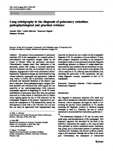
Results Among the 49 patients, 29 had a positive captopril scintigraphy based on standard criteria. Six of the positive cases did not have RAS. Six scintigraphically negative patients had RAS ( bilateral in 4 ). By using the new ER method the number of false negative remained unchanged, but the number of false positives fell to 1. The sensitivity remained the same (79.3%). The specificity increased from 70 to 95% (P70%) and serum creatinine of 155 mmol/l. a. Baseline study shows delayed transit of tracer on the right side (upper left) with significant ipsilateral worsening in the captopril scintigraphy performed 2 h later (upper right). Expected and recorded curves were found to be different ( lower left): recorded curve fell outside the expected limits ( lower right). This pattern of lateralization is strongly suggestive of haemodynamically significant RAS. Most interestingly, no difference (70%) and chronic renal failure (serum creatinine 265 mmol/l ). The post-captopril renogram resulting in a plateauing curve on the right side with rising curve on the left (upper left). Bilateral improvement of renal function was observed under baseline condition (upper right) also in the severely depressed right side. Nevertheless, expected and recorded curves did differ significantly ( lower left) with recorded curve outside the reference limits ( lower right) suggesting a significant lateralization.
for accurate detection of haemodynamically significant RAS. The scintigraphic approach is less expensive (the cost of captopril-enhanced plus baseline scintigraphy is about one-half that of arteriography), non-invasive, useful to monitor patients undergoing revascularization and, when supported by the ER method of calculation, made free of any subjective interpretation. Moreover, it is feasible in patients with advanced renal failure, who are particularly susceptible to the toxic effects of contrast media. In this context the application of the method is expecially useful in bilateral stenosis and in the presence of small or severely compromised kidneys. These are conditions which lead to possible misinterpretation of data, increasing the number of false positive results when applying the standard criteria of analysis of the dynamic curves in captoprilenhanced scintigraphy. In a low prevalence disease such as RVH, the lack of false-positive results is the main advantage of the method. This is relevant for patients with moderate to high risk of RVH, as suspected on clinical ground. The so-called false-negative cases missed with scintigraphic evaluation rarely benefit from revascularization procedures. This supports the idea that even a severe stenosis does not necessarely imply RVH. Taking into account the several risks associated with the procedures of contrast angiography and revascu-
larization (including nephrotoxicity and cholesterol embolism), our policy is to only subject patients found to be positive with captopril-enhanced scintigraphy as examined by method to arteriography plus angioplasty (in a single session). Indeed, the benefits of dilatation procedures in cases of haemodynamically ineffective RAS, i.e. not associated with activation of the renin–angiotensin system, is still a matter for debate [6 ], and autopsy studies [20] confirm that the arteriographic ‘gold standard’ identifies anatomical rather than functional conditions. Thus the so-called scintigraphically false-negative cases often represent cases in which haemodynamic conditions are not affected. The expected renogram method helps to identify these conditions confidently, allowing the planning of any therapeutic intervention with a greater knowledge of the risk–benefit balance [21 ]. Acknowledgements. The authors are indebted to Giulio Cesano MD for his assistance in preparation of the manuscript.
References 1. Schwartz CJ, White TA. Stenosis of renal artery: an unselected necropsy study. Br Med J 1964; 2: 1415–1451 2. Derkx FHM, Schalekamp MADH. Renal artery stenosis and hypertension. Lancet 1994; 344: 237–239 3. Working group on renovascular hypertension. Detection, evalu-
2086
4.
5. 6.
7.
8. 9.
10.
11.
ation and treatment of renovascular hypertension. Arch Intern Med 1994; 147: 820–829 Pickering TG. Renovascular hypertension. Medical evaluation and non-surgical treatment. In: Laragh JH, Brenner BM, ed. Hypertension: Pathophysiology, Diagnosis, and Management. Raven Press, New York, 1990; 1539–1560 Pedersen EB. Angiotensin-converting enzyme inhibitor renography. Pathophysiological, diagnostic and therapeutic aspects in renal artery stenosis. Nephrol Dial Transplant 1994; 9: 482–492 Prigent A. The diagnosis of renovascular hypertension: the role of captopril renal scintigraphy and related issues. Eur J Nucl Med 1993; 7( 20): 625–644 McLean AG, Hilson AJ, Scoble JE et al. Screening for renovascular disease with captopril-enhanced renography. Nephrol Dial Transplant 1992; 7: 211–215 Mann SJ, Pickering TG, Sos TA et al. Captopril renography in the diagnosis of renal artery stenosis; accuracy and limitations. Am J Med 1991; 90: 30–40 Fommei E, Ghione S, Hilson AJW et al. and the European multicenter study Group. Captopril radionuclide test in renovascular hypertension. Communications at the 8th International Symposium on Radionuclides in Nephrourology, Chester, May 1992. Nucl Med Commun 1992; 13: 370 Sfakianakis GN, Bourgoigne JJ, Jaffe D, Kryiakides G, PerezStable E, Duncan RC. Single-dose captopril scintigraphy in the diagnosis of renovascular hypertension. J Nucl Med 1987; 28: 1383–1392 Itoh K, Shinohara M, Togashi M, Koyanagi T. Captopril renal scintigraphy in a patient with bilateral renal artery stenosis. J Nucl Med 1989; 30: 2042–2045
D. Roccatello and G. Picciotto 12. Holley KE, Hunt JC, Brown AL, Kincaid OW, Sheps SG. Renal artery stenosis. A clinical-pathologic study in normotensive and hypertensive patients. Am J Med 1964; 37: 14–22 13. Eyler WR, Clark MD, Garman JE, Rian RL, Meininger DE. Angiography of the renal areas including a comparative study of renal arterial stenoses in patients with and without hypertension. Radiology 1962; 78: 879–881 14. Dustan HP, Humphries AW, De Wolf VG, Page JH. Normal pressure in patients with renal artery stenosis. JAMA 1964; 187: 1028–1032 15. Roccatello D, Picciotto G, Rabbia C, Pozzato M, DeFilippi PG, Piccoli G. Prospective study on captopril renography in hypertensive patients. Am J Nephrol 1992; 12: 406–411 16. Bourgoigne JJ, Rubbert K, Sfakianakis GN. Angiotensinconverting enzyme-inhibited renography for the diagnosis of ischemic kidneys. Am J Kidney Dis 1994; 24: 665–673 17. Maxwell MH, Bleifer KH, Franklin F, Varady PD. Cooperative study of renovascular hypertension: demographic analysis of the study. JAMA 1972; 220: 1195–1204 18. Dubovsky EV, Russel CD, Japanwalla M. Bilateral response to captopril is nonspecific. Eur J Nucl Med 1992; 8: 575 19. Prigent A, Froissart M, Azizi M, Plouin PF, Paillard M. Split and global renal function after percutaneous transluminal renal angioplasty in patients with unilateral renal artery stenosis. Eur J Nucl Med 1994; 212 (8): 795 20. Choudri AH, Cleland JGF, Rowlands PC, Tran TL, McCarty M, Al-Kutoubi MAO. Unsuspected renal artery stenosis in peripheral vascular disease. Br Med J 1964; 2: 1415–1421 21. Main J, Wilkinson R. Angioplasty for atheromatous renal artery stenosis. Do the benefits outweigh the risks? J Nephrol 1990; 3: 143–145 Received for publication: 29.3.96 Accepted in revised form: 20.5.97