Review Article Indian J Med Res 128, October 2008, pp 353-372
Carcinogenicity of hexavalent chromium A.L. Holmes*,**, S.S. Wise*,** & J.P. Wise, Sr.*,**,†
*
Wise Laboratory of Environmental & Genetic Toxicology, **Maine Center for Toxicology & Environmental Health, University of Southern Maine, 96 Falmouth St., Portland, ME 04104 † Department of Applied Medical Sciences, University of Southern Maine, 96 Falmouth St., Portland, ME 04104, USA
Received April 1, 2008
Hexavalent chromium (Cr(VI)), a commonly used industrial metal, is a well known human lung carcinogen. Epidemiology and animal studies suggest that the particulate Cr(VI) compounds, specifically the water insoluble compounds, are the more potent carcinogens; however, the carcinogenic mechanism remains unknown. Here we summarize recent Cr(VI)-induced human tumour, in vivo, cell culture and in vitro studies and put the data into context with three major paradigms of carcinogenesis: multistage carcinogenesis, genomic instability, and epigenetic modifications. Based on these studies, we propose a mechanism for chromate carcinogenesis that is primarily driven by the genomic instability paradigm.
Key words Chromate - chromium - chromosome instability - epigenetic changes - genomic instability - hexavalent chromium lead chromate - mutations - zinc chromate
Introduction/overview
workers was performed in 1948. Machle and Gregorius showed that 21.8 per cent of the chromate workers deaths were due to respiratory cancer compared to only 1.4 per cent in their reference population1. Large studies on chromate production workers have also been performed in Baltimore, Maryland; Plainsville, Ohio; and Great Britain2,3. Altogether, these studies indicate that these workers have about a 2-80 fold increased relative risk of developing lung cancer and that exposure to Cr(VI) and not Cr(III) was responsible for this effect2-4. Lung cancer risk also increased with exposure time. These studies indicate that insoluble calcium chromate is more carcinogenic than soluble sodium chromate because lung cancer rates decreased after factories changed to a low or no lime production process2.
For more than 100 years, numerous epidemiological studies have been performed on workers exposed to hexavalent chromium (Cr(VI)) to determine its carcinogenicity. Major studies pinpointing Cr(VI) as a human lung carcinogen have been performed on workers involved in chromate production, chromate pigment production and chromium plating. Chromate production involves combining trivalent chromite ore with soda ash and sometimes lime and heating the mixture to high temperatures causing the chromite to oxidize to soluble sodium chromate. If lime is used in the production process, insoluble calcium chromate is also produced. The first epidemiology study on chromate production 353
354
INDIAN J MED RES, OCTOBER 2008
Chromate pigment production involves reacting sodium chromate with zinc or lead to produce insoluble zinc chromate and lead chromate, respectively. Zinc chromate and lead chromate are the predominate chromium compounds used in pigments but insoluble barium and strontium chromate can also be used. Studies in these workers also found that insoluble chromate exposure increases lung cancer risk and that such risks increase with exposure time2,3,5,6. Animal studies support the epidemiologic findings including both the observations that chromate exposure causes lung cancer and that the water insoluble (particulate) compounds are the more potent carcinogenic form. The majority of studies investigating soluble Cr(VI) compounds administered through inhalation, intrabronchial implantation, or intratracheal instillation either showed no effect or small but non significant increases in lung tumour formation 7 . However, numerous studies investigating particulate Cr(VI) compounds such as calcium chromate, strontium chromate, lead chromate and zinc chromate observed significant increases in lung tumours. For example, Levy et al8 showed that intrabronchial implantation of soluble sodium chromate induced only 1 bronchial carcinoma in 100 rats whereas intrabronchial implantation of two different doses of strontium chromate induced 43 and 62 bronchial carcinomas in 99 rats. They reported no increase in bronchial carcinomas after intrabronchial implantation of particulate lead chromate 8, however, three studies injecting lead chromate either subcutaneously or intramuscularly induced a significant increase in sarcomas at the injection site indicating that this compound is also a carcinogen9-11. Cell culture data also indicate that the particulate compounds have greater potency. Working with C3H/ 10T1/2 cells, Patierno et al 12 showed that only particulate lead chromate could induce neoplastic transformation while soluble sodium chromate could not. Thus, epidemiology, whole animal and cell culture studies all indicate that the particulate compounds are the most potent carcinogenic forms of Cr(VI) with respect to lung cancer. Interestingly, recent work has shown that soluble Cr(VI) administered chronically in drinking water can induce oral and intestinal tumours13. Thus, it appears that while soluble Cr(VI) may only be weakly carcinogenic to the lungs, it may pose a more significant carcinogenic risk if ingested. Studies have not been done of particulate Cr(VI) after oral exposure, but given the low pH of the stomach, it is likely that
these exposures would ultimately become exposures to soluble Cr(VI) as particulate chromates dissolve at low pH. The focus of this review is on chromate-induced lung cancers as the drinking water data are very recent and no studies have been done to clarify the mechanism for these tumours. Characteristics of chromate-induced lung tumours Human pathology studies show that Cr(VI) deposits and persists at bronchial bifurcations where Cr(VI)associated cancers occur14,15, which is consistent with a particulate chromate exposure. The majority of lung cancers in chromate exposed workers are squamous cell carcinomas, often with multiple tumours15. In addition, chromium levels in workers with lung cancer are much higher than in workers without lung cancer 15 . Interestingly, one study found no correlation between chromium accumulation in the lungs of workers and duration of chromium exposure or multiple lung tumours but malignancy stage was significantly associated with chromium accumulation 16 . The association between malignant stage and chromium accumulation was also confirmed in animal studies where a pellet of strontium chromate was inserted into the bronchus of rats17. Molecular studies of chromate-induced lung tumours reveal that the tumours exhibit minimal mutations in key oncogenes and tumour suppressor genes but do exhibit genomic instability and epigenetic changes (Table I). For example, there were no significant differences in Bcl-2 and p53 expression between lung tumours from chromate workers, unexposed individuals and individuals with pneumoconiosis 18. Few p53 mutations and no ras mutations were observed in tumours from chromate workers19,20. By contrast, these tumours exhibited both chromosome instability (CIN) and microsattelite instability (MIN). Hirose et al21 found a statistically significant increase in microsatellite instability. Of 38 chromate-induced lung tumours analyzed, 30 exhibited instability in two or more microsatellite markers (79%) while MIN was observed in only 4 of 26 tumours from non-exposed individuals (15%) 21,22 . MIN also increased with longer exposure time21 and was strongly correlated with decreased expression of hMLH122. But interestingly, MIN was not significantly associated with hMSH2 repression22. In fact, 100 per cent of the tumours without MIN had repressed hMSH2 expression, while only 60 per cent of the tumours with extensive MIN exhibited hMSH2 repression. In
HOLMES et al: CARCINOGENICITY OF HEXAVALENT CHROMIUM
355
Table I. Characterization of Cr-induced tumours Study population
Assay
Summary of findings
19 lung tumours from 18 chromateexposed workers Unexposed with lung cancer Individuals with pneumoconiosis 20 lung tumours from 19 chromateexposed workers
Immunohistochemistry for cyclin D1, bcl-2, p53
38 lung tumours from 32 workers
PCR-SSCP analysis
38 lung tumours from 28 chromateexposed workers 26 lung tumours from individuals without chromate exposure
Analyzed six microsatellite instability markers
35 lung tumours from 26 chromateexposed workers 26 lung tumours from individuals without chromate exposure
Immunohistochemistry for MLH1 and MSH2 COBRA for MLH1 promotor methylation Microsatellite instability analysis
10 lung tumour specimens from chromate workers 90 biopsy specimens from 25 chromate workers
Microscopic X-ray fluorescence analyzer with transmitted X-ray mapping imaging
38 lung tumours from 31 chromate workers 40 lung tumours from 40 non-exposed individuals
Methylation-specific PCR for p16 promoter Immunohistochemistry for p16
31 lung tumours from 26 chromate workers 38 adenocarcinomas from non-exposed individuals 46 SCC from non-exposed individuals 89 healthy individuals
PCR genotyping methods for SP-B intron-4 polymorphisms
Cyclin D1 expression significantly increased in chromate workers Bcl-2 and p53 expression not significantly different from controls 20% of chromate-induced tumours had p53 point mutations Fewer p53 mutations in chromate-induced tumours than in lung tumours without chromate exposure No association between mutations and length of chromate exposure No point mutations in ras observed in chromate cancers. RER statistically higher in chromate vs. nonchromate lung cancer No significant difference in frequency of LOH but LOH frequency still 50-70% Period of chromate exposure in workers with RER were longer than those without RER Workers with longer exposures to Cr(VI) had higher frequency of MIN Chromate-induced tumours showed a statistically significant decrease in expression of hMSH2 and hMLH1 compared to non-chromate lung cancer Only the repression rate for hMLH1 was significantly correlated to MIN 62.5% of the chromate lung cancer exhibited hMLH1 gene methylation No correlation between chromium accumulation and chromate exposure, MIN, smoking index or presence of multiple lung carcinoma Amount of chromium accumulation significantly increased according to progression of malignancy 85.7% of chromate lung cancers had decreased p16 expression Workers with less than 15 years chromate exposure had no p16 methylation but workers with greater than 15 years exposure had higher levels of p16 methylation Reduced p16 expression was correlated with methylation at p16 SP-B variations were significantly higher in chromate workers with lung cancer compared to unexposed individuals with lung cancer or healthy individuals
PCR-SSCP analysis
addition, MIN was not significantly associated with chromium accumulation in the lung 16. These data suggest there may be a mechanism for MIN that does not involve loss of mismatch repair function. Hirose et al 21 also found between 50-75 per cent loss of heterozygosity (LOH) in 6 different loci they
Ref 18
19
20 21
22
16
23
24
examined, however, it was not significantly different compared to tumours without chromate exposure. The lack of statistical significance may suggest that LOH is required for all lung cancers as previous studies have shown that most lung cancers exhibit chromosome instability25.
356
INDIAN J MED RES, OCTOBER 2008
Chromate-induced tumours also exhibited changes in gene expression of cyclin D1 and p1618,23. Cyclin D1 expression was significantly increased in chromateinduced tumours but was not observed in the normal epithelia adjacent to the tumour18. P16 expression was decreased in 86.7 per cent of chromate-induced lung tumours which was associated with promoter methylation23. P16 promoter methylation was linked to exposure time with methylation at the promoter only found in workers exposed to chromate for 15 or more years23. Another study investigated variations in the surfactant protein B (SP-B) gene. Removal of foreign objects, such as chromate particles, from the upper respiratory tracts requires SP-B and gene variations in SP-B could enhance worker’s susceptibility to chromate lung cancer24. Ewis et al24 found that there was a significant increase in insertion or deletion variations in the SP-B gene in workers with chromateinduced lung tumours compared to unexposed individuals and chromate workers without lung cancer24. Potential mechanisms of chromate-induced carcinogenesis The mechanism of chromate-induced carcinogenesis remains unknown. Three well-accepted general paradigms of carcinogenesis include multistage carcinogenesis, genomic instability, and epigenetic modification. Using these three paradigms, we summarize how data collected over the past seven years support or refute these as models for chromate-induced carcinogenesis. Multi-stage carcinogenesis Multistage carcinogenesis is a multistep process which involves a series of cellular and molecular changes, as a result of the progressive accumulation of mutations and alterations in protooncogenes and tumour suppressor genes26. Thus, for a chemical to have a carcinogenic mechanism that follows this paradigm, it must be capable of inducing a significant number of mutations in target genes. Only one study investigated Cr(VI)-induced mutation frequency in vivo (Table II). This study used the Big Blue transgenic mouse 27 . The authors administered soluble potassium dichromate via intratracheal instillation and found increased mutation frequency in the lung that was dose- and timedependent 27. The majority of mutations were G:C targeted base substitutions with a few deletions27.
Cell culture studies also show that Cr(VI) can induce mutations (Table II). Numerous studies using a shuttle-vector mutagenesis assay found a concentrationdependent increase in mutations after exposure to 10200 uM soluble Cr(VI)28-31. In these studies a SV-40 based plasmid with the supF gene was treated extracellularly with Cr(VI) and then transfected into and replicated by a human fibroblast cell line immortalized with SV-40. Mutant clones were then selected for with Escherichia coli. These studies found that Cr(VI)-induced mutations were the result of Cr(III)DNA adducts and were not caused by reactive oxygen species or short-lived Cr(V) and Cr(IV) intermediates28,31. They also found that while Cr(III) forms both binary and ternary DNA adducts, it is the ternary DNA adducts that are the most mutagenic and these are generated within 15-60 min after exposure28,30. Further, they found that Cys-Cr(III)-DNA adducts and ascorbate-Cr(III)-DNA adducts were 5.3 and 31 times more mutagenic than binary lesions, respectively28,30. One study using cysteine as a reducing agent found that single base substitutions of GC to AT or TA were the predominant mutations 28 and another study using ascorbate found a similar level of base substitution and deletion mutations30. Three cell culture studies considered mutagenesis in an existing cellular chromosomal locus (Table II). One study investigating both soluble and particulate Cr(VI) compounds found that both potassium chromate (soluble) and barium chromate (particulate) induced mutations in transgenic V79 cells containing the bacterial gpt reporter gene. The mutation frequency of potassium chromate peaked at three-fold above background, while the mutation frequency of barium chromate peaked at 3.5-fold 32 . However, the significance of this increased mutation frequency is uncertain because statistical comparisons were apparently not conducted. In contrast to the in vivo study, 30-50 per cent of the mutations were deletions32. The second study found that the amount of ascorbate present in Chinese hamster ovary cells plays a role in mutation frequency at the hprt locus33. When cells containing 15 µM ascorbate were exposed to Cr(VI), there was no increase in mutations but if the cells were preloaded with 1.4 mM ascorbate the mutation frequency increased 19.2-fold after exposure to 40 µM Cr(VI). The third study found that exposure to 6 µM sodium chromate for 24 h in Chinese hamster ovary cells induced a 3.5-fold increase in mutation frequency and mutation frequency was attenuated in cells deficient in
HOLMES et al: CARCINOGENICITY OF HEXAVALENT CHROMIUM
357
Table II. Chromium (VI) - induced mutations Treatment
Assay (s)
Model system
Summary of effects
Potassium dichromate 1.7-6.75 mg/kg Intratracheal instillation
LacI gene mutagenesis assay
C57BL/6 Big Blue transgenic mice
Potassium chromate 10-100 µM 1h 2 mM cysteine
Shuttle vector mutagenesis assay
Cr-treated plasmids in HF/SV cells
Potassium chromate 25-200 µM 1h 2 mM cysteine Sodium chromate 10-200 µM 30 min 1 mM ascorbate
Shuttle vector mutagenesis assay
Cr-treated plasmids in HF/SV cells
Dose and time-dependent increase in mutation frequency in the mouse lung (4.2 fold increase after 4 wk) - 64% G:C to A:T transitions - 26% G:C to T:A transversions - few deletions GSH depletion decreased mutant frequency Concentration-dependent increase in mutations Cr(III)-DNA adducts and not oxidative damage were responsible for mutations Highest mutagenic lesions occurred 15-60 min after exposure Cys-Cr(III)-DNA adducts were more mutagenic than binary Cr(III)-DNA adducts Single base substitutions were most prevalent mutations Cr-DNA adducts induced mutations Adducts arrest replication
Shuttle vector mutagenesis assay
Cr-treated plasmids in HF/SV cells
Potassium chromate 25-200 µM 1h 0.2-1 mM ascorbate
Shuttle vector mutagenesis assay
Cr-treated plasmids in HF/SV cells
Potassium chromate 5-50 µM 2h Barium chromate 0.05-0.25 µg/cm2 24 h Potassium chromate 0-40 µM 1-6 h 15 µM or 1.4 mM ascorbate Sodium chromate 0-6 µM 24 h
Generation of gptmutants, PCR for deletions
Transgenic, V79 derived, cell line (G12)
hprt mutagenesis assay
CHO and V79 cells
hprt mutagenesis assay
CHO cells: AA8 UV-5 (XPD-) UV-41 (XPF-)
nucleotide excision repair (NER)34. These data suggest that proficient NER is required for the induction of mutations after Cr(VI) exposure. Considered together, these studies suggest that Cr(VI) induces mutations, specifically targeting G:C
Concentration-dependent increase in mutations and replication-blocking lesions Ascorbate-Cr(III)-DNA lesions were more mutagenic and more efficient at inhibiting replication than binary lesions Similar number of base substitutions and deletions Concentration-dependent increase in mutations Increased stability of Cr intermediates decreased mutation frequency and did not induce the formation of more potent mutation-inducing lesions 3-fold increase in mutation frequency at a dose that induced 40% survival ~50% complete gpt deletions 3.5-fold increase in mutation frequency at a dose that induced 75% survival ~30% complete gpt deletions No mutations observed in cells not pre-loaded with ascorbate Preloading with ascorbate induced a concentrationdependent increase in mutations at the hprt locus Concentration-dependent increase in mutation frequency with 6 µM inducing a 3.5 fold increase in mutation frequency NER-deficient cells showed attenuated mutagenesis
Ref 27
28
29
30
31
32
33
34
base substitutions. However, the data concerning Cr(VI)-induced deletion mutations are inconsistent. The in vivo study using the Big Blue transgenic mouse found few deletion mutations, however, the Big Blue mouse system is insensitive to these types of mutations27. The
358
INDIAN J MED RES, OCTOBER 2008
inconsistency in the in vitro data may be due to the different reducing agents used in the experiments. The studies that used cysteine as a reducing agent observed few deletion mutations while the studies that used ascorbate observed comparable numbers of deletion and substitution mutations. In addition, the one cell culture experiment that investigated mutation spectrum found that Cr(VI) induced 30-50 per cent deletion mutations. Thus, Cr(VI) appears to induce deletion mutations except when cysteine is the sole reducing agent. Interestingly, the data for Cr(VI)-induced tumours appear to contradict the whole animal and cell culture studies. Mutations appear to be infrequent in chromateinduced tumours, though these can be experimentally induced. This discrepancy may indicate that Cr(VI) is only weakly mutagenic, with mutations only occurring at very high doses. Because of the technical challenges of detecting mutations, experimental mutagenesis studies often use very high doses that do not reflect likely exposure scenarios. Indeed for the in vitro Cr(VI) studies, the concentrations utilized were often between 25 and 200 µM for short periods of time which is not likely to be an environmentally or occupationally relevant exposure. Exposing cells to this concentration range would result in very little cell survival. For example, exposure to 5 µM sodium chromate for 24 h induces close to 100 per cent cell death in human lung fibroblasts35,36. Studies on Cr(VI)-induced toxicity in human lung fibroblasts for shorter periods of time have not been done, but only about 5 per cent of HCT116+ch3 cells (a human colon cancer cell line) survived a 3 h treatment with 20 µM sodium chromate37. In addition, the majority of the mutation studies were performed using a shuttle vector system, which does not fully mimic the effects of Cr(VI) in a cell or the ability of DNA repair systems to repair the damage. These differences may contribute to the dramatically higher mutation frequency in the shuttle vector mutagenesis assays compared to the cellular chromosomal locus mutagenesis assay and cause Cr(VI) to appear to be a more potent mutagen in cell culture than it may actually be. The role of the reducing agent is an interesting factor. The published data suggest that the levels of these reducing agents can affect the mutation spectrum and potency 30,31,33 . However, the full interpretation is complicated by limited data of the levels of these agents in human lung cells. For example, Reynolds et al33 preloaded cells with 1.4 mM ascorbate. This level is comparable to ascorbate levels in freshly purified human
lymphocytes, however, these ascorbate levels are dramatically higher than those reported in human lung tissue. Specifically, in adults, lung tissue ascorbate levels range from 0.045-0.065 mg/g which is approximately 256 µM ascorbate38. Reynolds et al33 found that Cr(VI) increased mutation frequency only occurred in cells preloaded with 1.4 mM ascorbate, a level 5.5-times higher than human lung tissue levels. Cr(VI) was not mutagenic in cells with ascorbate levels lower than 1.4 mM. Thus, the reason mutation levels were low in Cr(VI)-induced human lung tumours may be due to the fact that ascorbate levels in the lung are not high enough for Cr(VI)-induced mutations to occur. Such a conclusion would be consistent with the low mutation rates in cell culture systems without ascorbate. Lymphocytes, although high in ascorbate probably turn over too quickly for mutations to be expressed in vivo, which likely explains the absence of significant frequencies of Cr(VI)-induced cancer in these potential target cells. Interestingly, Reynolds et al33 found that Cr(VI) was not mutagenic in cells without a high dose of ascorbate, while both Klein et al32 and Brooks et al34 reported that Cr(VI) was mutagenic in these cells. More specifically, Klein et al32 found a 3-fold increase in mutation frequency at 40 µM potassium chromate for 2 h while Reynolds et al33 found no increase in mutation frequency with the same treatment for 3 h in cells that were not pre-loaded with ascorbate. The discrepancy could be due to the different cell lines used. Klein et al32 used a CHO cell line that contains the bacterial gpt reporter gene, while Reynolds et al33 used the endogenous hprt gene in CHO cells to detect mutations. Therefore, the difference in mutation frequency could be due to differences in Cr(VI)induced mutations in exogenous versus endogenous genes. However, Brooks et al34 used the same CHO cell line as Reynolds et al33 and found a 3.5-fold increase in mutations after a 24 h exposure to Cr(VI) as opposed to 3 h in Reynolds et al suggesting that Cr(VI) is mutagenic without excess ascorbate. Given that two studies were positive and the other negative, it seems likely that ascorbate is not required for Cr(VI) mutagenesis, though it may accelerate the process. If Cr(VI) is indeed a weak mutagen, then the mutation frequency that occurs after occupational Cr(VI) exposure may not be high enough to induce multiple mutations in key genes. For example, based on the chromosomal locus mutagenesis assay in cells that were not loaded with high ascorbate, there was a 2-4 fold increase in mutation frequency above
HOLMES et al: CARCINOGENICITY OF HEXAVALENT CHROMIUM
background 32,34 . Assuming that the spontaneous mutation rate in dividing cells is 1.4 x 10⁻¹⁰ mutations per base pair per cell generation and there are ~70,000 genes in the human genome, it is estimated that only one mutation would arise spontaneously in a normal cells lifespan 39. Therefore, if Cr(VI) increases the normal mutation frequency by 2-4 fold, this would only produce 2-4 mutations in a cell over its lifespan. The likelihood of those 2-4 mutations randomly mutating a key tumour suppressor gene or oncogene seems low. In addition, tumour suppressor genes often require mutations in both alleles in order to be inactivated further decreasing the probability of losing these genes. On the other hand, it may be that the human tumour studies have not considered enough genes and Cr(VI) simply does not mutate p53 and ras. If this is true there may be a higher mutation rate in other genes. This possibility seems unlikely given that ras is mutated in 20-30 per cent of lung cancers40 and p53 is mutated in 50-90 per cent of lung cancers41. These data would suggest that Cr(VI)-induced tumours are unusual compared to other lung tumours. The multistage carcinogenesis paradigm requires the stepwise acquisition of mutations in multiple key tumour suppressor and oncogenes. Considering the above studies, it appears that Cr(VI) is not a potent mutagen and is unlikely to induce sufficient mutations for this paradigm to apply. Therefore, we suggest that the multistage carcinogenesis does not fit well for chromate carcinogenesis. Genomic instability The genomic instability paradigm argues that disruption of the control of genomic stability results in a cascade of changes in the whole genome 42. This control can be disrupted by interfering with DNA repair, kinetochore assembly, checkpoints, centrosome duplication, microtubule dynamics and numerous other cellular maintenance processes42. In particular, there are two types of genomic instability; microsatellite instability (MIN) and chromosome instability (CIN). MIN is characterized by changes in the lengths of microsatellites which are series of repetitive non-coding DNA sequences that are abundant in the human genome43. DNA polymerases often slip while replicating microsatellites resulting in insertion or deletion loops. In normal cells, these insertion/deletion loops are repaired by mismatch repair (MMR). If cells acquire defective MMR, they are unable to repair these lesions. Unrepaired insertion/deletion loops result in changes
359
in the lengths of microsatellites which is carried through to the whole genome increasing the mutation rate by more than 200-fold43. MIN has been observed in Cr(VI)-induced tumours21. Seventy nine per cent of the lung tumours from chromate workers exhibited MIN at two or more loci and 18 per cent of the tumours exhibited MIN at all five markers tested21. In contrast, only 15 per cent of lung tumours from patients without Cr(VI) exposure exhibited MIN at two or more loci 21 . MIN was associated with length of chromate exposure, with workers with longer exposures exhibiting a higher frequency of MIN21. MIN was also associated with hMLH1 repression22. No animal studies or cell culture studies have directly investigated the ability of Cr(VI) to induce MIN so it is uncertain if these effects are early or late events in tumourigenesis. This absence of data is largely due to the absence of any sufficient in vivo or in vitro tests for mismatch repair. Two cell culture studies investigated the effects of alterations in MMR gene expression. One study reported that MMR-deficient carcinoma cells exhibited decreased cytotoxicity, apoptosis and DNA double strand breaks37. In addition, MMR-deficient cells were unable to induce replication arrest due to Cr-DNA adducts37. The second study reported that depletion of MLH1 or MSH2 in normal lung cells decreased the formation of CREST(-) micronuclei and DNA double strand breaks after potassium chromate exposure33. These data suggest that proficient MMR plays a role in Cr(VI)-induced genotoxicity and cytotoxicity. This observation contrasts with the tumour data because deficient MMR is required for MIN, while proficient MMR is required for double strand break formation which may be a key step in the mechanism of Cr(VI)induced carcinogenesis. CIN occurs in the majority of lung cancers and these tumours can have both MIN and CIN25,44. CIN includes both numerical and structural changes. Numerical CIN involves the loss and gain of whole chromosomes. Structural CIN involves chromosome translocations and breaks. Exposure to Cr(VI) has been shown to induce both numerical and structural CIN. Numerical CIN has not been investigated in vivo or in chromate-induced tumours. Using cell culture models, five studies assessed numerical CIN after Cr(VI) exposure by counting metaphase chromosomes (Table IIIa). Two studies found that aneuploidy increased in normal human lung fibroblasts after a 30 h
360
INDIAN J MED RES, OCTOBER 2008 Table IIIa. Genomic instability: Microsatellite instability and numerical chromosome instability
Treatment
Assay(s)
Model system
Summary of effects
Potassium dichromate 0-30 µM 3h
Clonogenic survival Western blotting Flow cytometry H2A.X foci formation
After Cr(VI) exposure, MMRdeficient cells exhibited: - increased clonogenic survival - decreased apoptosis - inability to block replication as a result of Cr-DNA adducts - decreased DNA double strand breaks
37
Potassium chromate 0.2-5 µM 1-6 h 1.4 mM ascorbate Potassium dichromate 0.25-1 µM 30 h Potassium dichromate 0.25-1 µM 30 h
H2A.X foci formation Micronucleus assay
A549 human lung carcinoma cells Colon cancer cell lines: HCT116 (MLH1-/-) DLD1 (MSH6-/-) HCT116+ch3 (MLH+) DLD1+ch2 (MSH6+) Mouse embryonic fibroblasts: Mlh-/-, Mlh+/+ Pms-/-, Pms+/+ IMR90 human lung fibroblasts
Depletion of MLH1 or MSH2 decreased micronuclei and double strand break formation
33
Chromosome counting
MRC-5 human lung fibroblasts
45
Chromosome counting Anaphase-telophase assay
MRC-5 human lung fibroblasts
Lead chromate 0.1-1 µg/cm2 24-120 h
Chromosome counting Clonogenic aneuploidy Mitotic stage analysis Centrosome analysis
WTHBF-6 human lung fibroblasts
Lead chromate 0.1-1 µg/cm2 24-120 h
Chromosome counting Chromosome damage Clonogenic aneuploidy Mitotic stage analysis Western blot for Mad2 expression
WTHBF-6 human lung fibroblasts
Lead chromate 1-10 µg/cm2 120 h
Transformation assay Chromosome damage Centrosome analysis
BEP2D human lung epithelial cells
Increase in aneuploid cells Increase in hypodiploid cells and no increase in hyperdiploid cells Increase in aneuploid cells, specifically hypodiploid cells Increase in chromatin bridges, lagging chromosomes and lagging fragments Concentration- and time-dependent increase in: - aneuploid cells including hypodiploid and polyploid cells - centrosome number in both interphase and mitotic cells - abnormal mitotic figures including disorganized anaphase and mitotic catastrophe Aneuploid cells were able to survive and form colonies Concentration- and time-dependent increase in premature centromere division, centromere spreading and premature anaphase Lead chromate disrupts mitotic progression with an increase of cells in anaphase Decreased Mad2 expression Time- and concentration-dependent increase in tetraploid cells and tetraploid cells persist and form colonies SAC bypass due to chromium and not lead ions or the particle Chronic exposure to lead chromate induced loss of contact inhibition and anchorage-independent growth Foci cells exhibited aneuploidy and centrosome amplification
exposure to soluble chromate with a specific increase in hypodiploid cells but no increase in hyperdiploid cells45,46. Two studies found that chronic exposure to particulate chromate induced concentration- and time-
Ref
46
17
48
49
dependent increases in aneuploidy in normal human lung fibroblasts. However, in contrast to the soluble chromate study, these studies reported increases in both hypodiploid and tetraploid cells47,48. These particulate
HOLMES et al: CARCINOGENICITY OF HEXAVALENT CHROMIUM
chromate-induced aneuploid cells were able to survive and form colonies47,48. The fifth study showed that human lung epithelial cells transformed with lead chromate also had increased aneuploidy further indicating that the aneuploid phenotype persists49. There are a number of mechanisms that can give rise to numerical CIN, including centrosome amplification, spindle assembly checkpoint bypass, malfunctions in sister chromatid cohesion and abnormalities in kinetochore structure or function42. Centrosome amplification can induce aneuploidy through multipolar spindle formation and division50. Two studies considered centrosome amplification after Cr(VI) exposure 47,49 . Holmes et al 47 reported a concentration- and time-dependent increase in centrosome amplification in human lung fibroblasts exposed to particulate chromate. Centrosome amplification was also observed in human lung epithelial cells transformed by particulate chromate indicating that centrosome amplification may be an early event in Cr(VI)-induced carcinogenesis and that this phenotype persists49. One study considered spindle assembly checkpoint bypass as a potential mechanism of Cr(VI)-induced aneuploidy48. This study reported spindle assembly checkpoint bypass after chronic particulate chromate exposure with concentration- and time-dependent increases in premature anaphase, premature centromere division, centromere spreading and the total number of cells in anaphase. In addition, expression levels of Mad2, an important protein involved in the regulation of the spindle assembly checkpoint, were suppressed confirming spindle assembly checkpoint bypass on a molecular level48. This study also showed that Cr(VI) and not the lead cation or internalized particles were responsible for the spindle assembly checkpoint bypass48. The observed increase in centromere spreading may also indicate that chromium induces malfunction in sister chromatid cohesion. Consistent with spindle assembly checkpoint bypass and chromosome segregation malfunctions, Seoane et al.46 showed increased chromatin bridges, lagging chromosomes and lagging fragments after a 30 h soluble chromate exposure in human lung fibroblasts. The induction of translocations is the gold standard for evaluating and measuring structural CIN but so far no studies have investigated the ability of Cr(VI) to cause translocations. The majority of work investigating structural chromosomal effects has considered the induction of chromosomal aberrations which can lead to structural CIN (Table IIIb). Two different assays have
361
been used to assess chromosome damage: The chromosome damage assay and the micronucleus assay. The chromosome damage assay is a more sensitive assay and provides information on the kind of damage formed. The micronucleus assay is less sensitive but is quicker. A wealth of data using the chromosome damage assay shows that both particulate and soluble Cr(VI) induce chromosomal aberrations in human lung fibroblasts and human lung epithelial cells35,51-58. Furthermore, these studies show that the lead cation and internalized particles do not contribute to Cr(VI)-induced clastogenicity and that all of the effects are due to partial dissolution of chromate particles releasing extracellular Cr(VI). Three studies have investigated chromosome damage in vivo using the micronucleus assay. The National Toxicology Program (NTP) report, 2007 showed that soluble chromate administered orally through drinking water induced a concentration-dependent increase in micronuclei in normochromatic erythrocytes in am3C57BL/6 male mice but only a small increase in micronuclei in B6C3F1 mice and no increase in BALB/c mice59. A second study in the NTP report showed no increase in micronuclei in normochromatic erythrocytes after exposure to soluble chromate for 3 months through drinking water in F344/N rats and B6C3F1 mice59. The third study showed that exposure to a single dose of soluble chromate delivered by intraperitoneal injection increased micronuclei frequency in polychromatic erythrocytes in BDF1 mice60. The results from these in vivo studies are inconsistent and have some shortcomings in regard to Cr(VI)-induced lung cancer, especially since the NTP report was focused on the effects of Cr(VI) in drinking water. First, these studies used soluble chromates and administration routes that do not directly expose the lung. In addition, the authors investigated micronuclei formation in erythrocytes which are not the target cells in chromate-induced lung tumours. One cell culture based study using the micronucleus assay is consistent with the chromosome damage studies confirming that Cr(VI) induces chromosome aberrations33. Even though the in vivo data are inconsistent, the cell culture data clearly show that particulate and soluble chromate induce chromosome damage in human lung cells. Cr(VI)-induced chromosome aberrations are most likely caused by Cr(VI)-induced DNA double strand breaks (Table IIIc). No in vivo studies have specifically investigated double strand break formation. Several recent cell culture studies demonstrated that both
362
INDIAN J MED RES, OCTOBER 2008 Table III b. Genomic instability: Structural chromosome instability manifested as chromosome damage
Treatment
Assay(s)
Model system
Summary of effects
Ref
Lead chromate 0.1-5 µg/cm2 24 h Sodium chromate 1-10 µM 24 h Lead chromate 0.1-5 µg/cm2 24 h Sodium chromate 1-10 µM 24 h Lead chromate 0.1-5 µg/cm2 24 h Sodium chromate 1-5 µM 24 h Barium chromate 0.1-5 µg/cm2 24 h Lead chromate 0.1-5 µg/cm2 24 h Barium chromate 0.1-5 µg/cm2 24 h Lead chromate 0.1-5 µg/cm2 24 h Sodium chromate 1-2.5 µM 24 h Lead chromate 0.1-5 µg/cm2 24 h
Chromosome damage
Primary human lung cells
Lead chromate and sodium chromate induced chromosome damage in a concentration-dependent manner
35
Chromosome damage
Primary human lung cells and WTHBF-6 human lung fibroblasts
Chromosome damage levels were similar in primary human lung cells and WTHBF-6 (hTERT+) cells WTHBF-6 cells are as a useful lung cell model
51
Chromosome damage
WTHBF-6 human lung fibroblasts
Cr(VI) is the proximate clastogen Lead ions are not involved in the clastogenicity of lead chromate
52
Chromosome damage
WTHBF-6 human lung fibroblasts
53
Chromosome damage
WTHBF-6 human lung fibroblasts
Barium chromate induced concentration-dependent increases in chromosome damage Barium chromate was more genotoxic than lead chromate
Chromosome damage Chromium particle uptake
WTHBF-6 cells
Chromosome damage Neutral comet assay H2A.X foci formation
WTHBF-6 human lung fibroblasts
Lead chromate 0.1-1 µg/cm2 24-72 h Sodium chromate 0.5-2.5 µM 24-72 h
Chromosome damage
WTHBF-6 human lung fibroblasts
Lead chromate 0.5-50 µg/cm2 24 h Sodium chromate 0.5-10 µM 24 h Sodium dichromate 1.7-20.9 mg/kg in rats 3.1-27.9 mg/kg in mice 3 months orally Sodium dichromate 2.8-8.7 mg/kg 3 months orally
Chromosome damage
BEP2D lung epithelial cells
Micronucleus assay
F344/N rats and B6C3F1 mice
No increase in micronuclei in normochromatic erythrocytes
59
Micronucleus assay
B6C3F1, BALB/c, and am3-C57BL/6 mice
Concentration-dependent increase in micronuclei in normochromatic erythrocytes in am3-C57BL/6 male mice
59
Chromosome damage was due to external dissolution of lead chromate particles Particle-cell interaction was not required for chromosome damage induction Concentration-dependent increase in chromosome damage Concentration-dependent increase in DNA double strand breaks ATM is phosphorylated in response to DSB Lead chromate induced a concentration-dependent increase in chromosome damage which persisted over time Sodium chromate-induced chromosome damage decreased over time Lead chromate and sodium chromate induced similar amounts and spectrum of chromosome damage in human lung epithelial cells
54
55
56
57
58
Contd....
Treatment
Potassium dichromate 50 mg/kg i.p. injection 1 dose Potassium chromate 0.2-5 µM 1-6 h 1.4 mM ascorbate
HOLMES et al: CARCINOGENICITY OF HEXAVALENT CHROMIUM
363
Assay(s)
Ref
Model system
Micronucleus assay
40 BDF1 mice
Micronucleus assay
IMR90 human lung fibroblasts
soluble and particulate Cr(VI) induce DNA double strand breaks measured by gamma-H2A.-X foci formation or with the single cell gel electrophoresis assay (comet assay)33,37,56,61-64. These studies show that the breaks only form during S and G2 phases as a result of either excision repair of crosslinks/ternary adducts or collapsed replication forks due to repeated cycles of futile MMR attempting to repair these lesions33,34,63. In addition, failure of homologous recombination repair of DNA double strand breaks leads to more complex and increased chromosome damage and neoplastic transformation61,65. By contrast, failed NHEJ repair has no effect on chromosomal aberrations66.
Summary of effects
Small (NS) increase in micronuclei in B6C3F1 mice and no increase in BALB/c mice Increased frequency of micronuclei in 60 polychromatic erythrocytes Preloading with ascorbate increased CREST-negative micronuclei formation Depletion of MLH1 or MSH2 decreased micronuclei
33
include altered DNA methylation or acetylation, protein phosphorylation status changes, growth stimulation, and altered gene expression or signaling pathways. Epigenetic changes, including growth stimulation and enhanced survival, changes in phosphorylation and methylation status and altered gene expression and signaling pathways, have been considered after Cr(VI) exposure.
Epigenetic modifications
Five recent studies have considered the potential role of growth stimulation, escape from growth arrest and apoptosis inhibition after Cr(VI) exposure. All were cell culture studies (Table IVa). Two studies investigated the hypothesis that the cation released from the partial dissolution of particulate Cr(VI) compounds caused Cr(VI) damaged cells to survive. These studies used lead chromate as a model compound in human lung fibroblasts and found that lead did not promote survival of Cr(VI)damaged cells or stimulate the growth of Cr(VI)-damaged cells36,68. Three studies investigated the hypothesis that Cr(VI) could inhibit apoptosis. One study looked at direct inhibition by soluble Cr(VI) in human lung epithelial cells69. It reported that soluble Cr(VI) induced NF-kB activation which inhibited apoptosis possibly through the inhibition of p53 activation69. Two studies considered effects on apoptosis in cells that survived Cr(VI) treatments 70,71 . One study found that human skin fibroblasts which survived a highly toxic dose of sodium chromate were resistant to apoptosis and had at least a 2fold change in gene expression in genes involved in DNA repair, apoptosis and cell cycle regulation70. Son et al71 generated Cr(VI)-resistant clones from human kidney cells after three consecutive chromium trioxide treatments. These clones were resistant to the cytotoxicity of chromium trioxide and grew at a similar rate in media containing high concentrations of chromium oxide as cells in treatment-free media71.
Epigenetic modifications in cancer are changes in cellular functions that occur without altering the genetic material resulting in tumourigenesis67. Examples of epigenetic changes that could drive tumour formation
Two studies investigated the potential effects of Cr(VI) on phosphorylation (Table IVb). One was in cell culture and the other was in vitro. The cell culture study showed that tyrosine phosphorylation increased in a
The Cr(VI)-induced tumour and cell culture studies show that Cr(VI) induces genomic instability but further work needs to be performed to elucidate the specific mechanism. Studies show that the Cr(VI)-induced tumours exhibit MIN implying MMR inactivation, however, further work needs to be performed on the mechanism of MMR inactivation. Current data suggest that proficient MMR is required to produce double strand breaks and no studies have demonstrated how or why Cr(VI) would target MMR genes. Cell culture studies show that Cr(VI) induces aneuploidy, centrosome amplification and spindle assembly checkpoint bypass but these phenotypic changes still need to be confirmed in the tumours. In addition, data suggest that Cr(VI) induces structural chromosome instability because it induces chromosome damage and DNA double strand breaks but this needs to be confirmed by investigating the ability of Cr(VI) to induce translocations. However, considering all of the data, the genomic instability paradigm is a possible model for Cr(VI)-induced carcinogenesis.
364
INDIAN J MED RES, OCTOBER 2008 Table IIIc. Genomic instability: Structural instability manifested as DNA double strand breaks
Treatment
Assay(s)
Model system
Summary of effects
Potassium chromate 0.2-5 µM 1-6 h 1.4 mM ascorbate
H2A.X foci formation
IMR90 human lung fibroblasts
33
Potassium dichromate 0-30 µM 3h
H2A.X foci formation
Lead chromate 0.1-5 µg/cm2 24 h
Neutral comet assay H2A.X foci formation
A549 lung carcinoma cells Colon cell lines: HCT116 (MLH1-/-) DLD1 (MSH6-/-) HCT116+ch3 (MLH+) DLD1+ch2 (MSH6+) Mouse embryonic fibroblasts: Mlh-/-, Mlh+/+ Pms-/-, Pms+/+ WTHBF-6 human lung fibroblasts
Preloading with ascorbate increased H2A.X foci formation Depletion of MLH1 or MSH2 decreased double strand break formation Double strand break formation occurred in S phase Cr(VI) induced DNA double strand breaks Cells deficient in MMR exhibited decreased DNA double strand breaks
55
Lead chromate 0.1- 1 µg/cm2 120 h
Transformation assay
Concentration-dependent increase in DNA double strand breaks ATM is phosphorylated in response to double strand breaks Lead chromate induced loss of cell contact inhibition and anchorage independence in Mre11-deficient cells
Sodium chromate 0-6 µM 1-24 h
Western blot In vitro kinase assay PS translocation assay Cell growth analysis Cell cycle analysis
Sodium chromate 3-6 µM 1 or 3 h
Neutral comet assay H2A.X foci formation Cell cycle analysis
Potassium chromate 10-40 µM 30 min - 24 h
Comet assay H2A.X foci formation Flow cytometry S-phase arrest
Lead chromate 0.1-1 µg/cm2 24 h
Chromosome damage
Lead chromate 0.1-10 µg/cm2 24 h
Chromosome damage
WTHBF-6 human lung fibroblasts ATLD2 (Mre11-) human skin fibroblasts BJhTERT human skin fibroblasts Normal human skin fibroblasts ATM null human skin fibroblasts
ATM is activated after Cr exposure and phosphorylates p53 at Sre-15 and Chk2 at Thr-68. ATM-deficient cells were resistant to Cr apoptosis but exhibited a prolonged growth arrest Normal human skin Cr(VI) induced DNA double strand fibroblasts, breaks ATM-/- human skin Cr(VI)-induced DNA double fibroblasts strand breaks are S phase-dependent SV40-immortalized human Cr(VI) induced DNA double strand skin fibroblasts from A-T breaks heterozygote and from ATM is activated after exposure to A-T patient Cr and is required for S-phase HeLa cells checkpoint activation CH ovary cells: AA8 (WT) Cells deficient in RAD51C or irs1SF (XRCC3-)1SFwt8 XRCC3 exhibit increased (XRCC3+) chromosome damage and chromatid CH lung cells: V79 (WT) exchanges after treatment irs3(RAD51C-) irs3#6 with lead chromate (RAD51C+) CH ovary cell: Ku80-deficient cells exhibit similar CHO-K1 (WT), levels of chromosome damage xrs6 (Ku80-), compared to wild-type 2E (Ku80+)
time-dependent manner in human lung tumour cells after exposure to 300 µM Cr(VI) for 5-60 min 72 . More prolonged exposures reduced tyrosine phosphorylation levels back to basal levels72. The data suggest that the increased phosphorylation was due to the production of hydrogen peroxide and the hydroxyl radical during
Ref
37
61
62
63
64
65
66
the reduction of Cr(VI)72. The in vitro study also showed that Cr(VI) and Cr(V) induced phosphorylation by facilitating the transfer of a phosphate to the hydroxyl group of serine, threonine and tyrosine by alkali hydrolysis73. However, in contrast to the cell culture study, this in vitro study showed that Cr(VI) and more
HOLMES et al: CARCINOGENICITY OF HEXAVALENT CHROMIUM
efficiently Cr(V) induced the non-enzymatic phosphorylation of bovine serum albumin (BSA)73. Studies performed in Cr(VI)-induced tumours showed that methylation of the p16 promoter increased with greater than 15 years of exposure to chromate23 suggesting that methylation may be an important mechanism for Cr(VI)induced lung cancer. Cr(VI)-induced methylation has not been considered in experimental animals. These effects have not been considered in vivo. Only one cell culture study has investigated the potential effects of Cr(VI) on methylation (Table IVb). It considered Cr(VI) effects on methylation in a transgenic Chinese hamster lung cell line with a bacterial gpt reporter gene. The study investigated both particulate and soluble chromate and reported partial methylation in the gpt gene after soluble Cr(VI) exposure, but no methylation changes after particulate Cr(VI) exposure32. These observations may reflect real differences between the two types of chromate or alternatively may be related to exposure time as soluble chromate treatment was only for 2 h while the barium chromate treatment was for 24 h32.
365
Three studies have utilized microarrays to investigate gene expression changes after Cr(VI) exposure (Table IVc). One study considered expression in an in vivo model and evaluated the expression of 56 genes in lung and liver tissue after 3 days of intratracheal instillation of soluble chromate in male Sprague-Dawley rats74. Genes induced in the lung that are involved in Cr(VI) reduction, stress response, apoptosis, cell cycle control and DNA repair were altered. No changes were observed in the liver. Two studies considered gene expression changes after soluble chromate exposure in cultured cells. Ye and Shi 75 found changes in genes involved in carcinogenesis such as Src, MAPK and its related proteins, cell cycle regulation, checkpoint suppressor 1, wnt-13 and carcinoma-associated antigen GA7332 after a 2 h exposure to 300 µM soluble chromate in human lung tumour cells. Andrew et al76 considered 1200 genes and found expression changes in 44 genes after exposing immortalized human lung epithelial cells to 10 µM Cr(VI).
Table IVa. Epigenetic changes: Escape from growth arrest/apoptosis or growth stimulation Treatment
Assay(s)
Model system
Summary of effects
Ref
Lead chromate 0.1-5 µg/cm2 24 h
Clonogenic survival
WTHBF-6 human lung fibroblasts
Lead ions do not promote Cr(VI)-damaged cells to survive Cr(VI) ions are responsible for lead chromateinduced cytotoxicity
36
Lead chromate 0.1-5 µg/cm2 24 h Sodium chromate 1-10 µM 24 h
Clonogenic survival Clastogenicity Growth curves Cell cycle analysis Mitotic index
WTHBF-6 human lung fibroblasts
Lead ions do not stimulate Cr(VI)-damaged cells to grow Cr(VI) ions cause growth inhibition and arrest
68
Sodium dichromate 1-20 µM 36 h
Morphological changes indicative of apoptosis PARP cleavage TUNEL analysis
BEAS-2B lung epithelial cells: IKK = normal NF-kB activity; KM = low NF-kB activity
Cr(VI) induced NF-kB activation NF-kB activation prevented cells from undergoing apoptosis NF-kB deficient cells activated p53 whiles NF-kB proficient cells did not
69
Sodium chromate 1-9 µM 4-24 h
Phosphatidylserine translocation assay Clonogenic survival Growth curves Microarray
BJ-hTERT derived B-5Cr (cells that survived 99% clonogenic lethality)
B-5 Cr cells were resistant to apoptosis and exhibited increased clonogenic survival 2 fold difference in regulation of genes involved in DNA repair, apoptosis and cell cycle regulation
70
Chromium trioxide 5-100 µM 1-7+ wk
Generation of resistant clones Clonogenic survival Growth curve Toxicity analysis curve
293 human kidney cells
Generated Cr(VI)-resistant clones after consecutive treatments with chromium trioxide Cr(VI)-resistant cells grew at the same rate regardless of whether chromium trioxide was present or absent in the media Resistance was specific to Cr(VI)
71
Sodium chromate 1-10 µM 24 h
366
INDIAN J MED RES, OCTOBER 2008 Table IVb. Epigenetic changes: Phosphorylation and methylation changes
Treatment
Assay(s)
Model system
Summary of effects
Ref
Potassium dichromate 300 µM 5-60 min
Western blot for phosphotyrosine expression
A549 human lung carcinoma cells
Time-dependent increase in tyrosine phosphorylation after Cr(VI) exposure Tyrosine phosphorylation is the result of H2O2 and OH radical production Tyrosine phosphorylation returned to basal level after prolonged exposure
72
Potassium dichromate Cr(V) species 100-500 µM 2 min - 18 h
In vitro BSA phosphorylation
Bovine serum albumin + radiolabeled ATP
Concentration-dependent non-enzymatic phosphorylation of BSA by Cr(VI) and Cr(V) Cr(V) compounds were more efficient at phosphorylating BSA compared to Cr(VI) Phosphate transferred by alkali hydrolysis to hydroxyl groups of serine/ threonine and tyrosine
73
Potassium chromate 5-50 µM 2h Barium chromate 0.05-0.25 µg/cm2 24 h
Southern blot for the detection of methylation variants
Transgenic, V79 derived, Exposure to potassium chromate for 2 h cell line (G12) induced partial methylation at gpt locus
32
Exposure to barium chromate for 24 h induced no methylation changes Table IVc. Epigenetic changes: Gene expression changes
Treatment
Assay(s)
Model system
Summary of effects
Ref
Sodium dichromate 1.25-2.5 mg/kg intratracheal instillation 3 days
Microarray
Sprague-Dawley rats
No change in gene expression in the liver Expression of 56 genes increased in lung after Cr exposure Genes involved in Cr(VI) metabolism, stress response, protein and DNA repair, signal transduction, apoptosis and cell cycle regulation were altered
74
Potassium dichromate 300 µM 2h
Microarray
A549 human lung carcinoma cells
Activation of genes involved in tumourigenesis included NEN-1, cystatin M, carcinoma-associated antigen GA733-2, breast tumour anti-antigen, checkpoint suppressor 1, wnt-13 and cytochrome c-like polypeptide
75
BEAS-2B human lung epithelial cells
Chromium altered the expression of 44 genes Changes in mRNA expression induced changes at the protein level
76
Sodium dichromate 1200 gene nylon 10 µM array 4h
Alteration of cell signaling pathways is another epigenetic mechanism. Three major MAPK pathways are the ERK, JNK and p38 pathway. These pathways elicit a variety of responses depending on the activating signals but they range from cell growth and proliferation to apoptosis and differentiation. Many cell culture studies have investigated the effects of Cr(VI) on the MAPK pathways (Table IVd). These effects have not been considered in vivo. These studies found that JNK, p38 and to a lesser degree ERK was activated in a concentration and time-dependent manner by Cr(VI)77-80. p38 and JNK were activated in response to oxidative stress while ERK activation was not affected by
oxidative stress77. Activation of JNK, p38 and ERK were not responsible for Cr(VI)-induced cytotoxicity and therefore may promote cell survival and subsequently carcinogenesis77,78. Another study investigating the effects of lower levels of Cr(VI) on the JNK pathway reveal that Cr(VI) and not reactive oxygen species specifically activated JNK through the Lck/Fyn-CasCrk signaling cascade; however, there was no connection between JNK activation and carcinogenesis81. This study also showed that in addition to activating JNK, Lck can also activate STAT3 which induces the transactivation of IL-6. Activation of IL-6 for prolonged periods of time may induce chronic
HOLMES et al: CARCINOGENICITY OF HEXAVALENT CHROMIUM
367
Table IVd. Epigenetic changes: MAPK and NFkB signaling Treatment
Assay(s)
Model system
Summary of effects
Potassium dichromate 10-80 µM 1-12 h
Clonogenic survival Kinase activity assay
CL3 non-small cell lung carcinoma cells
Potassium dichromate 10-80 µM 1-12 h Sodium dichromate 10-80 µM 1-12 h
MTT assay Clonogenic survival Annexin V apoptosis assay Kinase activity assay Western blot Co-immunoprecipitation
CL3 non-small cell lung carcinoma cells
Potassium dichromate 0.2-200 µM 1h Potassium dichromate 10 µM 5-120 min
Western blot
Normal human small airway epithelial cells
Concentration- and time-dependent increases in JNK, p38 and only small in creases in ERK p38 and JNK were induced in response to oxidative stress while ERK was unaffected by oxidative stress Activation of JNK, p38 or ERK were not responsible for Cr(VI)-induced cytotoxicity Cr (VI)-activated JNK was not involved in apoptosis ERK, JNK and p38 were not involved in Cr (VI)-induced cytotoxicity Concentration- and time-dependent increase in the activation of JNK and c-Jun No increase in p38 activation ASK1 was activated by dissociation from its regulatory partner and was responsible for JNK activation Increased activation of p38, JNK and ERK
Kinase activity assays
A549 human lung carcinoma cells
Potassium dichromate 10 µM 1-72 h Soluble chromate 0.02-5 µg/ml 1 min -12 h
Western blot Immunofluorescence
BEAS-2B human lung epithelial cells and primary HBE cells
Mobility shift assay Reporter gene activity assay
RAW264.7 mouse macrophages and mouse JB6 skin fibroblasts
Potassium dichromate 0.1-200 µM 3-16 h
Electrophoretic mobility shift assay Western blot DNA fragmentation assay Non-radioactive ELISA Electrophoretic mobility shift assay
BEAS-2B human lung epithelial cells and A549 human lung carcinoma cells
Potassium dichromate 12.5-800 µM 6h Lead chromate Luciferase assay 10-50 µg/ml for NF-kB and AP-1 0.5-12 h
A549 human lung carcinoma cells
A549 human lung carcinoma cells
RAW264.7 mouse macrophages
inflammation and assist in the progression of lung cancer82. Hodges et al79 showed that JNK and c-Jun were activated in a time-dependent manner after exposure to sodium dichromate however, there was no effect on p38. The authors also showed that ASK1 was an upstream activator of JNK179. Studies suggest that NF-kB was
Nontoxic doses of Cr(VI) induced both JNK and reactive oxygen species Cr(VI) induced Fyn and Lck (but not Src and Cas) and were required for JNK activation Effects are specific to Cr(VI) and not due to ROS Cr(VI) activated Lck inducing prolonged activation of STAT3 and transactivation of IL-6 Jak was not activated NF-kB and AP-1 activation was timeand concentration-dependent OH radical scavengers inhibited Cr(VI) activation of NF-kB and AP-1 Inhibitors of p38 but not Erk reduced AP-1 activation Concentration-dependent activation of NF-kB and p53 Concentration-dependent decrease in cell proliferation and increase in apoptosis DNA binding of NF-kB increased in a concentration- dependent manner Reactive oxygen scavengers inhibited NF-kB activation NF-kB and AP-1 were activated by lead chromate
Ref 77
78
79
80
81
82
83
84
85
86
also activated by both soluble and particulate Cr(VI) and it was activated in response to ROS via the p38 pathway83-86. However, the activation of NF-kB may not play a role in tumourigenesis because activation of NFkB was associated with a decrease in cell proliferation and an increase in apoptosis84.
368
INDIAN J MED RES, OCTOBER 2008
Altogether these data show that chromate has the ability to induce epigenetic changes. However, at this time the consequences of these effects and their impacts on Cr(VI)-carcinogenesis remain unknown. For example, the effects on methylation may be meaningful if they were to silence a gene like p16 or MLH1 for lengthy periods of time while Cr causes DNA damage and chromosome instability. On the other hand, the effect on methylation may be merely transient, as suggested by the soluble and particulate data, and only occur briefly after the first couple hours of exposure and have no long term effects. It is also possible that some of these changes may inhibit tumour progression rather than promote it. For example, the activation of NF-kB may induce apoptosis rather than cell survival. More research needs to be performed in order to better elucidate the roles of epigenetic modification in chromate-induced carcinogenesis. Thus, epigenetic changes may have some importance
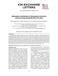
as a factor in Cr(VI) carcinogenesis, but given the potency of Cr(VI) as a clastogenic agent and the uncertain impact of the epigenetic changes, it does not appear to be sufficient to explain Cr(VI) carcinogenesis on its own. Conclusion Based on the recent chromate literature discussed above, we suggest that the paradigm that best describes chromate-induced lung cancer is genomic instability. In the Fig., we propose a potential mechanism of particulate chromate-induced carcinogenesis. Specifically, particulate Cr(VI) (1) dissolves outside the cell into the chromate anion (2A) and cation (2C). The cation enters into the cell via a channel protein (3-4) and intact particles enter into the cell via phagocytosis (2B) but both appear to have no effect36,52,55. The chromate anion, on the other hand, is the proximate genotoxic agent and enters into the cell
Fig. Proposed mechanism of Cr(VI)-induced carcinogenesis. Particulate Cr(VI) (1) partially dissolves outside the cell producing a chromate anion (2A) and a cation (2C). The cation enters into the cell through a channel protein (3,4) and intact particles are phagocytosed into the cell (2B). Both appear to have no adverse effect on the cell. The chromate anion enters the cell through an anion transporter (5) and is rapidly reduced to Cr(III) generating Cr(V), Cr(IV) and reactive oxygen species in the process (6). Cr(III) and possibly Cr(V) and Cr(IV) form ternary Cr-DNA adducts (7A) leading to a stalled DNA replication fork (8). These ternary adducts can be repaired by crosslink repair involving nucleotide excision repair (9A) or mismatch repair (9B) or possibly both. Both pathways cause a DNA double strand break during the repair process (10). The failure of base excision repair to repair oxidative damage could also contribute to DNA double strand break formation, but this is likely to be a minor component as it requires failure of repair (7B, 11). These DNA double strand breaks induce a prolonged G2 arrest (12) leading to both centrosome amplification and spindle assembly checkpoint bypass (13). Centrosome amplification and spindle assembly checkpoint bypass both lead to numerical chromosome instability (14) and ultimately neoplastic transformation and cancer (17). The failure to properly repair the DNA double strand breaks (15) results in structural chromosome instability (16) which also contributes to neoplastic transformation and cancer (17). Lastly, we propose that failure of mismatch repair (18) is the result of chromosome instability and mismatch repair failure leads to microsatellite instability (19) which may also contribute to neoplastic transformation and cancer (17). (1BER = Base excision repair; 2CLR = Crosslink repair; 3NER = Nucleotide excision repair; 4MMR = Mismatch repair; 5DSBs = Double strand breaks; 6HR = Homologous recombination; 7SAC = Spindle assembly checkpoint).
HOLMES et al: CARCINOGENICITY OF HEXAVALENT CHROMIUM
via an anion transporter (5)52. Once inside the cell, Cr(VI) is rapidly reduced to Cr(III), forming Cr(V), Cr(IV) and reactive oxygen species in the process (6)87. Cr(III), as well as Cr(V) and Cr(IV), can form ternary Cr-DNA adducts (7A) which lead to stalled DNA replication forks (8)28-31. The Cr-DNA adducts can be repaired through crosslink repair which involves proficient nucleotide excision repair and results in a DNA double strand break (9A-10)34. Mismatch repair also tries to repair Cr-DNA adducts but fails to repair them and undergoes a series of futile repair cycles that ultimately fail and collapse the replication fork also leading to a DNA double strand break (9B-10)33. A third pathway in the formation of double strand breaks could be the result of failure of base excision repair to properly repair oxidative damage (7B, 11, 10). Next, we propose that the DNA double strand breaks lead to a G2 arrest (12). With chronic exposure, this G2 arrest becomes prolonged and induces both centrosome amplification and spindle assembly checkpoint bypass (13) 47,48 . Centrosome amplification and spindle assembly checkpoint bypass induce numerical chromosome instability ultimately leading to neoplastic transformation and cancer (14,17)47-49. In addition, misrepair of double strand breaks can cause structural chromosome instability also contributing to neoplastic transformation and cancer (15-17)49,61. Interestingly, chromate-induced tumours exhibit MIN as well as CIN21, however, the formation of Cr(VI)induced DNA double strand breaks requires proficient, functional MMR33. Therefore, we propose that failure of MMR leading to MIN is a later change in the cells and probably a consequence of chromosome instability (18-19). Epigenetic changes and mutagenesis may contribute to this carcinogenic mechanism. For example, the activation of MAPK pathways may promote the survival of cells with Cr(VI)-induced CIN or DNA methylation may alter expression of genes and promote the growth of cells with Cr(VI)-induced CIN into a tumour. Mutagenesis may have some contribution if exposures are high enough. A mutation in a gene involved in MMR in the later stages of tumour progression could result in the MIN phenotype observed in the tumours. DNA methylation of MMR genes could also contribute to the later acquisition of the MIN phenotype. More work is needed to understand how the epigenetic changes are playing a role and if significant mutagenesis occurs in relevant genes at likely exposure levels.
369
References 1.
Machle W, Gregorius V. Cancer of the respiratory system in the United States chromate-producing industry. Public Health Rep 1948; 63 : 1114-27.
2.
The final standard on hexavalent chromium. Effective and practical protection for workers. Washington, DC: Occupational Safety & Health Administration; 2006. p. 20210.
3.
Langard S. One hundred years of chromium and cancer: A review of epidemiological evidence and selected case reports. Am J Ind Med 1990; 17 : 189-215.
4.
Leonard A, Lauwerys RR. Carcinogenicity and mutagenicity of chromium. Mutat Res 1980; 76 : 227-39.
5.
Davies JM. Lung-cancer mortality of workers in chromate pigment manufacture: An epidemiological survey. J Oil Col Chem Assoc 1979; 62 : 157-63.
6.
Davies JM. Lung cancer mortality among workers making lead chromate and zinc chromate pigments at three English factories. Br J Ind Med 1984; 41 : 158-69.
7.
IARC Monographs on the evaluation of carcinogenic risks to humans: Chromium, nickel and welding, vol. 49. Lyons, France: International Agency for Cancer Research; 1990.
8.
Levy LS, Martin PA, Bidstrup PL. Investigation of the potential carcinogenicity of a range of chromium containing materials on rat lung. Br J Ind Med 1986; 43 : 243-56.
9.
Maltoni C. Occupational carcinogenesis. Predictive value of carcinogenesis bioassays. Ann N Y Acad Sci 1976; 271 : 43143.
10. Maltoni C, Lefemine G, Chieco P, Carretti D. Vinyl chloride carcinogenesis: current results and perspectives. Med Lav 1974; 65 : 421-44. 11. Furst A, Schlauder M, Sasmore DP. Tumourigenic activity of lead chromate. Cancer Res 1976; 36 : 1779-83. 12. Patierno SR, Banh D, Landolph JR. Transformation of C3H/ 10T1/2 mouse embryo cells to focus formation and anchorage independence by insoluble lead chromate but not soluble calcium chromate: relationship to mutagenesis and internalization of lead chromate particles. Cancer Res 1988; 47 : 3815-23. 13. NTP Technical report abstract on the toxicology and carcinogenesis. Studies of sodium dichromate dihydrate F344/ N rats and B6C3F1 mice (drinking water studies). National Toxicology Program Toxicity Report Series Number 27, Research Triangle Park, NC, TR-546, National Institute of Health, Public Health Services, US Department of Health and Human Services; 2008. 14. Ishikawa Y, Nakagawa K, Satoh Y, Kitagawa T, Sugano H, Hirano T, et al. Characteristics of chromate workers’ cancers, chromium lung deposition and precancerous bronchial lesions: an autopsy study. Br J Cancer 1994; 70 : 160-6. 15. Ishikawa Y, Nakagawa K, Satoh Y, Kitagawa T, Sugano H, Hirano T, et al. “Hot Spots” of chromium accumulation at bifurcations of chromate workers’ bronchi. Cancer Res 1994; 54 : 2342-6. 16. Kondo K, Takahashi Y, Ishikawa S, Uchihara H, Hirose Y, Yoshizawa M, et al. Microscopic analysis of chromium accumulation in the bronchi and lung of chromate workers. Cancer 2003; 98 : 2420-9.
370
INDIAN J MED RES, OCTOBER 2008
17. Takahashi Y, Kondo K, Ishikawa S, Uchihara H, Fujino H, Sawada N, et al. Microscopic analysis of the chromium content in the chromium-induced malignant and premalignant bronchial lesions of the rat. Environ Res 2005; 99 : 267-72.
32. Klein CB, Su L, Bowser D, Leszczynska J. Chromate-induced epimutations in mammalian cells. Environ Health Perspect 2002; 110 : 739-43.
18. Katabami M, Dosaka-Akita H, Mishina T, Honma K, Kimura K, Uchida Y, et al. Frequent cyclin D1 expression in chromateinduced lung cancers. Hum Pathol 2000; 31 : 973-9.
33. Reynolds M, Stoddard L, Bespalov I, Zhitkovich A. Ascorbate acts as a highly potent inducer of chromate mutagenesis and clastogenesis: linkage to DNA breaks in G2 phase by mismatch repair. Nucleic Acids Res 2007; 35 : 465-76.
19. Kondo K, Hino N, Sasa M, Kamamura Y, Sakiyama S, Tsuyuguchi M, et al. Mutations of the p53 gene in human lung cancer from chromate-exposed workers. Biochem Biophy Res Commun 1997; 239 : 95-100.
34. Brooks B, O’Brien TJ, Ceryak S, Wise Sr JP, Wise SS, Wise Jr JP, et al. Excision repair is required for genotoxin-induced mutagenesis in mammalian cells. Carcinogenesis 2008; 29 : 1064-9.
20. Ewis AA, Kondo K, Lee J, Tsuyuguchi M, Hashimoto M, Yokose T, et al. Occupational cancer genetics: Infrequent ras oncogenes point mutation in lung cancer samples from chromate workers. Am J Ind Med 2001; 40 : 92-7.
35. Wise Sr. JP, Wise SS, Little JE. The cytotoxicity and genotoxicity of particulate and soluble hexavalent chromium in human lung cells. Mutat Res 2002; 517 : 221-9.
21. Hirose T, Kondo K, Takahashi Y, Ishikura H, Fujino H, Tsuyuguchi M, et al. Frequent microsatellite instability in lung cancer from chromate-exposed workers. Mol Carcinog 2002; 33 : 172-80. 22. Takahashi Y, Kondo K, Hirose T, Nakagawa H, Tsuyuguchi M, Hashimoto M, et al. Microsatellite instability and protein expression of the DNA mismatch repair gene, hMLH1, of lung cancer in chromate-exposed workers. Mol Carinog 2005; 42 : 150-8. 23. Kondo K, Takahashi Y, Hirose Y, Nagao T, Tsuyuguchi M, Hashimoto M, et al. The reduced expression and aberrant methylation of p16INK4a in chromate workers with lung cancer. Lung Cancer 2006; 53 : 295-302. 24. Ewis AA, Kondo K, Dang F, Nakahori Y, Shinohara Y, Ishikawa M, et al. Surfactant protein B gene variations and susceptibility to lung cancer in chromate workers. Am J Ind Med 2006; 49 : 267-73. 25. Masuda A, Takahashi T. Chromosome instability in human lung cancers: possible underlying mechanisms and potential consequences in the pathogenesis. Oncogene 2002; 21 : 688497. 26. Volgelstein B, Kinzler KW. The multistep nature of cancer. Trends Genet 1993; 9 : 138-41. 27. Cheng L, Sonntag DM, de Boer J, Dixon K. Chromium(VI)induced mutagenesis in the lungs of Big Blue transgenic mice. J Environ Pathol Toxicol Oncol 2000; 19 : 239-49. 28. Zhitkovich A, Song Y, Quievryn G, Voitkun V. Non-oxidative mechanisms are responsible for the induction of mutagenesis by reduction of Cr(VI) with cysteine: Role of ternary DNA adducts in Cr(III)-dependent mutagenesis. Biochemistry 2001; 40 : 549-60.
36. Holmes AL, Wise SS, Xie H, Gordon N, Thompson WD, Wise Sr JP. Lead ions do not cause human lung cells to escape chromate-induced cytotoxicity. Toxicol Appl Pharmacol 2005; 203 : 167-76. 37. Peterson-Roth E, Reynolds M, Quievryn G, Zhitkovich A. Mismatch repair proteins are activators of toxic responses to chromium-DNA damage. Mol Cell Biol 2005; 25 : 3596-607. 38. Yavorsky M, Almaden P, King CG. The vitamin C content of human tissues. J Biol Chem 1934; 106 : 525-9. 39. Loeb LA. A mutator phenotype in cancer. Cancer Res 2001; 61 : 3230-9. 40. Aviel-Ronen S, Blackhall FH, Shepherd FA, Tsao MS. K-ras mutations in non-small-cell lung carcinoma: a review. Clin Lung Cancer 2006; 8 : 30-8. 41. Mitsuuchi Y, Testa JR. Cytogenetics and molecular genetics of lung cancer. Am J Med Genet 2002; 115 : 183-8. 42. Lengauer C, Kinzler KW, Vogelstein B. Genetic instabilities in human cancers. Nature 1998; 396 : 643-9. 43. Karran P. Microsatellite instability and DNA mismatch repair in human cancer. Semin Cancer Biol 1996; 7 : 15-24. 44. Wistuba II, Bryant D, Behrens C, Milchgrub S, Virmani AK, Ashfaq R, et al. Comparison of features of human lung cancer cell lines and their corresponding tumours. Clin Cancer Res 1999; 5 : 991–1000. 45. Guerci A, Seoane A, Dulout FN. Aneugenic effects of some metal compounds assessed by chromosome counting in MRC-5 human cells. Mutat Res 2000; 469 : 35-40. 46. Seoane AL, Guerci AM, Dulout FN. Malsegregation as a possible mechanism of aneuploidy induction by metal salts in MRC-5 human cells. Environ Mol Mutagen 2002; 40 : 200-6.
29. Zhitkovich A, Quievryn G, Messer J, Motylevich Z. Reductive activation with cysteine represents a chromium(III)-dependent pathway in the induction of genotoxicity by carcinogenic chromium(VI). Environ Health Perspect 2002; 110 : 729-31.
47. Holmes AL, Wise SS, Sandwick SJ, Lingle WL, Negron VC, Thompson WD, et al. Chronic exposure to lead chromate causes centrosome abnormalities and aneuploidy in human lung cells. Cancer Res 2006; 66 : 4041-8.
30. Quievryn G, Peterson E, Messer J, Zhitkovich A. Genotoxicity and mutagenicity of chromium(VI)/ascorbate-generated DNA adducts in human and bacterial cells. Biochemistry 2003; 42 : 1062-70.
48. Wise SS, Holmes AL, Xie H, Thompson WD, Wise Sr JP. Chronic exposure to particulate chromate induces spindle assembly checkpoint bypass in human lung cells. Chem Res Toxicol 2006; 19 : 1492-8.
31. Quievryn G, Messer J, Zhitkovich A. Lower mutagenicity but higher stability of Cr-DNA adducts formed during gradual chromate activation with ascorbate. Carcinogenesis 2006; 27 : 2316-21.
49. Xie H, Holmes AL, Wise SS, Huang S, Peng C, Wise Sr JP. Neoplastic transformation of human bronchial cells by lead chromate particles. Am J Respir Cell Mol Biol 2007; 37 : 54452.
HOLMES et al: CARCINOGENICITY OF HEXAVALENT CHROMIUM 50. D’Assoro AB, Lingle WL, Salisbury JL. Centrosome amplification and the development of cancer. Oncogene 2002; 21 : 6146-53. 51. Wise SS, Elmore LW, Holt SE, Little JE, Antonucci PG, Bryant BH, et al. Telomerase-mediated lifespan extension of human bronchial cells does not affect hexavalent chromium-induced cytotoxicity or genotoxicity. Mol Cell Biochem 2004; 255 : 103-11. 52. Wise SS, Holmes AL, Ketterer ME, Hartsock WJ, Fomchenko E, Katsfis SP, et al. Chromium is the proximate clastogenic species for lead chromate-induced clastogenicity in human bronchial cells. Mutat Res 2004; 560 : 79-89. 53. Wise SS, Schuler JHC, Katsifis SP, Wise Sr JP. Barium chromate is cytotoxic and genotoxic to human lung cells. Environ Mol Mutagen 2004; 42 : 274-8. 54. Wise SS, Schuler JHC, Holmes AL, Katsifis SP, Ketterer ME, Hartsock WJ, et al. A comparison of two carcinogenic particulate hexavalent chromium compounds: Barium chromate is more potent than lead chromate in human lung cells. Environ Mol Mutagen 2004; 44 : 156-62. 55. Xie H, Holmes AL, Wise SS, Gordon N, Wise Sr JP. Lead chromate-induced chromosome damage requires extracellular dissolution to liberate chromium ions but does not require particle internalization or intracellular dissolution. Chem Res Toxicol 2004; 17 : 1362-7. 56. Xie H, Wise SS, Holmes AL, Xu B, Wakeman T, Pelsue SC, et al. Carcinogenic lead chromate induces DNA double-strand breaks and activates ATM kinase in human lung cells. Mutat Res 2005; 586 : 160-72. 57. Holmes AL, Wise SS, Sandwick SJ, Wise Sr. JP. The clastogenic effects of chronic exposure to particulate and soluble Cr(VI) in human lung cells. Mutat Res 2006; 610 : 813. 58. Wise SS, Holmes AL, Wise Sr. JP. Particulate and soluble hexavalent chromium are cytotoxic and genotoxic to human lung epithelial cells. Mutat Res 2006; 610 : 2-7. 59. NTP Technical report on the toxicity studies of sodium dichromate dihydrate administered in drinking water to male and female F344/N rats and B6C3F1 mice and male BALB/c and am3-C57BL/6 mice. National Toxicology Program Toxicity Report Series Number 27, National Institutes of Health, Public Health Service, US Department of Health and Human Services, Research Triangle Park, NC, 2007. 60. Balansky RM, D’Agostini F, Izzotti A, De Flora S. Less than additive interaction between cigarette smoke and chromium(VI) in inducing clastogenic damage in rodents. Carcinogenesis 2000; 21 : 1677-82. 61. Xie H, Wise SS, Wise Sr. JP. Deficient repair of particulate chromate-induced DNA double strand breaks leads to neoplastic transformation. Mutat Res 2008; 649 : 230-8.
371
64. Wakeman TP, Kim WJ, Callens S, Chiu A, Brown KD, Xu B. The ATM-SMC1 pathway is essential for activation of the chromium(VI)-induced S-phase checkpoint. Mutat Res 2004; 554 : 241-51. 65. Stackpole MM, Wise SS, Goodale BC, Duzevik EG, Munroe RC, Thompson WD, et al. Homologous recombination protects against particulate chromate-induced genomic instability in Chinese hamster cells. Mutat Res 2007; 625 : 145-54. 66. Camrye E, Wise SS, Milligan P, Gordon N, Goodale B, Stackpole M, et al. Ku80 deficiency does not affect particulate chromate-induced chromosome damage and cytotoxicity in Chinese hamster ovary cells. Toxicol Sci 2007; 97 : 348-54. 67. Weinberg RA. The biology of cancer. New York: Garland Science, Taylor & Francis Group, LLC; 2007. 68. Wise SS, Holmes AL, Moreland JA, Xie H, Sandwich SJ, Stackpole MM, et al. Human lung cell growth is not stimulated by lead ions after lead chromate-induced genotoxicity. Mol Cell Biochem 2005; 279 : 75-84. 69. Wang S, Chen F, Zhang Z, Jiang B, Jia L, Shi X. NF-kB prevents cells from undergoing Cr(VI)-induced apoptosis. Mol Cell Biochem 2004; 255 : 129-37. 70. Pritchard DE, Ceryak S, Ramsey KE, O’Brien TJ, Ha L, Fornsaglio JL, et al. Resistance to apoptosis, increased growth potential, and altered gene expression in cells that survived genotoxic hexavalent chromium [Cr(VI)] exposure. Mol Cell Biochem 2005; 279 : 169-81. 71. Son KH, Zhang M, Rucobo E, Nwaigwe D, Montgomery F, Leffert HL. Derivation and study of human epithelial cell lines resistant to killing by chromium trioxide. J Toxicol Environ Health A 2004; 67 : 1027-49. 72. Qian Y, Jiang BH, Flynn DC, Leonard SS, Wang S, Zhang Z, et al. Cr(VI) increases tyrosine phosphorylation through reactive oxygen species-mediated reactions. Mol Cell Biochem 2001; 222 : 199-204. 73. Vasant C, Sankaramanivel S, Jana M, Rajaram R, Ramasami T. Non-enzymatic phosphorylation of bovine serum albumin by Cr(V) complexes: Role in Cr(VI)-induced phosphorylation and toxicity. Mol Cell Biochem 2005; 275 : 153-64. 74. Izzotti A, Cariglia C, Balansky R, D’Agostini F, Longobardi M, DeFlora S. Selective induction of gene expression in rat lung by hexavalent chromium. Mol Carcinog 2002; 35 : 7584. 75. Ye J, Shi X. Gene expression profile in response to chromiuminduced cell stress in A549 cells. Mol Cell Biochem 2001; 222 : 189-97. 76. Andrew AS, Warren AJ, Barchowsky A, Temple KA, Klei L, Soucy NV, et al. Genomic and proteomic profiling of responses to toxic metals in human lung cells. Environ Health Perspect 2003; 111 : 825-38.
62. Ha L, Ceryak S, Patierno SR. Chromium (VI) activates ataxia telangiectasia mutated (ATM) protein. J Biol Chem 2003; 278 : 17885-94.
77. Chuang SM, Liou GY, Yang JL. Activation of JNK, p38 and ERK mitogen-activated protein kinases by chromium(VI) is mediated through oxidative stress but does not affect cytotoxicity. Carcinogenesis 2000; 21 : 1491-500.
63. Ha L, Ceryak S, Patierno SR. Generation of S phase-dependent DNA double strand breaks by Cr(VI) exposure: involvement of ATM in Cr(VI) induction of γH2AX. Carcinogenesis 2004; 25 : 2265-74.
78. Chuang SM, Yang JL. Comparison of roles of three mitogenactivated protein kinases induced by chromium(VI) and cadmium in non-small-cell lung carcinoma cells. Mol Cell Biochem 2001; 222 : 85-95.
372
INDIAN J MED RES, OCTOBER 2008
79. Hodges NJ, Smart D, Lee AJ, Lewis NA, Chipman JK. Activation of c-Jun N-terminal kinase in A549 lung carcinoma cells by sodium dichromate: role of dissociation of apoptosis signal regulating kinase-1 from its physiological inhibitor thioredoxin. Toxicology 2004; 197 : 101-12. 80. Tessier DM, Pascal LE. Activation of MAP kinases by hexavalent chromium, manganese and nickel in human lung epithelial cells. Toxicol Lett 2006; 167 : 114-21. 81. O’Hara KA, Klei LR, Barchowsky A. Selective activation of Src family kinases and JNK by low levels of chromium(VI). Toxicol Appl Pharmacol 2003; 190 : 214-23. 82. O’Hara KA, Vaghjiani RJ, Nemec AA, Klei LR, Barchowsky A. Cr(VI)-stimulated STAT3 tyrosine phosphorylation and nuclear translocation in human airway epithelial cells requires Lck. Biochem J 2007; 402 : 261-9.
83. Chen F, Ding M, Lu Y, Leonard SS, Vallyathan V, Castranova V, et al. Particulation of MAP kinase p38 and IkB kinase in chromium(VI)-induced NF-kB and AP-1 activation. J Environ Pathol Toxicol Oncol 2000; 19 : 231-8. 84. Lui K, Husler J, Ye J, Leonard SS, Cutler D, Chen F, et al. On the mechanism of Cr(VI)-induced carcinogenesis: Dose dependence of uptake and cellular responses. Mol Cell Biochem 2001; 222 : 221-9. 85. Kim YD, An SC, Oyama T, Kawamoto T, Kim H. Oxidative stress, hogg1 expression and NF-kB activity in cells exposed to low level chromium. J Occup Health 2003; 45 : 271-7. 86. Leonard SS, Roberts JR, Antonini JM, Castranova V, Shi X. PbCrO4 mediates cellular responses via reactive oxygen species. Mol Cell Biochem 2004; 255 : 171-9. 87. De Flora S, Wetterhahn KE. Mechanisms of chromium metabolism and genotoxicity. Life Chem Rep 1989; 7 : 169-244.
Reprint requests: Dr John Pierce Wise, Sr., University of Southern Maine, 96 Falmouth St., PO Box 9300 Portland, ME 04104, USA e-mail:
[email protected]