The Mediterranean Journal of Otology
CASE REPORT Case Report: Cochlear Implantation in a Renal Transplant Patient with Alport Syndrome Murat Sar›, Zahide Mine Yaz›c›, Ali Cemal Yumuflakhuylu, Ufuk Derinsu, Ça¤lar Batman
Department of Otorhinolaryngology Head and Neck Surgery (M. Sar›, Z.M. Yaz›c›, A.C. Yumuflakhuylu, C. Batman), Department of Audiology (U. Derinsu) Marmara University School of Medicine ‹stanbul, Turkey Correspondent Author: Ali Cemal Yumusakhuylu, MD Marmara Universitesi Hastanesi Tophanelioglu cad., No: 13/15, 34660 Altunizade, ‹stanbul/Turkey Tel: +90 216 327101 Fax: +90 216 3269637 E-mail :
[email protected] Submitted: May 12, 2008 Accepted: September 17, 2008 Mediterr J Otol 2008; 225-229
Copyright 2005 © The Mediterranean Society of Otology and Audiology
Selection criteria for cochlear implantation have been broadened in recent years. However, selection of patients who are using immunosuppresive drugs because of organ transplantation is not very clear. We intended to share one of our patients who was on chronic immunosuppression due to kidney transplantation. We didn’t experience wound infection, delay in wound healing or any complication in our patient. We concluded that cochlear implantation of carefully selected patients receiving chronic immunotherapy after organ transplantation may be safe and effective, but report of large series of cochlear implantation performed on organ transplant patients is needed for a definitive conclusion. 225
The Mediterranean Journal of Otology
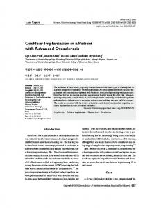
hearing loss in the right ear. High-resolution computurized tomography (HRCT) of the temporal bone was found normal. Magnetic resonance imaging (MRI) was found to be normal except non-ossifiying fibrosis of both cochlea (Figure 1). The transplant team agreed with the CIn operation. Her medical diseases; DM type 2, hypertension, were checked once again by internal medicine doctors, and it was reported that they were under control with regular therapy. In light of these evaluations CIn committee of Marmara University Medical Faculty decided her as a proper candidate for CIn.
Cochlear implants (CIs) are being used since 1980’s for rehabilitation of patients who has severe sensorineural hearing loss. As surgical and clinical experience and knowledge accumulates on the topic, selection criteria for cochlear implantation (CIn) have been broadened. However, selection of patients who are using immunosuppresive drugs either because of solid organ transplantation or chronic medical diseases is not very clear. Immunosuppression is known to be associated with impaired wound healing and increased risk of infection[1]. Performing CIn operation on patients who are put on chronic immunosuppresion due to organ transplantation bears its own risks such as wound infection, flap necrosis, extrusion of device and retarded healing[2]. Succesfull results of CI surgery in patients using chronic immunosuppressive medication was reported in recent years[3,4]. We are aiming to review our experience of CIn in a kidney transplant patient, who has also other comorbidities. CASE REPORT Medical history and preoperative evaluation The patient was a 60 years old female. She had Alport syndrome which explained her progressive sensorineural hearing loss since childhood. She has been wearing a hearing aid to her right ear for 15 years which she could not benefit lately. Before the CIn operation she has been treated with peritoneal dialysis for 6 months because of chronic renal failure due to Alport syndrome. She has received her CI 30 months after kidney transplantation. Her immunosuppressive maintenance therapy consisted of tacrolimus and prednisolone at the time of CIn surgery. Her medical history included diabetes mellitus type 2, hypertension and osteoporosis.
Figure-1: Magnetic resonance imaging showing non-ossifiying fibrosis of both cochlea.
Operation and postoperative follow-up She received one dose of 23-valent pneumococal polysaccharide vaccine (PPV23) (Pneumovax) one week before the operation. She received a preoperative dose of ceftriaxone disodium, 1 gr intravenously (IV) and prednisolone, 50 mg IV. The operation was performed through an extended postauricular incision on the right. After simple mastoidectomy, a posterior tympanotomy via facial recess approach was accomplished. A bed for the processor of the CI was also drilled to the temporal bone just posterior to the mastoidectomy cavity. Niche of the round window was widened. Med-El Pulsar electrode (Medical Electronics, Innsbruck, Austria) was inserted into the
She underwent neuro-otologic, auditory, radiologic, and psychologic evaluations. The preoperative audiogram was consistent with total hearing loss in the left ear and severe sensorineural 226
Case Report: Cochlear Implantation in a Renal Transplant Patient with Alport Syndrome
Preoperative and postoperative audiological evaluation
cochlea via round window approach, but because of non-ossifiying fibrosis only 6 electrodes could be carried forward. At this point operation team decided to insert Med-El Pulsar Split Array electrodes (Medical Electronics, Innsbruck, Austria). One more window was opened at the middle turn of the cochlea. Then electrodes of the Med-El Pulsar Split Array were successfully inserted into the cochlea via the previously mentioned windows. A small piece of muscle which was taken from temporal muscle was placed around the electrodes at the level where they entered to the cochlea. Electrodes were fixed to the posterior tympanotomy window with bone-cement in order to prevent detachment. Processor of the cochlear implant was placed into its bed on the temporal bone and fixed with 0 prolen and Dacron mesh. Ball electrode was placed under the temporal muscle. Then the entire periosteal flap was closed with absorbable sutures and skin with prolene sutures. Intraoperatively CI was checked out by audiologists and positive telemetry was obtained. After the operation electrodes were checked with Stenvers and Transorbitale x-ray graphies. Patient was monitored for 4 days in the hospital postoperatively. During hospital stay she received ceftriaxone disodium, 1 gr IV twice daily. Her immunosuppressive maintenance therapy; tacrolimus and prednisolone were not interrupted. She was discharged home with 1 week of oral cefixime, 400 mg twice daily.
Routine audiologic evaluation was carried out preoperatively. Air- and bone-conducted pure-tone audiometry was performed in the range of 250 to 8,000 Hz and 500 to 4,000 Hz, respectively (Figure 2). Speech reception thresholds could not be obtained bilaterally. Speech detection threshold was 90 dB in right ear, and in left ear there was no response. Tympanograms were bilaterally normal. Acoustic reflexes were absent in both ears in the 500-4,000 Hz range. Transient evoked otoacoustic emissions were absent bilaterally. Promontory stimulation test demonstrated good dynamic range, and decay was not observed in right ear. Preoperatively, no response was obtained in open-set and closed-set speech tests without lipreading. Three weeks after the operation, cochlear implant processor was programmed. Only 6 electrodes of the inferior branch of the split electrode could be programmed. No auditory sensation was determined by other electrodes. In first session, she perceived all the sounds as noise. The hearing thresholds with cochlear implant in the free field was shown in Figure 3. The patient specific auditory training program was planned and was given to her as a homework. After four days, at the second mapping the same 6 electrodes which were programmed at the first
Figure-3: The hearing thresholds with cochlear implant in the free field.
Figure-2: Air- and bone-conducted pure-tone audiometry.
227
The Mediterranean Journal of Otology
DISCUSSION
mapping were reprogrammed. One electrode from the inferior branch and all 5 electrodes of the superior branch of the electrode array revealed no auditory sensation. The auditory training program was prepared by the audiologist, the patient had to do the training at home with the help of her close relatives. The homework was checked every week at the clinic. Auditory performance was measured using pattern perception test which was devoloped at Marmara University Audiology Department. This test has four subtests:
Selection criteria for CIn are widened in several areas as the accumulation of experience and new data grow. CIn in patients who are at risk of poor healing because of either chronic co-morbidities or solid organ transplantation seems to be one of these areas. Mostly seen causes of morbidity in solid organ transplants are wound healing and infection[1]. These two are also the most common complications seen after CIn. Risk of wound infection has been studied by many authors among solid organ transplant patients who undergone surgery for prosthesis replacement; some of them has reported relatively higher rates of wound infection [2], whereas others has reported smaller [5] or even none [6]. Patterson et al reported no complications after CIn of 5 patients who have undergone liver, kidney and heart transplantation [4].
Subtest A: There are two words in each item one of them is monosyllabic, second word is trisyllabic. The task of the patient is to perceive the difference between monosyllabic and trisyllabic words. Subtest B: There are two words in each item one of them is monosyllabic, second word is disyllabic. The task of the patient is to perceive the difference between monosyllabic and disyllabic words.
We would like to draw attention that HRCT could not successfully show non-ossifying fibrosis, whereas MRI could yield efficacious information in preoperative assessment which enables to take proper precautions as in our case.
Subtest C: There are two words in each item one of them is disyllabic, second word is trisyllabic. The task of the patient is to perceive the difference between disyllabic and trisyllabic words.
Immunosuppressive maintenance therapy is a routine after organ transplantation. Timing of CIn and type of immunosuppressive regimen in transplant patients receiving immunotherapy is also important. At least 6 months should pass after transplantation because peak immunosuppression passes after 6 months[7]. Our patient was taking tacrolimus and prednisolone. Tacrolimus was found to be interfering with wound healing by decreasing wound nitric oxide synthesis which is necessary for regulation of wound
Subtest D: There are two words in each item both of them are disyllabic. The task of the patient is to perceive the difference between two disyllabic words. For each item, the tester reads the word that has to be detected by the subject from the answer sheet of this test and the subject is informed to discriminate between two words. Table 1 illustrates the speech perception test results of the patient at first and third month of the operation.
Table-1: Speech perception test results at first and third month of the operation. SPEECH TESTS (CLOSED SET)
PRE-OP.
POST-OP. ONE MONTH (%)
POST-OP. THREE MONTH (%)
Pattern Perception Test A
NR
100
100
Pattern Perception Test B
NR
100
100
Pattern Perception Test C
NR
88
100
Pattern Perception Test D
NR
52
84
228
Case Report: Cochlear Implantation in a Renal Transplant Patient with Alport Syndrome
fibroblast synthetic activity[8]. Glucocorticoids are known with their interference on re-epithelization, wound contraction, inflammation and collagen metabolism. Although she had also Alport syndrome, diabetes mellitus type 2 and hypertension during her postoperative follow-up, we did not experience wound infection, delay in wound healing or any complication. She experienced a normal postoperative convalescence period like other healthy cochlear implant patients.
2.
Tannenbaum DA, Matthews LS, Grady-Benson JC. Infection around joint replacements in patients who have a renal or liver transplantation. J Bone Joint Surg Am 1997; 79:36-43.
3.
Odabas› O, Mobley SR, Bolanos RA, et al. Cochlear implantation in patients with compromised healing. Otolaryngol Head Neck Surg 2000;123:738-741.
4.
Patterson DM, Telischi FF, Connell SS, et al. Cochlear implantation in organ transplantation. Laryngoscope 2008; 118:116-119.
5.
Deo S, Gibbons CL, Emerton M, Simpson AH. Total hip replacement in renal transplant patients. J Bone Joint Surg Br 1995; 77:299-302.
6.
Cuellar DC, Sklar GN. Penile prosthesis in the organ transplant recipients. Urology 2001; 57:138-141.
7.
Rubin RH, Wolfson JS, Cosimi AB, TolkoffRubin NE. Infection in the renal transplant recipient. Am J Med 1981; 70:405.
8.
Schaffer MR, Fuchs N, Proksch B, et al. Tacrolimus impairs wound healing. Transplantation 1998; 65:813-818.
CONCLUSION Patients who are taking immunotherapy either due to organ transplantation or chronic medical diseases are sometimes in need of CIn. Our aim was to evaluate the potential surgical and postoperative risks and complications in a kidney transplant patient who has also other co-morbidities. Although this article reports the short term results of a patient who had multiple comorbidities, we concluded that CIn of carefully selected patients receiving chronic immunotherapy after organ transplantation might be safe and effective, but report of large series of CIn performed on organ transplant patients is needed for a definitive conclusion. REFERENCES 1.
Flechner SM, Zhou L, Derweesh I, et al. The impact of sirolismus, mycophenolate mofetil, cyclosporine, asathioprine, and steroids on wound healing in 513 kidney-transplant recipients. Transplantation 2003; 76:1729-1734.
229