J Physiol Biochem DOI 10.1007/s13105-017-0567-z
MINI REVIEW
Cell source, differentiation, functional stimulation, and potential application of human thermogenic adipocytes in vitro Dinh-Toi Chu 1,2
&
Yang Tao 3 & Le Hoang Son 4 & Duc-Hau Le 5
Received: 3 March 2017 / Accepted: 29 May 2017 # University of Navarra 2017
Abstract Recent investigations have showed that the functional thermogenic adipocytes are present in both infants and adult humans. Accumulating evidence suggests that the coexistence of classical and inducible brown (brite) adipocytes in humans at adulthood and these adipocytes function to generate heat from energy resulting in reducing body fat and improving glucose metabolism. Human thermogenic adipocytes can be differentiated in vitro from stem cells, cell lines, or adipose stromal vascular fraction. Pre-activated human brite adipocytes in vitro can maintain their thermogenic function in normal or obese immunodeficient mice; therefore, they improve glucose homeostasis and reduce fat mass in obese animals. These key findings have opened a new door to use in vitro thermogenic adipocytes as a cell therapy to prevent obesity and related disorders. Thus, this paper intends to highlight our knowledge in aspects of in vitro human brite/brown adipocytes for the further studies.
* Dinh-Toi Chu
[email protected]
1
Institute for Research and Development, Duy Tan University, 03 Quang Trung, Da Nang, Vietnam
2
Faculty of Biology, Hanoi National University of Education, Hanoi, Vietnam
3
College of Food Science and Technology, Nanjing Agricultural University, Nanjing, Vietnam
4
VNU University of Science, Vietnam National University, Hanoi, Vietnam
5
VINMEC Research Institute of Stem Cell and Gene Technology, Hanoi, Vietnam
Keywords Human thermogenic adipocytes in vitro . Brown/ brite adipocytes . Brown/brite adipogenesis . Thermogenesis . Overweight and obesity
Introduction There are two types of adipocytes which regulate lipid metabolism by controlling energy balance in humans and other mammals [10, 35, 51]. They are white and brown (thermogenic) adipocytes locating in fat depots along the body, white fat cells store energy while brown cells dissipate energy as producing heat [10, 35, 51]. Therefore, over accumulation of lipid in white fat tissues induced by genetics and/ or diet is the cause of overweight and obesity [10, 11, 31]. Thermogenic adipocytes include classical and inducible brown adipocytes which share thermogenic function but are different in developmental origin [8, 12, 53]. In human, classical brown fat cells are naturally present in brown fat depots, but abundant in newborns and less in adults; wherever, brite adipocytes are found within the white adipose tissue (WAT) near vascular and neural innervation and appear in response to several stimuli such as cold. They are characterized to express intermediate UCP1 mRNA levels [8]. Thermogenic fat cells were firstly found in newborn infants [2, 26, 42], and lately in adult humans [13–15, 52, 59, 60]. In humans, brown adipose tissues (BATs) are a mix of both classical and inducible thermogenic cells [8, 28, 42, 56, 62]. However, there is some evidence showing that two subtypes of human brown fat cells may be developed from different stem cell populations as they do in rodents [1, 9, 26, 47, 48, 55, 58]. Activation of BATs by cold exposure, beta-adrenergic receptor agonist, or vitamin such as nicotinamide riboside (NR) could improve metabolic homeostasis and reduce
Chu et al.
weight gain in several experiments [7, 8, 14, 16, 20, 27, 30, 32, 37, 52, 59, 60]. In vitro, human brown/brite adipocytes can be developed from stem cells, cell lines, or stromal vascular fractions (SVFs) of adipose tissues [4–6, 18, 21–25, 29, 34, 38, 39, 43, 45, 46, 50, 61, 63]. As white adipocytes, differentiation process of brown/brite adipocytes in vitro needs a stimulation of peroxisome proliferator-activated receptor γ (PPARγ) [9, 41, 43]. Importantly, differentiated brown/brite adipocytes have a full thermogenic function in vitro [4–6, 18, 21–25, 29, 34, 38, 39, 43, 45, 46, 50, 61, 63], and they still keep their activities in lipid metabolism when transplanted into animal body as recently investigated [8, 40, 44, 57]. However, several studies indicate that murine classical brown and brite adipocytes come from different stem cell populations, that is myf5 positive (myf5+) myotomal precursors for classical brown adipocytes [1, 58], but both myf5 negative (myf5−) and positive (myf5+) precursors for brite fat cells [48, 54, 55]. Therefore, under the same adipogenic medium, murine stromal vascular fraction (SVF) of interscapular brown adipose tissue (iBAT) develops into classical brown adipocytes, but SVFs of inguinal or epididymal fat depots differ into brite adipocytes as indicated by the expression of specific markers for type of thermogenic cells [9, 48, 62]. Furthermore, brown and brite adipocytes in vitro have different responses to browning reagents such as norepinephrine (NE) and triiodothyronine (T3) [9, 28, 33].
Cell sources for human brown/brite adipocytes in vitro In what follows, we discuss the cell sources for inducing human brown/brite adipocytes in vitro, as well as the adipogenic medium and stimulators of human brown/brite adipocytes
Table 1 Cell sources for induction of human brown/brite adipocyte in vitro
in vitro. Summary about these features should be referenced in Tables 1 and 2.
Stromal vascular fraction of human adipose depots Stromal vascular fractions (SVFs) can be directly differentiated into thermogenic adipocytes after being isolated from human fat tissues [4, 21, 38, 39]. In a report published in 2014, Lee et al. used SVFs of cervical fat to induce white or brite adipocytes in vitro [38]. In 2016, Barquissau and Maude et al. used SVFs from subcutaneous abdominal fat of healthy people to develop brite adipocytes in vitro [4, 23]. Furthermore, SVFs of supraclavicular adipose tissues have been used to develop classical brown adipocytes in vitro in works done by several groups [3, 36, 56].
Human adipose tissue stem cells Human adipose tissue stem cells (hASCs) are popularly established from a cell population of adult white adipose tissues [4, 5, 18, 22, 23, 34, 43, 46, 50]. These stem cells are employed for inducing brite adipocytes in vitro in several experiments [4, 5, 18, 34, 40, 43, 46]. These white adipose tissues may be subcutaneous or visceral fat deports of healthy people or patients [18, 46]. Moreover, hASCs for development of thermogenic adipocytes can be established from SVFs of prepubic and umbilical fat pad of a child [4, 22, 49], or white fat tissues of infants [23, 50]. In another report, hASCs were established from cell fraction of patients’ mediastinal brown adipose tissues, and those stem cells could differentiate into brown adipocytes in vitro, so they were named Bbrown adipose derived stem cells—BADSCs^ [57].
Cell type
Origin
Induced adipocyte
Ref
hASCs hASCs hASCs hASCs BADSCs hiPSCs bmMSCs SVFs SVFs SVFs PAZ6 cell line SGBS cell line
Human subcutaneous white fat Prepubic fat Umbilical fat White adipose tissues Mediastinal adipose tissues Human fetal brain and spinal cord tissues Human bones Human cervical fat Human subcutaneous white fat Human supraclavicular fat Infant brown adipose tissues Human adipose tissues
Brite Brite Brite Brite or white Brown Brite or white Brite or white Brite or white Brite or white Brown Brown Brite
([18]; [5]; [43]; [34]) ([4]; [22]; [49]) [49] [23] [57] ([45]; [25]) ([39]; [61]) [38] ([56]; [46]; [4]; [23]) ([56]; [36]; [3]) ([63]; [29]; [6]; [24]) [24]
Cell source, differentiation and stimulation of in vitro human brite/brown adipocytes Table 2
Adipogenic medium and stimulators of human brown/brite adipocytes in vitro
Cell type
Adipocyte Brown/brite adipogenic medium Basic medium
Stimulators Adipogenic reagents
Ref
Browning reagents
hASCs hASCs
Brite Brite
DMEM/F-12, FBS, hFGF2, P/S INS, IMBX, DEX, transferrin DMEM/F-12, FBS, hFGF2, P/S INS, IMBX, DEX, transferrin
ROSI, T3 ISO, CL ROSI or GW7647, T3 ISO, CL
[18] [4]
hASCs
Brite
DMEM/F-12, FBS, hFGF2, P/S INS, IMBX, DEX, transferrin
ROSI, T3
([43]; [34]; [49])
hASCs hASCs
Brite Brite
DMEM/F-12, FBS, P/S INS, IMBX, DEX DMEM/F-12, FBS, hFGF2, P/S INS, IMBX, DEX
ROSI, T3, cPGI2 ROSI, T3
[22] [23]
hASCs BADSCs
Brite Brown
DMEM/F-12, FBS, DMEM/F-12, FBS
INS, IMBX, DEX, indomethacin ROSI, T3 INS, IMBX, DEX ROSI, T3, FNDC5
[46] [57]
hiPSCs
Brite
DMEM/F-12, FBS
INS, IMBX, DEX
ROSI, T3
FSK
[25]
bmMSCs
Brite
DMEM/F-12, FBS, P/S
INS, IMBX, DEX
ROSI, T3, BMP7
FSK
[39]
bmMSCs
Brite
DMEM/F-12, FBS, P/S
T3
FSK
[61]
SVFs
Brite
INS, IMBX, DEX, ascorbate-2 phosphate, indomethacin INS, IMBX, DEX,
ROSI, T3
CL
[38]
SVFs SVFs
Brite Brite
DMEM/F-12, FBS, biotin, P/S DMEM/F-12, FBS, hGGF2, P/S INS, IMBX, DEX, transferrin DMEM/F-12, FBS, hFGF2, P/S INS, IMBX, DEX
ROSI or GW7647, T3 ROSI, T3
SVFs SVFs
Brown Brown
DMEM/F-12, FBS DMEM/F-12, FBS
ROSI, T3 ROSI
FSK ISO
PAZ6 cell line
Brown
DMEM/F-12, FBS
PIO, T3
PAZ6 cell line
Brown
PAZ6 cell line
Brown
DMEM/F-12, FBS, Hepes, P/S DMEM/F-12, FBS
ISO, NE, E, ([63]; [29]) CGP FSK [24]
SGBS cell line Brite
DMEM/F-12, FBS, Hepes, P/S
INS, IMBX, DEX INS, IMBX, DEX, transferrin, GH, IGF-I, pantothenate, biotin INS, IMBX, DEX
INS, IMBX, DEX, pantothenate, ROSI, T3 biotin INS, IMBX, DEX, pantothenate, PIO, T3 biotin INS, IMBX, DEX, pantothenate, ROSI, T3 biotin, cortisol
[4] [23] [56] ([36]; [3])
[6] FSK
[24]
Concentration of each chemical in the adipogenic medium varies among the studies; therefore, when selecting an adipogenic mix to induce brown/brite adipocytes in vivo, researchers should optimize the concentration of each chemical in the mix to have a good adipogenic medium in their lab condition
Human induced pluripotent stem cells Human-induced pluripotent stem cells (hiPSCs), established from neural stem cells or hASCs, could be differentiated into white or brown/brite adipocytes under suitable conditions [25, 45]. hiPSCs are firstly simulated to form embryoid bodies (EBs) by supplementing 20% KSR (proliferative medium) into DMEM/F12 medium [45], then treated by 1 nM retinoic acid for 3 days to generate white adipose progenitors (WAPs) or 5 days for brown-like adipose progenitors (BAPs) [25, 45]. WAPs or BAPs are finally differentiated into white or brown adipocytes in specific adipogenic media [25, 45].
Bone marrow-derived mesenchymal stem cells Bone marrow-derived mesenchymal stem cells (bmMSCs) are established from adult human bone marrow, and they have been used for formation of thermogenic adipocytes in some
experiments [39, 61]. Under stimulation of brown adipogenic medium, bmMSCs can differentiate into brite adipocytes with a full thermogenic function [61].
Cell lines Human thermogenic adipocytes can be induced from cell lines [6, 24, 29, 63]. In their reports, Ralf Jockers et al. showed that PAZ6 cell line could differentiate robustly into classical brown adipocytes in vitro [6, 24, 29, 63]. This cell line was established from cells of infant brown adipose tissues [6, 24, 29, 63]. The thermogenic adipocytes formatted from PAZ6 cell line expressed β-adrenoceptors and were functionally thermogenic [29, 63]. Another cell lines named human Simpson-Golabi-Behmel syndrome (SGBS) preadipocytes also have capacity to differentiate into thermogenic adipocytes [24]. SGBS cells are established from adipose SVFs of a patient with Simpson-Golabi-Behmel syndrome [19].
Chu et al.
Adipogenic differentiation of human brown/brite adipocytes in vitro Generally, adipogenic differentiation of both white and thermogenic adipocytes in vitro needs activation of peroxisome proliferator-activated receptor γ (PPARγ) by its agonists [9, 41, 43]. Interestingly, a long-term exposure of PPARγ to its synthetic ligands promotes formation and function of thermogenic adipocytes in vitro [17, 18, 48]. Therefore, almost media for differentiation and development of human brown/brite adipocytes in vitro contains a PPARγ agonist such as rosiglitazone (ROSI) [3–5, 18, 22, 23, 25, 34, 36, 38, 40, 43, 45, 46]. The most popular brown/brite adipogenic medium includes DMEM/F12, fetal bovine serum (FBS), penicillin and streptomycin (P/S), insulin (INS), 3 isobultyL-1 methylxanthine (IBMX), dexamethasone (DEX), and ROSI [3–5, 18, 22, 23, 25, 34, 36, 38, 40, 43, 45, 46]. This is also widely used for inducing thermogenic adipocytes in vitro from rodent cells [9, 48]. In this medium, a mix of DMEM/F12, FBS, and P/S is basic medium which supplies nutrition for cell maintenance, growth, proliferation, and activity; INS, IBMX, and DEX contribute to stimulate a process of adipogenic differentiation; and ROSI plays both as an adipogenic simulator and as a browning regent, it activates expression and activity of brown genes such as Ucp1, Pgc1α, and Pparα [3, 4, 36, 43]. Other PPARγ agonists including troglitazone (TRO), ciglitazone (CIG), and GW9662 can also induce brite adipocytes from hASCs [49], but it seems that they have weaker browning effect than ROS [49]. Pioglitazone (PIO), a selective nuclear PPARγ agonist, may induce human brown adipocytes in vitro from cell lines in some experiments [6, 29, 63]. Additionally, PPARα agonists such as GW7647 were also found to have a browning effect on human adipocytes [4]. Several other reagents are possibly added into above adipogenic medium for inducing human thermogenic adipocytes in specific experiments [18, 22, 36, 57]. These additional reagents may help to improve cell maintenance, growth, adipogeneic differentiation, or thermogenic activity such as human fibroblast growth factor 2 (hFGF2), human growth hormone (GH), pantothenate, insulin-like growth factor-1 (IGF-1), transferrin, indomethacin, cortisol, and biotin [3–6, 18, 22, 24, 34, 36, 43, 46]. Moreover, some regents having browning effects such as triiodothyronine (T3, one of thyroid hormones) [18, 34, 38, 43, 46], recombinant fibronectin type III domain containing 5 (FNDC, a precursor of irisin) [57], and bone morphogenetic protein 7 (BMP7) [39, 46] have been also supplemented into adipogenic medium for human thermogenic adipocytes. In a report, BMP7 was found to have capacity to induce fully functional brite adipocytes from hASCs in vitro [46]. Fibroblast growth factor-21 (FGF21) can have browning effect on human white adipocytes in vitro [38]. It
induces Ucp1 expression and increases fatty acid oxidation and oxygen consumption rate (OCR) in white fat cells in vitro [38]. Some works reported that carbaprostacyclin (cPGI2) could induce brown/brite adipogenesis of hASCs in vitro [22], but the browning effect of cPGI2 was weaker than ROSI [22].
Stimulation of thermogenic function of human brown/brite adipocytes in vitro To stimulate thermogenic function of human brown/brite adipocytes, several agonists of beta-adrenergic receptors (ARs) have being used [3, 4, 18, 36, 38]. Some non-selective βadrenoreceptor agonists in human such as isoproterenol (ISO), norepinephrine (NE), and epinephrine (E) are proved to increase expression and activity of Ucp1, lipolysis, beta oxidation, and OCR in human brown/brite adipocytes in vitro [3, 5, 18, 36, 38]. Specific β-AR agonists such CL316243 (CL), a selective agonist for β3-AR, could also increase the expression of both Ucp1 mRNA and protein in brite adipocytes [4, 18, 38]. CGP12177A (CGP), a β3-ARspecific partial agonist but an antagonist for β1- and β2AR, increased lipolysis in brown adipocytes reported by some groups [29, 63]. Mechanistically, the ARs’ agonists increase cAMP signaling to trigger and enhance thermogenesis in Ucp1-expressing adipocytes. Other cAMP signaling inducers such as Forskolin (FSK) may be used to increase the expression and activity of Ucp1, lipolysis, and heat production in human brown/brite adipocytes in vitro [8, 24, 25, 29, 39, 44, 56, 61, 63]. In some experiments, FSK were found to increase OCR in both white and thermogenic adipocytes in vitro [24, 39, 56].
Potential applications of human brown/brite adipocytes in vitro Drug screening The success in inducing functional human brown/brite adipocytes in vitro provides good cell models for searching drugs which may stimulate the induction and function of thermogenic adipocytes in human body [18]. Some examples have been reported such as in a model of brite/white adipocytes in vitro, FGF21 was identified as a brite adipokine which could promote a thermogenic program in white fat tissues [38]. Another work using brite adipocytes differentiated from hASCs showed that cardiac natriuretic peptides (NPs) could stimulate Bbrowning^ of white fat cells to increase heat production [5].
Cell source, differentiation and stimulation of in vitro human brite/brown adipocytes
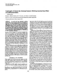
Fig. 1 Schematic illustration for cell source, differentiation, stimulation, and application of human thermogenic adipocytes in vitro. Human brown/brite adipocytes can be differentiated from cell lines, stem cells, or adipose SVFs. Those cell lines and stem cells are established from several human materials such as adipose tissues, brain, and bone
marrow. In vitro, human thermogenic adipocytes are activated by several cAMP inducers such as β adrenoreceptor (e.g., isoproterenol— ISO, norepinephrine—NE) and Forskolin. The differentiated human brown/brite adipocytes are useful tools for drug screening, and therapy development to prevent obesity and its related disorders
Potential use as cell therapy
Human thermogenic adipocytes are popularly differentiated by an adipogenic medium containing DMEM/F12, FBS, P/S, INS, IBMX, DEX and ROSI (Table 2), and the maintain of ROSI along differentiation process is needed for inducing and increasing the formation of thermogenic adipocytes. However, the concentration of each drug in that adipogenic medium varies among the studies. The activation of human brown/brite adipocytes in vitro requires the stimulations of cAMP signaling by several cAMP inducers such as β adrenoreceptor agonists or Forskolin (Table 2 and Fig. 1). Human thermogenic adipocytes in vitro are useful tools for drug screening and potential cell therapies for obesity and its related disorders (Fig. 1).
Stem cells derived from human adipose tissues can differentiate into metabolically functional thermogenic adipocytes in vitro [8, 44, 46, 57]. Importantly, these induced brite/ brown adipocytes in vitro can maintain their thermogenic function in vivo after being transplanted into animal models [8, 40, 44, 57]. In a NOD-SCID mouse model of diet induced obesity (DIO), transplantation of differentiated human brown adipocytes within a 3D adipose extracellular matrix scaffold significantly reduced blood glucose level and body weight of animals [57]. This is supported by another observation where Min et al. reported that transplantation of pre-activated brite adipocytes differentiated from human adipose SVFs could improve systemic glucose tolerance in both normal and obese NOD-SCID mice [8, 44]. Furthermore, Lee et al. found that administration of human thermogenic adipocytes had antiobesity effect because these adipocytes reduced obesityinduced inflammation in liver, hyperlipidemia, Pparγ expression, but increased Pparα levels in adipose tissues [40]. These results predict new cell therapies based on thermogenic adipocytes to prevent obesity and its related disorders in humans.
Conclusions There are several cell sources for inducing human brown/brite adipocytes in vitro including adipose SVFs, stem cells (hASCs, hiPSCs, BADSCs, and bmMSCs), and cell lines (PAZ6 and SGBS) (Table 1 and Fig. 1). Those cell sources may come from various types of human tissues such as fat, brain, and bone marrow (Table 1 and Fig. 1).
Abbreviations ARs, adrenergic receptors; BAT, brown adipose tissue; bmMSCs, bone marrow-derived mesenchymal stem cells; BMP7, bone morphogenetic protein 7; CIG, ciglitazone; CL, CL316243; DEX, dexamethasone; DMEM/F-12, Dulbecco’s Modified Eagle Medium/Nutrient Mixture F-12; E, epinephrine; FBS, fetal bovine serum; FGF21, fibroblast growth factor-21; FSK, Forskolin; GH, human growth hormone; hASCs, human adipose tissue stem cells; hFGF2, human fibroblast growth factor 2; hiPSCs, human induced pluripotent stem cells; IBMX, 3 isobultyL-1 methylxanthine; IGF-1, insulin-like growth factor-1; INS, insulin; ISO, isoproterenol (ISO); NE, norepinephrine; PIO, pioglitazone; PPARγ, peroxisome proliferator-activated receptor γ; P/S, penicillin and streptomycin; ROSI, rosiglitazone; SVF, stromal vascular fraction; T3, triiodothyronine; TRO, troglitazone; WAT, white adipose tissue
Chu et al. Compliance with ethical standards Ethical statement This article does not contain any studies with human participants or animals performed by any of the authors. Conflict of interest The authors declare that they have no conflict of interest.
References 1.
2. 3. 4.
5.
6.
7.
8.
9.
10.
11.
12. 13. 14. 15.
16.
17.
18.
19.
Atit R et al (2006) β-catenin activation is necessary and sufficient to specify the dorsal dermal fate in the mouse. Dev Biol 296(1):164– 176 Barbara C, Nedergaarb J (2004) Brown adipose tissue: function and physiological significance. Physiol rev 84(1):277–359 Barclay JL et al (2015) Effects of glucocorticoids on human brown adipocytes. J Endocrinol 224(2):139–147 Barquissau V et al (2016) White-to-brite conversion in human adipocytes promotes metabolic reprogramming towards fatty acid anabolic and catabolic pathways. Molecular Metabolism 5(5):352– 365 Bordicchia M et al (2012) Cardiac natriuretic peptides act via p38 MAPK to induce the brown fat thermogenic program in mouse and human adipocytes. J Clin Invest 122(3):1022–1036 Brydon L et al (2001) Functional expression of MT2 (Mel1b) melatonin receptors in human PAZ6 adipocytes. Endocrinology 142(10):4264–4271 Cantó C et al (2012) The NAD+ precursor nicotinamide riboside enhances oxidative metabolism and protects against high-fat dietinduced obesity. Cell Metab 15(6):838–847 Chu D-T, Tao Y (2017) Human thermogenic adipocytes: a reflection on types of adipocyte, developmental origin, and potential application. J Physiol Biochem 73(1):1–4 Chu D-T et al (2014) Expression of adipocyte biomarkers in a primary cell culture models reflects preweaning adipobiology. J Biol Chem 289(26):18478–18488 Chu D-T, Tao Y, Taskén K (2017a) OPA1 in lipid metabolism: function of OPA1 in lipolysis and thermogenesis of adipocytes. Horm Metab res 49(4):276–285 Chu D-T et al (2017b) C57BL/6J mice as a polygenic developmental model of diet-induced obesity. Physiological Reports 5(7): e13093 Chu-Dinh T, Chu DT (2014) 4-1BB and the epigenetic regulations of this molecule. Medical Epigenetics 2(3):80–85 Cunningham S et al (1985) The characterization and energetic potential of brown adipose tissue in man. Clin Sci 69(3):343–348 Cypess AM et al (2009) Identification and importance of brown adipose tissue in adult humans. N Engl J med 360(15):1509–1517 Cypess AM et al (2013) Anatomical localization, gene expression profiling and functional characterization of adult human neck brown fat. Nat med 19(5):635–639 Cypess AM et al (2015) Activation of human brown adipose tissue by a β3-adrenergic receptor agonist. Cell Metab 21(1):33– 38 Digby JE et al (1998) Thiazolidinedione exposure increases the expression of uncoupling protein 1 in cultured human preadipocytes. Diabetes 47(1):138–141 Elabd C et al (2009) Human multipotent adipose-derived stem cells differentiate into functional brown adipocytes. Stem Cells 27(11): 2753–2760 Fischer-Posovszky P et al (2008) Human SGBS cells—a unique tool for studies of human fat cell biology. Obesity Facts 1(4):184– 189
20.
Fu T et al (2014) MicroRNA 34a inhibits beige and brown fat formation in obesity in part by suppressing adipocyte fibroblast growth factor 21 signaling and SIRT1 function. Mol Cell Biol 34(22):4130–4142 21. Gesta S, Tseng Y-H, Kahn CR (2007) Developmental origin of fat: tracking obesity to its source. Cell 131(2):242–256 22. Ghandour RA et al (2016) IP-receptor and PPARs trigger the conversion of human white to brite adipocyte induced by carbaprostacyclin. Biochimica et Biophysica Acta (BBA) Molecular and Cell Biology of Lipids 1861(4):285–293 23. Giroud M et al (2016) Let-7i-5p represses brite adipocyte function in mice and humans. Scientific Reports 6:28613 24. Guennoun A et al (2015) Comprehensive molecular characterization of human adipocytes reveals a transient brown phenotype. J Transl med 13(1):135 25. Hafner A-L et al (2016) Brown-like adipose progenitors derived from human induced pluripotent stem cells: identification of critical pathways governing their adipogenic capacity. Scientific Reports 6: 32490 26. Harms M, Seale P (2013) Brown and beige fat: development, function and therapeutic potential. Nat med 19(10):1252–1263 27. Houtkooper RH, Pirinen E, Auwerx J (2012) Sirtuins as regulators of metabolism and healthspan. Nature reviews. Molecular Cell Biology 13(4):225–238 28. Jespersen, Naja Z., et al., A classical brown adipose tissue mRNA signature partly overlaps with brite in the supraclavicular region of adult humans. Cell Metab, 2013. 17(5): p. 798–805. 29. Jockers R et al (1998) Desensitization of the β-adrenergic response in human brown adipocytes. Endocrinology 139(6):2676–2684 30. Jukarainen S et al (2015) Obesity is associated with low NAD+/ SIRT pathway expression in adipose tissue of BMI-discordant monozygotic twins. The Journal of Clinical Endocrinology & Metabolism 101(1):275–283 31. Jura M et al (2016) Mest and Sfrp5 are biomarkers for healthy adipose tissue. Biochimie 124:124–133 32. Khan NA et al (2014) Effective treatment of mitochondrial myopathy by nicotinamide riboside, a vitamin B3. EMBO Molecular Medicine 6(6):721–731 33. Klaus S et al (1995) Functional assessment of white and brown adipocyte development and energy metabolism in cell culture. Dissociation of terminal differentiation and thermogenesis in brown adipocytes. J Cell Sci 108(10):3171–3180 34. Klepac K et al (2016) The Gq signalling pathway inhibits brown and beige adipose tissue. Nat Commun 7:10895 35. Koppen A, Kalkhoven E (2010) Brown vs white adipocytes: the PPARγ coregulator story. FEBS Lett 584(15):3250–3259 36. Lee P et al (2011) Inducible brown adipogenesis of supraclavicular fat in adult humans. Endocrinology 152(10):3597–3602 37. Lee P et al (2014a) Irisin and FGF21 are cold-induced endocrine activators of brown fat function in humans. Cell Metab 19(2):302– 309 38. Lee P et al (2014b) Functional thermogenic beige adipogenesis is inducible in human neck fat. Int J Obes 38(2):170–176 39. Lee MH et al (2016a) ECM microenvironment unlocks brown adipogenic potential of adult human bone marrow-derived MSCs. Scientific Reports 6:21173 40. Lee C-W, Hsiao W-T, Lee OK-S (2016b) Mesenchymal stromal cell-based therapies reduce obesity and metabolic syndromes induced by a high-fat diet. Transl res 182:61–74.e8 41. Lefterova MI et al (2014) PPARγ and the global map of adipogenesis and beyond. Trends in Endocrinology & Metabolism 25(6): 293–302 42. Lidell ME et al (2013) Evidence for two types of brown adipose tissue in humans. Nat med 19(5):631–634
Cell source, differentiation and stimulation of in vitro human brite/brown adipocytes 43.
Loft A et al (2015) Browning of human adipocytes requires KLF11 and reprogramming of PPARγ superenhancers. Genes dev 25: 1281–1294 44. Min SY et al (2016) Human ‘brite/beige’ adipocytes develop from capillary networks, and their implantation improves metabolic homeostasis in mice. Nat med 22(3):312–318 45. Mohsen-Kanson T et al (2014) Differentiation of human induced pluripotent stem cells into brown and ahite adipocytes: role of Pax3. Stem Cells 32(6):1459–1467 46. Okla M et al (2015) BMP7 drives human adipogenic stem cells into metabolically active beige adipocytes. Lipids 50(2):111–120 47. Peirce V, Carobbio S, Vidal-Puig A (2014) The different shades of fat. Nature 510(7503):76–83 48. Petrovic N et al (2010) Chronic peroxisome proliferator-activated receptor γ (PPARγ) activation of epididymally derived white adipocyte cultures reveals a population of thermogenically competent, UCP1-containing adipocytes molecularly distinct from classic brown adipocytes. J Biol Chem 285(10):7153–7164 49. Pisani D et al (2011) Differentiation of human adipose-derived stem cells into Bbrite^ (brown-in-white) adipocytes. Front Endocrinol 2: 87 50. Rodriguez A-M et al (2004) Adipocyte differentiation of multipotent cells established from human adipose tissue. Biochem Biophys res Commun 315(2):255–263 51. Saely CH, Geiger K, Drexel H (2012) Brown versus white adipose tissue: a mini-review. Gerontology 58(1):15–23 52. Saito M et al (2009) High incidence of metabolically active brown adipose tissue in healthy adult humans: effects of cold exposure and adiposity. Diabetes 58(7):1526–1531
53.
Sanchez-Gurmaches J, Guertin DA (2014a) Adipocyte lineages: tracing back the origins of fat. Biochim Biophys Acta (BBA) Mol Basis dis 1842(3):340–351 54. Sanchez-Gurmaches J, Guertin DA (2014b) Adipocytes arise from multiple lineages that are heterogeneously and dynamically distributed. Nat Commun 5:4099 55. Seale P et al (2008) PRDM16 controls a brown fat/skeletal muscle switch. Nature 454(7207):961–967 56. Shinoda K et al (2015) Genetic and functional characterization of clonally derived adult human brown adipocytes. Nat med 21(4): 389–394 57. Silva FJ et al (2014) Metabolically active human brown adipose tissue derived stem cells. Stem Cells 32(2):572–581 58. Timmons JA et al (2007) Myogenic gene expression signature establishes that brown and white adipocytes originate from distinct cell lineages. Proc Natl Acad Sci 104(11):4401–4406 59. van Marken Lichtenbelt WD et al (2009) Cold-activated brown adipose tissue in healthy men. N Engl J med 360(15):1500–1508 60. Virtanen KA et al (2009) Functional brown adipose tissue in healthy adults. N Engl J med 360(15):1518–1525 61. Wang Y-L et al (2016) Concomitant beige adipocyte differentiation upon induction of mesenchymal stem cells into brown adipocytes. Biochem Biophys res Commun 478(2):689–695 62. Wu J et al (2012) Beige adipocytes are a distinct type of thermogenic fat cell in mouse and human. Cell 150(2):366–376 63. Zilberfarb V et al (1997) Human immortalized brown adipocytes express functional beta3-adrenoceptor coupled to lipolysis. J Cell Sci 110(7):801