The Journal of Immunology
Expansion of Functional Endogenous Antigen-Specific CD4ⴙCD25ⴙ Regulatory T Cells from Nonobese Diabetic Mice1 Emma L. Masteller,* Matthew R. Warner,* Qizhi Tang,* Kristin V. Tarbell,2† Hugh McDevitt,† and Jeffrey A. Bluestone3* CD4ⴙCD25ⴙFoxp3ⴙ regulatory T cells (Treg) are critical for controlling autoimmunity. Evidence suggests that Treg development, peripheral maintenance, and suppressive function are dependent on Ag specificity. However, there is little direct evidence that the Treg responsible for controlling autoimmunity in NOD mice or other natural settings are Ag specific. In fact, some investigators have argued that polyclonal Ag-nonspecific Treg are efficient regulators of immunity. Thus, the goal of this study was to identify, expand, and characterize islet Ag-specific Treg in NOD mice. Ag-specific Treg from NOD mice were efficiently expanded in vitro using IL-2 and beads coated with recombinant islet peptide mimic-MHC class II and anti-CD28 mAb. The expanded Ag-specific Treg expressed prototypic surface markers and cytokines. Although activated in an Ag-specific fashion, the expanded Treg were capable of bystander suppression both in vitro and in vivo. Importantly, the islet peptide mimic-specific Treg were more efficient than polyclonal Treg in suppressing autoimmune diabetes. These results provide a direct demonstration of the presence of autoantigen-specific Treg in the natural setting that can be applied as therapeutics for organ-specific autoimmunity. The Journal of Immunology, 2005, 175: 3053–3059. he CD4⫹CD25⫹ regulatory T cells (Treg)4 have been shown to be essential for controlling autoimmunity (1). This is apparent in NOD mice, a mouse model of type 1 diabetes in which depletion of Treg results in exacerbated T cellmediated destruction of the insulin-producing islet cells of the pancreas. An unanswered question in understanding how CD4⫹CD25⫹ Treg control autoimmunity is whether there is an Ag-specific Treg repertoire that is responsible for maintaining organ-specific tolerance (2). Early studies by Seddon and Mason (3) in rat models suggested that organ-specific tolerance was dependent on Treg that specifically recognize autoantigens expressed by the target organ. Subsequent studies showed that the transfer of polyclonal CD4⫹CD25⫹ Treg was sufficient to prevent diabetes in NOD mice (4, 5). The process was inefficient, however, and required the infusion of high numbers of Treg possibly due to the low frequency of islet-specific cells. Recently, using a TCR transgenic (Tg) model, we and others showed that small numbers of islet
T
*Diabetes Center, Department of Medicine, University of California, San Francisco, CA 94143; and †Department of Microbiology and Immunology, Stanford University School of Medicine, Stanford, CA 94395 Received for publication April 12, 2005. Accepted for publication June 21, 2005. The costs of publication of this article were defrayed in part by the payment of page charges. This article must therefore be hereby marked advertisement in accordance with 18 U.S.C. Section 1734 solely to indicate this fact. 1 This work was supported in part by a grant from the Juvenile Diabetes Research Foundation Center Grant 4-2004-372; and National Institutes of Health Grants DERC P30 DK63720 and U19 AI56388. 2 Current address: Laboratory of Cellular Physiology and Immunology, and the Chris Browne Center for Immunology and Immune Disease, The Rockefeller University, New York, NY 10021. 3 Address correspondence and reprint requests to Dr. Jeffrey A. Bluestone, Diabetes Center, University of California, 513 Parnassus Avenue, Box 0540, San Francisco, CA 94143-0540. E-mail address:
[email protected] 4
Abbreviations used in this paper: Treg, regulatory T cell; Ct, threshold cycle; GAD, glutamic acid decarboxylase; GITR, glucocorticoid induced tumor necrosis factor receptor; HEL, hen egg lysozyme; mIgG, mouse IgG; Teff, effector T cell; Tg, transgenic. Copyright © 2005 by The American Association of Immunologists, Inc.
Ag-specific BDC2.5 TCR Tg⫹ Treg were much more effective than polyclonal Treg in blocking and reversing diabetes in NOD mice (6, 7). Although an artificial system, these studies provide strong support for the hypothesis that Ag specificity is critical for optimal Treg function and imply that effective clinical therapy will benefit from the ability to identify and expand relevant Ag-specific Treg from polyclonal populations. Treg have been shown to possess a diverse TCR repertoire that is biased toward self Ags; however, little is known about the Ag specificity of Treg arising under natural conditions (8). The low numbers of endogenous Treg have limited their study and therapeutic use. We recently described a method for the in vitro expansion of polyclonal Treg using a combination of IL-2 and beads coated with anti-CD3 and anti-CD28 mAb (6). This technique was used to expand Ag-specific Treg from BDC2.5 TCR Tg⫹ mice that express a TCR specific for an islet Ag. These expanded Treg were shown to be efficacious in preventing and reversing diabetes in NOD mice (6). In the present study, we examined whether natural Treg that have the BDC2.5 specificity exist in conventional NOD mice using a novel protocol that selectively expands Ag-specific Treg. The expansion protocol was adapted from the one described above by substituting the anti-CD3 mAb with a rMHC class II I-Ag7 presenting the BDC2.5 TCR mimotope peptide 1040-31 (p31)4 (9, 10). We show that not only do these cells exist in the unmanipulated Treg repertoire, but that these rare Ag-specific Treg can be expanded and used to prevent autoimmune diabetes in Tregdeficient NOD mice. These results have important implications not only for the basic understanding of Treg biology, but may also lead to an effective clinical therapy in autoimmune diseases.
Materials and Methods Mice NOD mice were purchased from Taconic Farms; BDC2.5 TCR Tg⫹ mice were obtained from D. Mathis (Joslin Clinic, Harvard University, Boston, MA) (11); and glutamic acid decarboxylase (GAD)286 TCR Tg⫹ mice (12), 0022-1767/05/$02.00
3054 NOD.CD28⫺/⫺ mice (13), and NOD.TCR␣⫺/⫺ mice (The Jackson Laboratory) were housed and bred under specific pathogen-free conditions at the University of California San Francisco Animal Barrier Facility in accordance with institutional guidelines.
Reagents Anti-CD3⑀ (145-2C11), anti-CD28 (PV-1), anti-CD4 (GK1.5), and antiglucocorticoid induced tumor necrosis factor receptor (GITR) (DTA-1; a gift from S. Sakaguchi, Kyoto University, Kyoto, Japan) were produced and purified in our laboratory. Anti-CD4 and anti-GITR were conjugated to FITC in our laboratory. R-PE-conjugated anti-CD25 (7D4) was purchased from Southern Biotechnology Associates. R-PE-conjugated anti-CTLA4 (UC10-4F10-11), biotinylated-anti-V2 TCR (B20.6), anti-V4 TCR (KT4), anti-V12 TCR (MR11-1), and anti-ICOS (7E 17G9) were purchased from BD Pharmingen. Allophycocyanin-conjugated anti-CD62L (MEL-14), anti-CD4 (GK1.5), and streptavidin were purchased from eBioscience. p31-I-Ag7-mouse(m)IgG2a and hen egg lysozyme (HEL)-I-Ag7mIgG2a were produced in our laboratory, as previously described (10). Peptides were produced by the University of California Biomolecular Resource Center. The 1040-31 peptide, specific for BDC2.5 TCR Tg⫹ T cells, consisted of amino acids YVRPLWVRME (9). The HEL11–25 protein consisted of amino acids AMKRHGLDNYRGYSL. GAD286 –300 was a gift from E. Sercarz (Torrey Pines Institute for Molecular Studies, San Diego, CA). Both latex and paramagnetic beads were used throughout the described studies with equal efficiency. For coating of latex beads, 1 ⫻ 108 5-m white sulfate latex beads (Interfacial Dynamics) were coated with 30 g of anti-CD28 and 3 g of either anti-CD3 or p31-I-Ag7-mIgG2a. For coating of paramagnetic beads, 1 ⫻ 108 Dynabeads M-450 Tosylactivated (Dynal Biotech) were coated with 50 g of anti-CD28 and 5 g of either anti-CD3 or p31-I-Ag7-mIgG2a, following the manufacturer’s instructions.
Cell sorting and in vitro expansion of T cells CD4⫹ T cells were enriched from spleen and lymph nodes (inguinal, axillary, brachial, submandibular, pancreatic, and mesenteric) by negative selection using an AutoMACS (Miltenyi Biotec) or Stemsep beads (StemCell Technologies). Cells were stained and sorted, as previously described, on a Mo-Flo (DakoCytomation) into CD4⫹CD25⫹CD62L⫹ Treg and CD4⫹CD25⫺CD62L⫹ T cell effector (Teff) populations to ⬎97% purity (6). Purified T cells at 106/ml were stimulated in flat-bottom plates at a 1:1 bead-cell ratio with anti-CD3 and anti-CD28- or p31-I-Ag7mIgG2a and anti-CD28-coated beads in medium supplemented with 2000 IU/ml human rIL-2 (Chiron). Complete DMEM was made, as previously described (6). Cultures were expanded with IL-2-supplemented medium when needed. Beads were removed at the end of the culture period before further experimentation. To remove coated latex beads, cultures were incubated with biotinylated anti-mouse IgG2a (Southern Biotechnology Associates) and biotinylated anti-hamster Ig (Vector Laboratories), washed, incubated with streptavidin microbeads (Miltenyi Biotec), washed, and run over an MS column (Miltenyi Biotec). Cells in the flow through fraction were collected. Dynabeads were removed using a Dynal MPC-L magnet (Dynal Biotech).
Flow cytometry For staining with peptide-I-Ag7 multimers, peptide-I-Ag7-mIgG2a was complexed with Alexa 488-conjugated protein A (Molecular Probes), as previously described (10), or complexed with biotinylated protein A (SigmaAldrich) and allophycocyanin-conjugated streptavidin (eBioscience) at a 3:1:0.25 molar ratio. Cells (106) were stained with 1 g of p31-I-Ag7mIgG2a for 2 h on ice. Abs against TCR V were added at the same time as the peptide-I-Ag7 multimers. Other Abs were added during the last 30 min of staining or when using peptide-I-Ag7/biotinylated protein A multimers, during the secondary staining along with allophycocyanin-conjugated streptavidin. Isotype-matched Abs were used for control staining for CTLA-4 and ICOS expression on expanded Treg. Freshly isolated CD4⫹CD25⫺ cells were used as negative control staining for GITR analysis. Flow cytometric analysis was performed on a FACSCalibur flow cytometer (BD Biosciences).
In vitro suppression assays The indicated numbers of expanded Treg cultures were added to 50,000 freshly isolated CD4⫹ T cells with 50,000 NOD.TCR␣-deficient spleen cells or NOD CD4⫹/CD8⫹-depleted spleen cells and either the indicated peptide at 0.1 M or anti-CD3 at 1 g/ml. Cultures were incubated for 72 h and pulsed with 1 Ci/well [3H]thymidine during the last 14 h.
Ag-SPECIFIC CD4⫹CD25⫹ Treg CONTROL DIABETES CFSE assays The p31-I-Ag7-expanded Treg, either unsorted or FACS sorted based on p31-I-Ag7 multimer binding, were labeled with 2.5 M CFSE and cultured with irradiated splenic APC and 1 M of either HEL or p31 peptide in the presence of 100 IU of IL-2/ml. Cells were collected at 72 h and analyzed by flow cytometry.
Real-time PCR analysis Total RNA was isolated from expanded T cells using QIAshredder and RNeasy Mini Kit spin columns (Qiagen). cDNA was synthesized from 50 – 600 ng of sample RNA using SuperScript III reverse transcriptase and oligo(dT)12-18 primer (Invitrogen Life Technologies). Real-time PCR contained 1.25–28.5 ng of cDNA. Primers and probes for Foxp3 and 18S rRNA were purchased as premixed reagents from Applied Biosystems. Real-time PCR was performed on an ABI PRISM 7900 HT Sequence Detection System using TaqMan Universal PCR Master Mix (Applied Biosystems). All samples and controls were run in duplicate, and average threshold cycle (Ct) values were used to normalize the signal levels in samples relative to an endogenous control (18S rRNA). The normalized sample values were then used to calculate the fold difference in expression of Foxp3 between expanded Treg and Teff. The expression ratios of Treg to Teff were calculated by the following formula: (Treg/Teff)Foxp3 ⫽ 2n, n ⫽ (TeffCtFoxp3 ⫺ TeffCt18S) ⫺ (TregCtFoxp3 ⫺ TregCt18S).
Cytokine ELISA To measure cytokine production, 1 ⫻ 106 p31-I-Ag7-expanded Treg were cultured with 5 ⫻ 106 irradiated APC, 100 nM 1040-31 peptide, and 1 g/ml anti-CD28. Culture supernatants were harvested at 48 h. Cytokine concentrations were determined by ELISA using Ab pairs purchased from BD Pharmingen.
Adoptive transfer The indicated numbers of p31-I-Ag7-expanded Treg cultures or expanded polyclonal Treg cultures were transferred to NOD.CD28⫺/⫺ mice via retroorbital injection. Nonfasting blood glucose levels in recipient mice were monitored using an Accu-Check glucometer (Roche Diagnostic Systems). Mice were considered diabetic after two readings above 250 mg/dL.
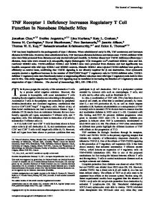
Results Recombinant peptide-MHC can specifically expand rare Agspecific Treg To selectively expand Ag-specific Treg, we adapted the technique used to expand polyclonal Treg by substituting the anti-CD3 mAb with a rMHC class II I-Ag7 presenting the BDC2.5 TCR mimotope peptide 1040-31 (p31) (10). The mimotope peptide was used because the endogenous BDC2.5 Ag is not yet identified (9). Initial experiments were conducted with BDC2.5 Tg⫹ Treg to determine whether p31-I-Ag7- and anti-CD28-coated beads could expand low frequency Ag-specific cells from a polyclonal population and whether Treg expanded in this manner retained suppression activity. Polyclonal CD4⫹CD25⫹ Treg from NOD mice were seeded with low frequencies of sorted BDC2.5 TCR Tg⫹ Treg at 0.1– 0.001% of the total population and cultured with p31-I-Ag7- and anti-CD28-coated beads in the presence of IL-2. The p31-I-Ag7coated beads were extremely efficient in expanding CD4⫹CD25⫹ BDC2.5 TCR Tg⫹ Treg. Although cultures initially seeded at 0.01 and 0.001% BDC2.5 TCR Tg⫹ Treg did not expand appreciably based on total cell number (Fig. 1A), flow cytometric analysis using p31-I-Ag7 multimers revealed that BDC2.5 TCR Tg⫹ Treg had expanded in all cultures. At the lowest seeding, BDC2.5 TCR Tg⫹ Treg grew from 0.001 to 34.3% of the population (Fig. 1B), reflecting greater than 12 cell divisions during the 10-day culture period and resulting in nearly a 5000-fold expansion of the Agspecific cells. Expanded CD4⫹CD25⫹ BDC2.5 Treg were then tested for suppressive activity in in vitro cultures using freshly isolated CD4⫹CD25⫺ BDC2.5 Tg⫹ cells as responders and the p31 peptide as an Ag-specific stimulus. Expanded CD4⫹CD25⫹ BDC2.5 Treg efficiently suppressed the response the BDC2.5 Tg⫹ responder
The Journal of Immunology
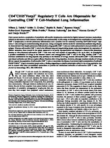
3055 cultured with p31-I-Ag7 beads, as described in Materials and Methods. Over a 2-wk period, Treg cultured in this manner generally expanded from 1- to 10-fold based on total cell number, with 4- to 5-fold expansion being the most typical. Expansion profiles of four representative cultures are shown in Fig. 2A. The lower fold expansion was usually seen when lower numbers of purified Treg (⬍0.5 ⫻ 106) were seeded into the wells. The reason for the greater expansion observed in some cultures remains unknown. There was no correlation with the age of mice, as starting populations of CD4⫹CD25⫹CD62L⫹ cells taken from 4- or 8-wk-old mice demonstrated the same range of expansion of 1- to 10-fold. Flow cytometry analysis demonstrated that after expansion with the p31-I-Ag7 beads, up to 10% of CD4⫹CD25⫹ cells stained positive for the p31-I-Ag7 multimer (Fig. 2B). Control cultures of CD4⫹CD25⫹ cells expanded with anti-CD3-coated beads did not
FIGURE 1. Peptide-I-Ag7 beads can expand low frequency Ag-specific BDC2.5 TCR Tg⫹ Treg in vitro. A, CD4⫹CD25⫹CD62L⫹ BDC2.5 TCR Tg⫹ T cells were seeded at the indicated frequencies into CD4⫹CD25⫹CD62L⫹ NOD cells and cultured with p31-I-Ag7- and antiCD28-coated beads along with IL-2. The fold expansion of total cell numbers relative to the initial cell input of BDC2.5 and NOD cells is shown. Cultures were quantitated by viable cell counting. B, Cultures initially seeded at 100 or 0.001% BDC2.5 Tg⫹ Treg were expanded for 10 days with p31-I-Ag7 beads, stained with either p31-I-Ag7 multimers or control HELI-Ag7 multimers, and analyzed by flow cytometry. C, Graded doses of BDC2.5 Tg⫹ Treg expanded with p31-I-Ag7 and anti-CD28 beads were cultured with 5 ⫻ 104 freshly isolated CD4⫹CD25⫺ BDC2.5 Tg⫹ responder cells and 5 ⫻ 104 irradiated APC. Responder cells alone and Treg alone are also shown. Cultures were stimulated with 100 nM BDC2.5 mimotope peptide p31 for 72 h. Proliferation was measured by [3H]thymidine incorporation.
cells in a dose-dependent manner (Fig. 1C). Thus, the expansion procedure using recombinant peptide-MHC class II as an Ag-specific stimulus resulted in a large expansion of rare Ag-specific Treg that retained suppressive function. Rare Ag-specific Treg can be expanded from wild-type NOD mice This approach was applied next to the expansion of Ag-specific CD4⫹CD25⫹ Treg from conventional NOD mice. Initial staining of freshly isolated Treg from NOD mice with the p31-I-Ag7 multimer failed to detect positively staining cells above background levels, indicating that, if present, these Ag-specific cells were present at low frequency (data not shown). Sorted CD4⫹CD25⫹CD62L⫹ cells from 4- to 8-wk-old NOD mice were
FIGURE 2. Ag-specific Treg can be expanded from wild-type NOD mice. A, Sorted CD4⫹CD25⫹CD62L⫹ from wild-type NOD mice were cultured with p31-I-Ag7 and anti-CD28 beads in the presence of IL-2. The expansion of four representative cultures over a 2-wk period is shown. B, CD4⫹CD25⫹CD62L⫹ cells cultured with either p31-I-Ag7 beads or anti-CD3 (2C11) beads were stained with anti-CD4 and either p31- or HEL-I-Ag7 multimers to identify Ag-specific cells. Staining of CD4⫹CD25⫺CD62L⫹ Teff cultured with p31-I-Ag7 beads is also shown. C, Cultures of p31-I-Ag7-expanded Treg were left unsorted or sorted into p31I-Ag7 multimer-positive and -negative populations using FACS. The resulting populations were CFSE labeled and stimulated with HEL or p31 peptide in the presence of irradiated APC and IL-2. Cultures were analyzed at 72 h by flow cytometry for CFSE intensity.
3056 stain positive for p31-I-Ag7 above background levels. In cultures of CD4⫹CD25⫺CD62L⫹ Teff expanded with p31-I-Ag7-coated beads under the same conditions, 40 –50% of the population stained positive for the p31-I-Ag7 multimer (Fig. 2B). The p31-IAg7-expanded Treg were also tested for specificity against peptides in proliferation assays. The p31-I-Ag7-expanded Treg responded to p31 peptide, but not control HEL peptide when cultured with APC and IL-2 (data not shown). As shown in Fig. 2B, in general, 90% of the p31-I-Ag7-expanded Treg population failed to stain for the p31-I-Ag7 multimer when analyzed by flow cytometry. It was possible that the unstained population was comprised of p31-I-Ag7-reactive cells with TCRs with too low of an affinity to be detected by flow cytometry (14). Alternatively, this population could be comprised of cells that were expanding in an Ag-nonspecific manner. To distinguish between these two alternatives, p31-I-Ag7-expanded Treg populations were sorted into p31-I-Ag7 multimer-positive and -negative populations using FACS. The resulting populations were then labeled with CFSE and stimulated with the polyclonal activator anti-CD3 mAb or with the p31 peptide or a control HEL peptide in the presence of APC and IL-2. All three populations, unsorted, p31-I-Ag7 multimer positive, and p31-I-Ag7 multimer negative, expanded equally well to anti-CD3 mAb when analyzed for CFSE dilution (data not shown). As shown in Fig. 2C, ⬎50% of the Treg from the unsorted population entered into cell cycle when stimulated with the p31 peptide compared with only ⬃14% when stimulated with the HEL peptide. The background proliferation with the HEL peptide probably reflected the residual presence of beads in the culture before cell sorting into p31-I-Ag7 multimer-positive and -negative populations. In the p31-I-Ag7 multimer-positive population, ⬎65% of the cells entered into the cell cycle when stimulated with the p31 peptide. Importantly, ⬎50% of the cells from the p31-I-Ag7 multimer-negative population entered into cell cycle when stimulated with the p31 peptide, demonstrating the presence of p31-reactive cells in this population. The expansion protocol using recombinant peptide-MHC class II was therefore very efficient in expanding Ag-specific Treg with TCRs with a broad range of affinities for Ag. Characterization of expanded Ag-specific Treg Expanded Ag-specific Treg were examined for the expression of the Treg lineage marker Foxp3 using quantitative real-time PCR. To ensure that p31-I-Ag7-reactive cells were analyzed, p31-I-Ag7expanded Treg and Teff were sorted into p31-I-Ag7 multimer-positive and -negative populations by FACS before analysis. In a representative experiment shown in Fig. 3A, expanded p31-I-Ag7 multimer⫹ Treg expressed ⬃3000-fold more Foxp3 relative to expanded p31-I-Ag7 multimer-positive Teff. Upon challenge with Ag and APC, p31-I-Ag7-expanded Treg expressed low levels of the proinflammatory cytokines IL-2, IL-4, and IFN-␥, and expressed high levels of the anti-inflammatory cytokine IL-10 (Fig. 3B). Peptide-I-Ag7-expanded Treg retained high levels of expression of CD62L and CD25 throughout the culture period (Fig. 4A). In contrast, peptide-I-Ag7-expanded Teff down-regulated CD62L and expressed relatively lower levels of CD25. Peptide-I-Ag7-expanded Treg also expressed high levels of CTLA-4, GITR, and ICOS (Fig. 4B). Peptide-I-Ag7-expanded Teff expressed levels of CTLA-4 comparable to peptide-I-Ag7-expanded Treg (data not shown). GITR was also up-regulated on the expanded Teff as compared with naive Teff, but remained lower than levels observed on expanded Treg. ICOS expression was higher on expanded Teff as compared with expanded Treg (data not shown). We then examined the V repertoire of the p31-I-Ag7-expanded
Ag-SPECIFIC CD4⫹CD25⫹ Treg CONTROL DIABETES
FIGURE 3. Expanded Ag-specific Treg express Foxp3 and IL-10. A, p31-I-Ag7-expanded cells were sorted into p31-I-Ag7 multimer-positive and -negative populations by FACS. Levels of Foxp3 mRNA expression were determined for the indicated populations by real-time PCR analysis. The expression levels of Foxp3 in p31-I-Ag7-expanded CD4⫹CD25⫹ Treg relative to p31-I-Ag7-expanded CD4⫹CD25⫺ Teff are shown. B, p31-I-Ag7expanded Treg were stimulated with p31 peptide in the presence of irradiated APC. Secreted cytokine levels were determined 48 h after stimulation by ELISA.
T cells. The BDC2.5 TCR expresses a TCR derived from the V4 family (11). However, when p31-I-Ag7-expanded Treg and Teff were costained with p31-I-Ag7 multimers and different TCR V reagents, neither population was monoclonal. Although there was a significant number of V4⫹ p31-I-Ag7 multimer⫹ T cells in the Treg culture, other TCR V were also present in significant numbers (Fig. 4C). For instance, V2 and V12 accounted for 10.2 and 13.7% of the p31-I-Ag7 multimer⫹ Treg, respectively, in this representative culture. TCR V4⫹ T cells were generally present in the p31-I-Ag7 multimer⫹ Teff population, but at a lower percentage. Together, these results suggest that broad repertoires of Treg and Teff reactive against the islet peptide mimic are resident in conventional NOD mice, and that the islet peptide mimic-reactive Treg repertoire is not identical with the islet peptide mimicreactive Teff repertoire. Suppression by Ag-specific Treg in vitro Previous studies have shown that CD4⫹CD25⫹ Treg can suppress proliferation of CD4⫹ effectors in vitro and that the suppressive effect is dependent on stimulation of CD4⫹CD25⫹ Treg through their TCR. Therefore, expanded p31-I-Ag7 Treg were examined for suppressive activity and specificity in vitro. Expanded p31-I-Ag7 Treg effectively suppressed the proliferation of Ag-specific CD4⫹ BDC2.5 TCR Tg⫹ cells in a dose-specific manner when cultures were stimulated with either anti-CD3 or 1040-31 peptide (Fig. 5A).
The Journal of Immunology
3057 ificity of the expanded Treg, expanded polyclonal Treg and p31-IAg7-expanded Treg were assessed for the ability to suppress BDC2.5 TCR Tg⫹ or GAD286 TCR Tg⫹ CD4⫹ cells (12) through either polyclonal T cell activation via anti-CD3 or through Agspecific T cell activation. Both polyclonal Treg and p31-I-Ag7-expanded Treg suppressed BDC2.5 TCR Tg⫹ responders when stimulated with anti-CD3, whereas only the p31-I-Ag7-expanded Treg suppressed cultures stimulated with the BDC2.5 1040-31 peptide (Fig. 5B). Similarly, both polyclonal Treg and p31-I-Ag7-expanded Treg suppressed the response of GAD286 TCR Tg⫹ CD4⫹ T cells when stimulated with anti-CD3 (Fig. 5C, bottom). However, neither the polyclonal Treg population nor the p31-I-Ag7-expanded Treg population suppressed GAD286 TCR Tg⫹ responders when stimulated with the GAD286 –300 peptide (Fig. 5C, top). Most significantly, p31-I-Ag7-expanded Treg were capable of suppressing GAD286 TCR Tg⫹ responders when the culture was stimulated with both the GAD286 –300 and the 1040-31 peptide (Fig. 5D). Collectively, these data demonstrate that the suppressive activity of the p31-I-Ag7-expanded Treg is dependent on Ag-specific stimulation through the TCR, although, once stimulated with cognate Ag, p31-I-Ag7-expanded Treg are capable of exerting bystander suppression.
FIGURE 4. Surface phenotype of expanded Ag-specific Treg. A, Cultures of p31-I-Ag7-expanded Treg or Teff were stained for expression of CD25 and CD62L. B, Surface staining of p31-I-Ag7-expanded Treg. Light lines indicate control staining. Heavy lines indicate staining of p31-I-Ag7expanded Treg populations. C, Cultures of p31-I-Ag7-expanded Treg and Teff were costained with p31-I-Ag7 multimers and the indicated TCR V Abs. Numbers shown represent the percentage of p31-I-Ag7 multimer-positive cells staining positive for the indicated TCR V.
In contrast, p31-I-Ag7-expanded CD4⫹CD25⫹ Teff failed to suppress freshly isolated BDC2.5 CD4⫹ T cells and resulted in augmentation of proliferation (data not shown). The p31-I-Ag7-expanded Treg, but not the Teff, were anergic to stimulation (both p31 peptide and anti-CD3) in the absence of CD28 costimulation or addition of IL-2 consistent with reports for freshly isolated Treg (Fig. 5B and data not shown). To further characterize the Ag spec-
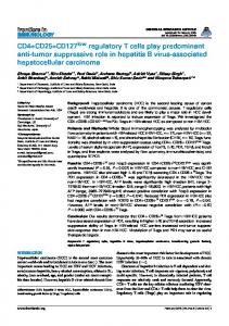
Expanded Ag-specific Treg prevent autoimmune diabetes We then tested the ability of small numbers of p31-I-Ag7-expanded Treg to suppress diabetes in CD28⫺/⫺ NOD mice. CD28⫺/⫺ NOD mice have normal numbers of Teff and Th1 responses and undergo an accelerated form of autoimmune diabetes due to a deficiency in Treg (4, 15). Previous studies have shown that delay or prevention of diabetes in this model required the transfer of high numbers of polyclonal Treg (8 –20 ⫻ 106) (4). In sharp contrast, the transfer of as few as 1.8 ⫻ 106 p31-I-Ag7-expanded Treg into 5- to 7-wk-old mice prevented the development of diabetes in the majority of mice for at least 15 wk of age (Fig. 6). The in vivo administered p31-I-Ag7-expanded Treg cultures were an admixture of ⬃10% high avidity cells, as determined by the ability to bind p31-I-Ag7 multimers, and lower avidity cells that were not detected by the p31-I-Ag7 multimers when analyzed by flow cytometry, but were capable of responding to p31 peptide presented by APC (see Fig.
FIGURE 5. p31-I-Ag7-expanded Treg suppress in an Ag-specific manner in vitro. A, 5 ⫻ 104 freshly isolated BDC2.5 CD4⫹CD25⫺ T cells were cultured with varying amounts of p31-I-Ag7-expanded Treg and 5 ⫻ 104 irradiated APC. Cultures were stimulated with either 100 nM 1040-31 peptide or 1 g/ml anti-CD3. Proliferation was determined, as described in Fig. 1C. B and C, 5 ⫻ 104 p31-I-Ag7 or 2C11-expanded Treg were cultured with freshly isolated BDC2.5 TCR Tg⫹ CD4⫹CD25⫺ responders (B) or GAD286 TCR Tg⫹ CD4⫹CD25⫺ responders (C), 5 ⫻ 104 irradiated APC and the 1040-31 peptide (B) or GAD286 –300 peptide (C), or anti-CD3. Proliferation was determined, as described above. D, p31-I-Ag7-expanded Treg were cultured with GAD286 TCR Tg⫹CD4⫹CD25⫺ responder cells, as described in C, and stimulated with a combination of the 1040-31 and GAD286 –300 peptides.
3058
FIGURE 6. Expanded Ag-specific Treg prevent diabetes in vivo. Fiveto 7-wk-old prediabetic CD28-deficient NOD mice were injected with 1.8 – 2 ⫻ 106 p31-I-Ag7-expanded Treg (n ⫽ 11), 2 ⫻ 106 (n ⫽ 6), or 8 ⫻ 106 (n ⫽ 2) 2C11-expanded Treg or left untreated (n ⫽ 6). Diabetes was determined by monitoring blood glucose levels.
2, B and C). Thus, the Ag-specific p31-I-Ag7-expanded cells were highly efficient in protecting the onset of diabetes induced by a fully functional polyclonal T cell response. Suppression of autoimmunity by the p31-I-Ag7-expanded Treg appeared to be organ specific. Although animals that received the p31-I-Ag7-expanded Treg were protected from diabetes, the mice continued to exhibit other autoimmune syndromes based on continued lymphocytic infiltration in the salivary and thyroid glands (data not shown).
Discussion The identification of CD4⫹CD25⫹Foxp3⫹ T cells as important regulators of tolerance has opened a major area of investigation in autoimmunity. The field has been limited, however, by the small numbers of circulating Treg and the inability to define their antigenic specificities. In this study, we demonstrated that Ag-specific CD4⫹CD25⫹Foxp3⫹ Treg reactive to an islet peptide mimic reside in the periphery of diabetes-susceptible NOD mice. Furthermore, we demonstrated that these rare Ag-specific cells can be selectively expanded in vitro from a polyclonal population, and that these expanded Treg retain phenotypic and functional characteristics of freshly isolated CD4⫹CD25⫹Foxp3⫹ Treg. The in vitro expanded Treg retained high expression of CD62L important for allowing trafficking to lymph nodes upon administration of these cells in vivo, and also retained expression of molecules such as Foxp3, GITR, and CTLA-4, implicated as important for Treg function. Activation of suppressive activity of the in vitro expanded Treg was Ag specific, but once activated, the Treg were capable of suppressing Teff with other specificities. This aspect is important for controlling diseases such as type 1 diabetes in which numerous self Ags are targeted by pathogenic effector cells. Most importantly, we showed that in vivo, expanded Ag-specific Treg are highly efficient at controlling organ-specific autoimmunity. These results support previous studies demonstrating that immune regulation by CD4⫹CD25⫹ Treg is dependent on the Ag specificity of the Treg (6, 7) and are not consistent with reports suggesting that Treg function in an Ag-nonspecific fashion by competing for T cell niches (16). Thus, efficient Treg function is likely to require Agspecific activation either for maintenance in the periphery or for function at the site of inflammation or the draining lymph node. The BDC2.5 mimotope-reactive Treg expanded from wild-type NOD mice described in these studies were not as efficient at controlling autoimmune diabetes as previously described Treg expressing a Tg BDC2.5 TCR and expanded with anti-CD3-coated beads (6). Several possibilities could explain this difference. One possi-
Ag-SPECIFIC CD4⫹CD25⫹ Treg CONTROL DIABETES bility is that the BDC2.5 TCR Tg⫹ Treg possess a TCR with much higher affinity for the expanding p31 peptide Ag as well as the endogenous islet Ag than the in vitro expanded Treg from wildtype NOD mice. As shown in Figs. 2C and 4C, the in vitro expansion method with recombinant peptide-MHC produces a population with a broad repertoire with varying affinity for the expanding p31 peptide Ag. Thus, it is possible that the p31-I-Ag7expanded Treg culture as a population possesses a lower affinity for self Ag or that only a subset of the expanded Treg responds to the endogenous self Ag. Differences in the methods of stimulation between the two studies (anti-CD3 vs peptide-MHC) are unlikely to be the cause of the variation in function because BDC2.5 TCR Tg⫹ Treg expanded with p31-I-Ag7 beads are comparable in function in vivo to BDC2.5 TCR Tg⫹ Treg expanded with anti-CD3 beads (E. Masteller and J. Bluestone, unpublished data). The ability to expand functional Ag-specific Treg has important implications for the development of Treg-based approaches for clinical therapy. The expansion of small numbers of autoantigenspecific Treg with restricted repertoires is likely to be clinically efficacious because of the ability to suppress polyclonal pathogenic T cell responses either by bystander cytokine production and/or recruitment of endogenous regulatory cells while avoiding panimmune suppression. Finally, it should be noted that many organspecific Ags have been identified that contribute to autoimmune diseases such as type 1 diabetes and multiple sclerosis. It is possible that currently available human MHC multimer reagents could be used to expand human organ-specific Treg from blood for effective treatment of autoimmune diseases (17).
Acknowledgments We thank Shuwei Jiang and Cliff McArthur for cell sorting, Paul Wegfarht for expert assistance with the mice, Kyung Eun Yoon for critical technical assistance, Dr. Craig Meagher for assistance with determining the histopathology, Dr. Abul Abbas and Dr. Mark Anderson for critical reading of the manuscript, and all Bluestone lab members for critical discussion.
Disclosures The authors have no financial conflict of interest.
References 1. Sakaguchi, S. 2004. Naturally arising CD4⫹ regulatory T cells for immunologic self-tolerance and negative control of immune responses. Annu. Rev. Immunol. 22: 531–562. 2. Bluestone, J. A., and Q. Tang. 2004. Therapeutic vaccination using CD4⫹CD25⫹ antigen-specific regulatory T cells. Proc. Natl. Acad. Sci. USA 101(Suppl. 2): 14622–14626. 3. Seddon, B., and D. Mason. 1999. Peripheral autoantigen induces regulatory T cells that prevent autoimmunity. J. Exp. Med. 189: 877– 882. 4. Salomon, B., D. J. Lenschow, L. Rhee, N. Ashourian, B. Singh, A. Sharpe, and J. A. Bluestone. 2000. B7/CD28 costimulation is essential for the homeostasis of the CD4⫹CD25⫹ immunoregulatory T cells that control autoimmune diabetes. Immunity 12: 431– 440. 5. Gregori, S., N. Giarratana, S. Smiroldo, and L. Adorini. 2003. Dynamics of pathogenic and suppressor T cells in autoimmune diabetes development. J. Immunol. 171: 4040 – 4047. 6. Tang, Q., K. J. Henriksen, M. Bi, E. B. Finger, G. Szot, J. Ye, E. L. Masteller, H. McDevitt, M. Bonyhadi, and J. A. Bluestone. 2004. In vitro-expanded antigenspecific regulatory T cells suppress autoimmune diabetes. J. Exp. Med. 199: 1455–1465. 7. Tarbell, K. V., S. Yamazaki, K. Olson, P. Toy, and R. M. Steinman. 2004. CD25⫹CD4⫹ T cells, expanded with dendritic cells presenting a single autoantigenic peptide, suppress autoimmune diabetes. J. Exp. Med. 199: 1467–1477. 8. Hsieh, C. S., Y. Liang, A. J. Tyznik, S. G. Self, D. Liggitt, and A. Y. Rudensky.
The Journal of Immunology
9.
10.
11. 12.
2004. Recognition of the peripheral self by naturally arising CD25⫹CD4⫹ T cell receptors. Immunity 21: 267–277. Judkowski, V., C. Pinilla, K. Schroder, L. Tucker, N. Sarvetnick, and D. B. Wilson. 2001. Identification of MHC class II-restricted peptide ligands, including a glutamic acid decarboxylase 65 sequence, that stimulate diabetogenic T cells from transgenic BDC2.5 nonobese diabetic mice. J. Immunol. 166: 908 –917. Masteller, E. L., M. R. Warner, W. Ferlin, V. Judkowski, D. Wilson, N. Glaichenhaus, and J. A. Bluestone. 2003. Peptide-MHC class II dimers as therapeutics to modulate antigen-specific T cell responses in autoimmune diabetes. J. Immunol. 171: 5587–5595. Katz, J. D., B. Wang, K. Haskins, C. Benoist, and D. Mathis. 1993. Following a diabetogenic T cell from genesis through pathogenesis. Cell 74: 1089 –1100. Tarbell, K. V., M. Lee, E. Ranheim, C. C. Chao, M. Sanna, S.-K. Kim, P. Dickie, L. Teyton, M. Davis, and H. McDevitt. 2002. CD4⫹ T cells from glutamic acid decarboxylase (GAD)65-specific T cell receptor transgenic mice are not diabetogenic and can delay diabetes transfer. J. Exp. Med. 196: 481– 492.
3059 13. Lenschow, D. J., K. C. Herold, L. Rhee, B. Patel, A. Koons, H. Y. Qin, E. Fuchs, B. Singh, C. B. Thompson, and J. A. Bluestone. 1996. CD28/B7 regulation of Th1 and Th2 subsets in the development of autoimmune diabetes. Immunity 5: 285–293. 14. Mallet-Designe, V. I., T. Stratmann, D. Homann, F. Carbone, M. B. Oldstone, and L. Teyton. 2003. Detection of low-avidity CD4⫹ T cells using recombinant artificial APC: following the antiovalbumin immune response. J. Immunol. 170: 123–131. 15. Tang, Q., K. J. Henriksen, E. K. Boden, A. J. Tooley, J. Ye, S. K. Subudhi, X. X. Zheng, T. B. Strom, and J. A. Bluestone. 2003. Cutting edge: CD28 controls peripheral homeostasis of CD4⫹CD25⫹ regulatory T cells. J. Immunol. 171: 3348 –3352. 16. Barthlott, T., G. Kassiotis, and B. Stockinger. 2003. T cell regulation as a side effect of homeostasis and competition. J. Exp. Med. 197: 451– 460. 17. Danke, N. A., D. M. Koelle, C. Yee, S. Beheray, and W. W. Kwok. 2004. Autoreactive T cells in healthy individuals. J. Immunol. 172: 5967–5972.