Meat Science 86 (2010) 103–109
Contents lists available at ScienceDirect
Meat Science j o u r n a l h o m e p a g e : w w w. e l s ev i e r. c o m / l o c a t e / m e a t s c i
Review
Cellular signaling pathways regulating the initial stage of adipogenesis and marbling of skeletal muscle Min Du a,⁎, Jingdong Yin b, Mei J. Zhu a a b
Department of Animal Science, University of Wyoming, Laramie, WY 82071, USA College of Animal Science and Technology, China Agricultural University, Beijing 100193, China
a r t i c l e
i n f o
a b s t r a c t
Article history: Received 26 January 2010 Received in revised form 19 March 2010 Accepted 8 April 2010
Due to extensive efforts to increase lean growth, intramuscular fat (marbling) is reducing in beef, pork and chicken breast, which impairs the eating quality of meat. Because fat is the major contributor to meat flavor, the presence of intramuscular fat is indispensible for the high eating quality of meat. However, up to now, our understanding of adipogenesis (formation of fat cells) in skeletal muscle is limited. Adipocyte differentiation in skeletal muscle initiates from multipotent mesenchymal stem cells, which are abundant in skeletal muscle at early developmental stages. In this review, the known cellular mechanisms regulating adipogenesis from multipotent cells are summarized, which include hedgehog, Wingless and Int (Wnt)/βcatenin, and bone morphogenesis protein (BMP) mediated signaling pathways, as well as AMP-activated protein kinase. Promoting adipogenesis within skeletal muscle will effectively increase intramuscular fat, improving the quality of meat. © 2010 The American Meat Science Association. Published by Elsevier Ltd. All rights reserved.
Keywords: Adipogenesis Meat Skeletal muscle Mesenchymal stem cells Signaling Marbling
Contents 1. Introduction . . . . . . . . . . . 2. Adipogenesis. . . . . . . . . . . 3. Hedgehog signaling . . . . . . . 4. Wnt/β-catenin signaling . . . . . 5. Bone morphogenetic proteins . . . 6. AMP-activated protein kinase . . . 7. Other signaling pathways involved 8. Conclusion. . . . . . . . . . . . Acknowledgement . . . . . . . . . . References . . . . . . . . . . . . . .
. . . . . . in . . .
. . . . . . . . . . . . . . . . . . . . . . . . . . . . . . . . . . . . . . . . . . adipogenesis. . . . . . . . . . . . . . . . . . . . . .
. . . . . . . . . .
. . . . . . . . . .
. . . . . . . . . .
. . . . . . . . . .
. . . . . . . . . .
. . . . . . . . . .
. . . . . . . . . .
. . . . . . . . . .
1. Introduction Intensive genetic selection of animals for their lean growth has dramatically reduced intramuscular fat (marbling) and impaired the eating quality of meat. Intramuscular fat is crucial for meat palatability (Hausman et al., 2009; Tong et al., 2008). In recent years, huge efforts have been exerted to enhance intramuscular fat but only obtained very limited success. Intramuscular fat can be enhanced through the enlargement of existing adipocytes (hypertrophy) and increase in the number of adipocytes (hyperplasia) (Du et al., 2010b). The majority of
⁎ Corresponding author. Tel.: +1 307 766 3429; fax: +1 307 766 2355. E-mail address:
[email protected] (M. Du).
. . . . . . . . . .
. . . . . . . . . .
. . . . . . . . . .
. . . . . . . . . .
. . . . . . . . . .
. . . . . . . . . .
. . . . . . . . . .
. . . . . . . . . .
. . . . . . . . . .
. . . . . . . . . .
. . . . . . . . . .
. . . . . . . . . .
. . . . . . . . . .
. . . . . . . . . .
. . . . . . . . . .
. . . . . . . . . .
. . . . . . . . . .
. . . . . . . . . .
. . . . . . . . . .
. . . . . . . . . .
. . . . . . . . . .
. . . . . . . . . .
. . . . . . . . . .
. . . . . . . . . .
. . . . . . . . . .
. . . . . . . . . .
. . . . . . . . . .
. . . . . . . . . .
. . . . . . . . . .
. . . . . . . . . .
. . . . . . . . . .
. . . . . . . . . .
. . . . . . . . . .
. . . . . . . . . .
. . . . . . . . . .
103 104 105 106 106 106 107 107 107 107
available studies on intramuscular fat focus on the conversion of preadipocytes to adipocytes, adipocyte lipid metabolism and hypertrophy through nutritional management (Hausman et al., 2009; Smith et al., 2009). Mechanisms regulating the initial stage of adipogenesis, the conversion from a multipotent mesenchymal stem cell to preadipocytes, are far less studied especially in livestock (Fig. 1). The poor understanding of such mechanisms limits our ability to effectively enhance marbling in beef cattle and other livestock. Both muscle cells and adipocytes (fat cells) are derived from mesenchymal stem cells (MSCs), which are abundant in the skeletal muscle at early developmental stages, especially during the fetal and neonatal stages, but wanes as animals become older. The majority of MSCs develop into myogenic cells, but a small portion of these cells differentiate into adipocytes which are the basis for intramuscular fat
0309-1740/$ – see front matter © 2010 The American Meat Science Association. Published by Elsevier Ltd. All rights reserved. doi:10.1016/j.meatsci.2010.04.027
104
M. Du et al. / Meat Science 86 (2010) 103–109
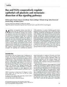
Fig. 1. Strategies to increase intramuscular fat (marbling) in beef cattle. Panel A: early stage development of intramuscular fat is characterized by adipocyte hyperplasia through adipogenesis from multipotent cells, while late stage development is characterized by hypertrophy; Panel B: Adipogenesis from mulitpotent cells within skeletal muscle is regulated by complex cell signaling pathways, and genetic, nutrition and other factors regulate adipogenesis via altering these pathways.
accumulation that produce marbling in offspring (Du et al., in 2010b). Adipogenesis is regulated by genetic, nutrition and environmental factors, all of which dictate the key signaling pathways regulating adipogenesis in skeletal muscle and, thus, marbling in resulting meat (Harper & Pethick, 2004). Japanese black cattle is well known for its extremely high marbling, which is apparently mainly due to increase in the number of intramuscular adipocytes, though an increase in adipocyte size was also detected when compared to European cattle (Gotoh et al., 2009). Because the association between the late stage adipogenesis, lipid metabolism and marbling has been the subject of several excellent previous reviews (Fernyhough, Okine, Hausman, Vierck, & Dodson, 2007; Hausman et al., 2009; Hocquette et al. 2010; Smith et al., 2009), in this review, we only discuss important cell signaling pathways regulating the initial stage of adipogenesis (Fig. 1). We first describe the key transcription factors regulating adipogenesis, followed by the discussion of important cell signaling pathways regulating these key transcription factors and the early stage of adipogenesis. 2. Adipogenesis In the bovine fetus, primary muscle fibers form within the first two months post-conception (Russell & Oteruelo, 1981), and the majority of muscle fibers form during the secondary myogenesis which occurs in the fetal stage between 2 and 7 months of gestation in cattle (Russell & Oteruelo, 1981). In fetal cattle and sheep, maternal nutrient fluctuation during late gestation reduces muscle fiber size but not number (Du et al., 2010a; Greenwood, Slepetis, Hermanson & Bell, 1999). However, this stage is crucial for intramuscular adipogenesis. Adipogenesis is initiated around mid-gestation in ruminant animals
(Feve, 2005; Gnanalingham, Mostyn, Symonds & Stephenson, 2005; Muhlhausler, Duffield, & McMillen, 2006), which partially overlaps with the period of secondary myogenesis (Du & Zhu, 2009). The mid to late gestation is a period critical for adipogenesis in cattle, and maternal nutritional management to enhance the number of MSCs committed to adipogenesis will increase the number of intramuscular adipocytes and thus marbling. Adipogenesis is regulated by several key transcription factors, including CAAT/enhancer binding proteins (C/EBPs) and peroxisome proliferator-activated receptor γ (PPARγ) (Hausman et al., 2009). C/ EBPβ and -δ are first induced by adipogenic stimuli and followed by an increase in PPARγ and C/EBPα expression. C/EBPα is induced during the initial phase of adipogenesis and directly binds to the PPARγ promoter to induce its expression (Clarke, Robinson, & Gimble, 1997; Wu et al., 1999). The expression of PPARγ further promotes the expression of C/EBPα, providing a self-reinforcing regulatory loop. Terminal differentiation of an adipocyte needs the concerted action of PPARγ and C/EBPs to turn on lipid synthesis and other adipocytespecific programs (Fernyhough et al., 2007; Hausman et al., 2009). PPARγ, but not C/EBPα alone can stimulate adipocyte differentiation, clearly showing the critical role of PPARγ in adipogenesis (Rosen et al., 2002). The basic helix–loop–helix protein, adipocyte determination and differentiation-dependent factor-1/sterol regulatory elementbinding protein-1 (ADD-1/SREBP-1), is another important protein induced during the early stages of adipogenesis (Kim & Spiegelman, 1996). Activation of PPARγ promotes terminal differentiation through the induction of a range of genes important for triglyceride uptake and storage, such as fatty acid-binding protein (aP2), acyl-CoA synthetase, fatty acid transport protein-1, lipoprotein lipase and others (Frohnert, Hui, & Bernlohr, 1999; Rosen & MacDougald, 2006).
M. Du et al. / Meat Science 86 (2010) 103–109
PPARγ has two isoforms, PPARγ1 and PPARγ2, with PPARγ2 possessing 30 extra amino acids in the N-terminal. As a nuclear receptor, PPARγ shares a common structure which includes several distinct domains: ligand independent activation domain, DNA binding domain, hinge domain, and ligand binding domain. The Ser 112 within the ligand independent activation domain is subject to phosphorylation by mitogen-activated protein kinase (MAPK) (Shao et al., 1998) which inhibits its ligand binding and reduces adipogenesis (Hu, Kim, Sarraf, & Spiegelman, 1996). PPARγ forms a heterodimer in partner with retinoid × receptor α (R × Rα) and binds to peroxisome proliferator response elements (PPREs) on the promoters of targeted genes (IJpenberg, Jeannin, Wahli, & Desvergne, 1997). Therefore, retinoid acids affect adipogenesis via R × Rα and its interaction with PPARγ (Ziouzenkova & Plutzky, 2008; Ziouzenkova et al., 2007). PPARγ is a ligand-activated transcriptional factor. In the inactive state, PPARγ is associated with co-repressors to silence its transcription activity. Binding of ligands leads to the replacement of co-repressors with co-activators possessing histone acetyl transferase activity such as cAMP response element-binding protein binding protein (CBP/p300). Acetylation of histones leads to local chromatin decondensation and gene expression. Fatty acids are ligands for PPARγ (Kliewer et al., 1995; Schopfer et al., 2005); oxidized fatty acids activate PPARγ with higher potency compared to the native fatty acids (Nagy, Tontonoz, Alvarez, Chen, & Evans, 1998). The potency of PPARγ to initiate adipogenesis also depends on the recruitment of other co-activators and/or corepressors. Peroxisome proliferator-activated receptor γ co-activator 1 (PGC-1α) is an important highly regulated co-activator, which binds to PPARγ in a ligand independent manner (Puigserver et al., 1998). The interaction between PPARγ and PGC-1α is crucial for the generation of brown adipose tissue (Nedergaard, Petrovic, Lindgren, Jacobsson, & Cannon, 2005). Brown adipogenesis receives lots of attention recently due to its role in obesity and base energy consumption. Brown fat is characterized by high mitochondrial density and the presence of uncoupled protein-1 (UCP-1) which uncouples oxidative phosphorylation leading to large amounts of heat generation. Brown fat is crucial for neonatal animals to cope with hypothermia shortly after birth. The amount of brown fat in animals peaks at birth and then brown fat converts to white fat postnatally. The brown fat differentiation is initiated by a transcription complex composed of C/EBPβ and PR domain containing 16 (PRDM16) (Kajimura et al., 2009), while the complex of PPARγ and PRDM16 is necessary for later stage brown fat differentiation and maintenance of brown fat identity (Seale et al., 2008). Muscle cells can be converted into brown fat by force expression of PRDM16 (Seale et al., 2008), enhancing
105
brown fat differentiation inside skeletal muscle is a promising method to enhance marbling. In short, PPARγ governs the adipogenic process and its expression leads to adipogenesis. The expression of PPARγ and its function in adipogenesis is regulated by several known signaling pathways, which are reviewed in the following. 3. Hedgehog signaling Hedgehog signaling pathway has fundamental roles in the formation of tissue patterns during the embryonic development. There are three different hedgehog proteins in mammals, which are Sonic, India and Desert hedgehogs. The Sonic hedgehog (SHH) is the best studied and our discussion will focus on SHH. SHH is synthesized as a precursor protein which undergoes proteolytic cleavage and lipid modifications to generate a mature morphogen. Secreted morphogen is recognized by a 12-transmembrane domain receptor, Patched (PTC). There are two patched homologues, PTCH1 and PTCH2, with PTCH1 as the primary mediator (Nieuwenhuis et al., 2006). The binding of SHH to PTCHs changes its structure to release an associated protein Smoothened (SMO). SMO then binds to glioma-associated oncogenes (GLIs) to induce gene expression. There are three GLIs, namely GLI1, GLI2 and GLI3. GLI1 and GLI2 primarily work as transcriptional-activators while GLI3 as a repressor (Park et al., 2000; Persson et al., 2002). The down-stream effectors of GLIs include myogenic regulatory factor, Myf5, to promote myogenesis (Borello et al., 2006; Gustafsson et al., 2002). GLIs enhance myogenesis through Pax3/7 activation (Feng, Adiarte, & Devoto, 2006). A growing body of evidences shows the role of SHH signaling in adipogenesis. Obesity down-regulates SHH signaling, including the expression of Smo, Gli1, Gli2 and Gli3 (Suh et al., 2006). Activation of SHH signaling inhibits adipogenesis in 3T3-L1 and C3H10T1/2 cells (Cousin, Fontaine, Dani, & Peraldi, 2007; Spinella-Jaegle et al. 2001; Suh et al. 2006; Zehentner, Leser & Burtscher, 2000). A decrease in Hedgehog signaling is necessary but not sufficient to trigger adipocyte differentiation (Fontaine, Cousin, Plaisant, Dani, & Peraldi, 2008). It appears that SHH signaling promotes stem cell differentiation to either myogenic cells or osteogenic cells, while SHH down-regulation enhances adipogenesis. Myogenic, osteogenic and adipogenic cells are all derived from the mesoderm. Mechanisms linking SHH signaling and adipogenesis remain poorly defined. SHH may elicit antiadipogenic effects by enhancing GATA binding protein 2 (GATA2) expression (Suh et al., 2006). GATA2 interacts directly with PPARγ and C/EBPα, which may deplete PPARγ involved in the promotion of adipogenesis (Hu & Davies, 2009; Schupp et al., 2009). GATA2 inhibits brown adipogenesis (Tsai et al., 2005). On
Fig. 2. Hedgehog, Wingless and Int (Wnt)/β-catenin, bone morphogenesis proteins (BMPs) and adipogenesis. C/EBPα: CAAT/enhancer binding protein α; COUP-TFII: chicken ovalbumin upstream promoter-transcription factor II; GATA2: GATA binding protein 2; Shn: Schnurri protein; PPARγ: peroxisome proliferator-activated receptor γ.
106
M. Du et al. / Meat Science 86 (2010) 103–109
the other hand, GATA proteins recruit MEF2 to promote myogenesis and other cell differentiation processes (Morin, Charron, Robitaille, & Nemer, 2000). Another mechanism for the role of SHH signaling in adipogenesis is mediated through chicken ovalbumin upstream promoter-transcription factor II (COUP-TFII, also called NR2F2). COUP-TFII binds to the promoters of PPARγ and C/EBPα to inhibit their expression (Fig. 2) (Okamura et al. 2009; Xu, Yu, Hsu, Eguchi, & Rosen, 2008). SHH induces expression of COUP-TFII (Krishnan et al., 1997), and a SHH response element has been identified in the COUP-TFII promoter (Krishnan, Elberg, Tsai, & Tsai, 1997).
4. Wnt/β-catenin signaling Wnt proteins are secreted glycoproteins crucial for the development of many cell types and tissues (Johnson & Rajamannan, 2006). The canonical Wnt pathway is β-catenin dependent and called the Wnt/βcatenin signaling pathway (Huelsken & Birchmeier, 2001). Binding of Wnt to Frizzled proteins activates Disheveled (DSH) family proteins (Johnson & Rajamannan, 2006), which then inhibits a complex of proteins that includes axin, glycogen synthesis kinase-3 β (GSK-3β), and anaphase-promoting complex (APC), leading to β-catenin accumulation (Katanaev, Ponzielli, Semeriva, & Tomlinson, 2005; Polesskaya, Seale, & Rudnicki, 2003). Without Wnt stimulation, the axin/GSK-3β/APC complex promotes the degradation of β-catenin through its phosphorylation by GSK-3β (Liu, Rubin, & Kimmel, 2005). Stabilized β-catenin enters the nucleus and interacts with members of the T cell factor/ Lymphoid enhancer factor (TCF/LEF) family of transcription factors to activate specific target genes (Dierick & Bejsovec, 1999; Hecht & Kemler, 2000). Therefore, β-catenin plays a pivotal role in canonical Wnt/βcatenin signaling (Armstrong & Esser, 2005; Mermelstein, Portilho, Mendes, Costa, & Abreu, 2007). In skeletal muscle, β-catenin regulates the expression of transcription factors Pax3 and Gli (Borycki, Brown, & Emerson, 2000; Capdevila, Tabin, & Johnson, 1998). Pax3 is essential for skeletal myogenesis and acts upstream of MyoD during skeletal muscle development, while Gli factors induce Myf-5 expression (Gustafsson et al., 2002; Ridgeway & Skerjanc, 2001). Myf5 is a direct target of Wnt/βcatenin (Borello et al., 2006). Activation of the Wnt signaling pathway enhances myogenesis and inhibits adipogenesis in cultured mesenchymal stem cells (Shang et al., 2007). Blocking the β-catenin pathway reduces myogenesis (Pan et al., 2005; Yamanouchi, Hosoyama, Murakami, & Nishihara, 2007). In our previous study in pregnant ewes which were fed 150% (over-nourished) or 100% (control) of nutrient requirements, the Wnt/β-catenin signaling was down-regulated in fetal muscle from over-nourished ewes compared to control ewes, which should at least partially be responsible for the down-regulation of myogenesis, but up-regulation of adipogenesis in the fetal muscle of over-nourished ewes (Tong et al., 2008; Zhu et al., 2008). Besides binding with TCF/LEF transcription factors to induce myogenesis, β-catenin also serves as a cofactor of forkhead transcription factors (FOXO) (Manolagas & Almeida, 2007). β-Catenin binds directly to FOXO and enhances FOXO transcription activity in mammalian cells (Essers et al., 2005). Inflammation and oxidative stress enhance the expression of FOXO, which cause a diversion of the limited pool of β-catenin from TCFmediated transcription to FOXO-mediated transcription (Du et al., 2010a). Reduced binding between TCF and β-catenin is observed after FOXO over-expression and cellular oxidative stress (Hoogeboom et al., 2008). In our over-nourished sheep study, an inflammatory response was observed in fetal skeletal muscle, which enhanced the formation of FOXO/ β-catenin complex, down-regulating myogenesis and up-regulating adipogenesis (Tong et al., 2009). Furthermore, Wnt/β-catenin signaling regulates adipogenesis through COUP-TFII. Wnt/β-catenin signaling activates the expression of COUP-TFII, which recruits the silencing mediator for retinoic acid receptor and thyroid hormone receptor (SMRT) co-repressor complex to inhibit the expression of PPARγ1 and γ2 (Fig. 2) (Okamura et al., 2009).
5. Bone morphogenetic proteins BMPs regulate diverse cellular processes such as cell differentiation, proliferation and apoptosis. BMPs belong to the transforming growth factor β (TGFβ) super-family. Two types of receptors are needed to activate BMP signaling, namely type 1 and type 2 receptors. Both receptors contain a domain with serine/threonine kinase activity. Upon binding to BMPs, the kinase activity of type 2 receptor is activated and phosphorylates type I receptor resulting the activation of intrinsic serine/threonine kinase which further phosphorylates Smad 1, 5 and/or 8, so called regulatory Smads. Phosphorylated Smads then partner with Smad 4 to form a heterodimer which in turn migrates to nuclei to initiate gene expression. There are around 15 BMPs identified in mammals. BMP-2 and 4 regulates adipogenesis and osteogenesis, while BMP-7 promotes brown adipogenesis (Tseng et al., 2008). The effect of BMPs on adipogenesis depends on the dosage. At low dosages, BMPs promote adipogenesis but at high dosages osteogenesis is enhanced instead. BMP-2, 4 and 7 all induce intracellular lipid accumulation and PPARγ expression, but only BMP-7 promote the expression of UCP-1, a marker of brown adipogenesis. In addition, the expression of PRDM16, a critical regulator of brown adipogenesis, was also enhanced by BMP-7 (Tseng et al., 2008). BMP-2 promotes the adipocytic lineage and the expression of genes associated with the fat-cell phenotype (Zehentner et al., 2000). Therefore, low levels of BMP-2 and 4 promote white adipogenesis while BMP-7 promotes brown adipogenesis. BMP-2 promotes adipogenesis through Smad1 and p38, with the activation of both needed for adipogenesis induced by BMP-2 (Hata et al., 2003). The Smad 1, 5 and 8 generally prefer GC-rich binding elements even better than the Smad binding elements (5′-GTCTAGAC-3′), a DNA sequence in certain promoters preferably bound by Smad 2 and 3 (Zawel et al., 1998). Due to the non-specific binding of Smad 1, 5 and 8 with their target sequence, it is thought that co-activators are crucial in selecting BMP targets. Schnurri (Shn) is one of such co-activators. In vertebrates, there are three Shn orthologs, namely Shn-1, -2 and -3. Shn-2 knockout mice have less white adipose tissue compared to wild-type mice (Jin et al., 2006). Shn-2 enters nuclei upon BMP-2 stimulation and then, forms a complex with Smad1/4 and C/EBPα to induce PPARγ2 expression (Fig. 2) (Jin et al., 2006). Certainly, our current understanding regarding the identification of gene targets by BMPs and its link to adipogenesis remains rudimentary, and more studies are apparently warranted. 6. AMP-activated protein kinase Different from the previously discussed signaling pathways induced by extracellular morphogens, AMP-activated protein kinase (AMPK) is an intracellular kinase involved in the regulation of adipogenesis. AMPK is ubiquitously expressed in all mammalian cells and in the lower eukaryotes as its homologues (Aschenbach et al., 2002; Hardie, 2008). AMPK is composed of a 63 kDa catalytic subunit (α), a 30 kDa β subunit and a 38–63 kDa γ subunit (Davies et al., 1994; Mitchelhill et al. 1994). All three subunits, in a ratio of 1α: 1β: 1γ, are required for a functional AMPK (Dyck et al., 1996; Woods et al., 1996). Each subunit has isoforms (α1, α2, β1, β2, γ1, γ2, and γ3) and their expression shows tissue specificity (Beri et al., 1994; Cheung, Salt, Davies, Hardie, & Carling, 2000; Hardie, Carling, & Carlson, 1998; Stapleton et al., 1997). The major role of AMPK is to regulate energy metabolism, which is allosterically activated by AMP through phosphorylation of Thr 172 on α subunit by upstream kinases (Hardie, 2008). In our previous studies in cattle, AMPK activity was positively associated with muscularity and negatively associated with the content of intramuscular adipocytes (Underwood et al., 2007; Underwood et al., 2007), indicating that AMPK switches MSCs in skeletal muscle from adipogenesis to myogenesis. Activation of AMPK by AICAR increases the expression of myogenic enhancer factor-2 (MEF2), enhancing myogenesis (Al-Khalili et al., 2004). On the other hand, activation of AMPK
M. Du et al. / Meat Science 86 (2010) 103–109
inhibits the expression of PPARγ and C/EBPs in 3T3-L1 cells and also in obese mice (Giri et al., 2006; Habinowski & Witters, 2001). Genistein inhibits adipocyte differentiation via activation of AMPK (Hwang et al., 2005). Over-nutrition in pregnant ewes inhibited AMPK activity in fetal skeletal muscle while enhancing expression of PPARγ, a marker of adipogenesis; in addition, activation of AMPK by AICAR inhibited adipogenesis in cultured 3T3-L1 cells (Dagon, Avraham, & Berry, 2006; Tong et al., 2008; Zhu et al., 2008). Above evidences clearly demonstrate that AMPK activation inhibits adipogenesis. However, the mechanisms linking AMPK to adipogenesis remains undefined, which might be associated with the role of AMPK in lipid metabolism. Inhibition of AMPK promotes lipid synthesis while reducing oxidation, which induces intracellular lipid accumulation. As ligands of PPARγ, such accumulation of lipids is expected to activate PPARγ to promote adipogenesis (Du et al., 2010b). 7. Other signaling pathways involved in adipogenesis There are other pathways implicated in the regulation of adipogenesis, of which Notch signaling is a major one. Notch signaling is a matrix dependent signaling pathway conserved throughout the animal kingdom (Bray, 2006). In an early study, Notch signaling pathway is identified to be crucial for adipogenesis in preadipocyte 3T3-L1 cells (Garces et al., 1997). However, in a later study, Notch pathway was observed to be unnecessary for adipogenesis (Nichols et al., 2004). Preadipocyte factor-1 (Pref-1, also called Dlk-1) inhibits Notch signaling and potentiates adipogenesis in C3H10T1/2 cells (Nueda, Baladron, Sanchez-Solana, Ballesteros, & Laborda, 2007), yet in another study, Pref-1 is observed to inhibit adipogenesis in 3T3-L1 cells (Wang, Kim, Kim, & Sul, 2006). These reports appear to be contradictory with each other. The possible reasons for such controversy may be due to the difference in treatments and cellular conditions, indicating that the effect of Notch signaling on adipogenesis is quite complicated. Other proteins, such as the retinoblastoma (pRb) family proteins, promote adipogenesis through direct interaction with C/EBPs (Chen, Riley, Chen, & Lee, 1996). Hormones such as insulin and IGF-1 also affect adipogenesis Tseng et al., 2005; Grohmann et al., 2005). In addition, myostatin, a member of transforming growth factor β family, is involved in the regulation of adipogenesis in bovine pre-adipocytes (Hirai et al., 2007a, b). Continued studies are likely to identify much more factors influencing the expression and function of PPARγ and adipogenesis. 8. Conclusion Available studies identified that extracellular morphogens, including hedgehog, Wnt/β-catenin, and BMP signaling pathways regulate the early stage of adipogenesis. AMPK, an intracellular kinase, also has crucial roles in this process. Future efforts to elucidate the mechanisms regulating adipogenesis should be focused on: 1) deepening our understanding of identified pathways on the initial stage of adipogenesis including co-activators and co-inhibitors involved in and their molecular interactions; 2) further efforts to identify additional pathways regulating adipogenesis; 3) examining the integration of these identified signaling pathways and their cross-talks to regulate adipogenesis; and 4) up to now, most of these knowledge about adipogenesis is obtained from rodent and cell line studies, studies using primary cultures of farm animals and in vivo studies in livestock are apparently needed, especially those studies directed at assessing the impact of genetic, physiological and nutritional factors on the early stage of adipogenesis in farm animals. Acknowledgement This work was supported by Research Initiative Grants 2008-3520618826 and 2006-55618-16914 from the USDA Cooperative State Research, Education and Extension Service.
107
References Al-Khalili, L., Chibalin, A. V., Yu, M., Sjodin, B., Nylen, C., Zierath, J. R., et al. (2004). MEF2 activation in differentiated primary human skeletal muscle cultures requires coordinated involvement of parallel pathways. American Journal of Physiology Cell Physiology, 286, C1410−C1416. Armstrong, D. D., & Esser, K. A. (2005). Wnt/beta-catenin signaling activates growthcontrol genes during overload-induced skeletal muscle hypertrophy. American Journal of Physiology Cell Physiology, 289, C853−C859. Aschenbach, W. G., Hirshman, M. F., Fujii, N., Sakamoto, K., Howlett, K. F., & Goodyear, L. J. (2002). Effect of AICAR treatment on glycogen metabolism in skeletal muscle. Diabetes, 51, 567−573. Beri, R. K., Marley, A. E., See, C. G., Sopwith, W. F., Aguan, K., Carling, D., et al. (1994). Molecular-cloning, expression and chromosomal localization of human AMPactivated. FEBS Letters, 356, 117−121. Borello, U., Berarducci, B., Murphy, P., Bajard, L., Buffa, V., Piccolo, S., et al. (2006). The Wnt/beta-catenin pathway regulates Gli-mediated Myf5 expression during somitogenesis. Development, 133, 3723−3732. Borycki, A., Brown, A. M., & Emerson, C. P., Jr. (2000). Shh and Wnt signaling pathways converge to control Gli gene activation in avian somites. Development, 127, 2075−2087. Bray, S. J. (2006). Notch signalling: A simple pathway becomes complex. Nature Reviews Molecular Cell Biology, 7, 678−689. Capdevila, J., Tabin, C., & Johnson, R. L. (1998). Control of dorsoventral somite patterning by Wnt-1 and beta-catenin. Developmental Biology, 193, 182−194. Chen, P. L., Riley, D. J., Chen, Y., & Lee, W. H. (1996). Retinoblastoma protein positively regulates terminal adipocyte differentiation through direct interaction with C/EBPs. Genes and Development, 10, 2794−2804. Cheung, P. C. F., Salt, I. P., Davies, S. P., Hardie, D. G., & Carling, D. (2000). Characterization of AMP-activated protein kinase gamma-subunit isoforms and their role in AMP binding. Biochemical Journal, 346, 659−669. Clarke, S. L., Robinson, C. E., & Gimble, J. M. (1997). CAAT/enhancer binding proteins directly modulate transcription from the peroxisome proliferator-activated receptor gamma 2 promoter. Biochemical and Biophysical Research Communications, 240, 99−103. Cousin, W., Fontaine, C., Dani, C., & Peraldi, P. (2007). Hedgehog and adipogenesis: Fat and fiction. Biochimie, 89, 1447−1453. Dagon, Y., Avraham, Y., & Berry, E. M. (2006). AMPK activation regulates apoptosis, adipogenesis, and lipolysis by eIF2alpha in adipocytes. Biochemical and Biophysical Research Communications, 340, 43−47. Davies, S. P., Hawley, S. A., Woods, A., Carling, D., Haystead, T. A., & Hardie, D. G. (1994). Purification of the AMP-activated protein kinase on ATP-gamma-sepharose and analysis of its subunit structure. European Journal of Biochemistry, 223, 351−357. Dierick, H., & Bejsovec, A. (1999). Cellular mechanisms of wingless/Wnt signal transduction. Current Topics in Developmental Biology, 43, 153−190. Du, M., & Zhu, M. J. (2009). Fetal programming of skeletal muscle development. In M. Du, & R. J. McCormick (Eds.), Applied muscle biology and meat science (pp. 81−96). Boca Raton, FL: CRC Press. Du, M., Yan, X., Tong, J. F., Zhao, J., & Zhu, M. J. (2010a). Maternal obesity, inflammation, and fetal skeletal muscle development. Biology of Reproduction, 82, 4−12. Du, M., Tong, J., Zhao, J. F., Zhao, J., Underwood, K. R., Zhu, M., Ford, S. P., & Nathanielsz, P. W. (2010b). Fetal programming of skeletal muscle development in ruminant animals. Journal of Animal Science, 88, E51−E60. Dyck, J. R., Gao, G., Widmer, J., Stapleton, D., Fernandez, C. S., Kemp, B. E., et al. (1996). Regulation of 5′-AMP-activated protein kinase activity by the noncatalytic beta and gamma subunits. Journal of Biological Chemistry, 271, 17798−17803. Essers, M. A., de Vries-Smits, L. M., Barker, N., Polderman, P. E., Burgering, B. M., & Korswagen, H. C. (2005). Functional interaction between beta-catenin and FOXO in oxidative stress signaling. Science, 308, 1181−1184. Feng, X., Adiarte, E. G., & Devoto, S. H. (2006). Hedgehog acts directly on the zebrafish dermomyotome to promote myogenic differentiation. Developmental Biology, 300, 736−746. Fernyhough, M. E., Okine, E., Hausman, G., Vierck, J. L., & Dodson, M. V. (2007). PPARγ and GLUT-4 expression as developmental regulators/markers for preadipocyte differentiation into an adipocyte. Domestic Animal Endocrinology, 33, 367−378. Feve, B. (2005). Adipogenesis: Cellular and molecular aspects. Best Practice and Research Clinical Endocrinology and Metabolism, 19, 483−499. Fontaine, C., Cousin, W., Plaisant, M., Dani, C., & Peraldi, P. (2008). Hedgehog signaling alters adipocyte maturation of human mesenchymal stem cells. Stem Cells, 26, 1037−1046. Frohnert, B. I., Hui, T. Y., & Bernlohr, D. A. (1999). Identification of a functional peroxisome proliferator-responsive element in the murine fatty acid transport protein gene. Journal of Biological Chemistry, 274, 3970−3977. Garces, C., Ruiz-Hidalgo, M. J., Font de Mora, J., Park, C., Miele, L., Goldstein, J., et al. (1997). Notch-1 controls the expression of fatty acid-activated transcription factors and is required for adipogenesis. Journal of Biological Chemistry, 272, 29729−29734. Giri, S., Rattan, R., Haq, E., Khan, M., Yasmin, R., Won, J. S., et al. (2006). AICAR inhibits adipocyte differentiation in 3T3L1 and restores metabolic alterations in dietinduced obesity mice model. Nutrition & Metabolism, 3, 31−51. Gnanalingham, M. G., Mostyn, A., Symonds, M. E., & Stephenson, T. (2005). Ontogeny and nutritional programming of adiposity in sheep: Potential role of glucocorticoid action and uncoupling protein-2. American Journal of Physiology: Regulatory, Integrative and Comparative Physiology, 289, R1407−R1415. Gotoh, T., Albrecht, E., Teuscher, F., Kawabata, K., Sakashita, K., Iwamoto, H., et al. (2009). Differences in muscle and fat accretion in Japanese Black and European cattle. Meat Science, 82, 300−308. Greenwood, P. L., Slepetis, R. M., Hermanson, J. W., & Bell, A. W. (1999). Intrauterine growth retardation is associated with reduced cell cycle activity, but not myofibre number, in ovine fetal muscle. Reproduction, Fertility, and Development, 11, 281−291.
108
M. Du et al. / Meat Science 86 (2010) 103–109
Grohmann, M., Sabin, M., Holly, J., Shield, J., Crowne, E., & Stewart, C. (2005). Characterization of differentiated subcutaneous and visceral adipose tissue from children: The influences of TNF-alpha and IGF-I. Journal of Lipid Research, 46, 93−103. Gustafsson, M. K., Pan, H., Pinney, D. F., Liu, Y., Lewandowski, A., Epstein, D. J., et al. (2002). Myf5 is a direct target of long-range Shh signaling and Gli regulation for muscle specification. Genes & Development, 16, 114−126. Habinowski, S. A., & Witters, L. A. (2001). The effects of AICAR on adipocyte differentiation of 3T3-L1 cells. Biochemical and Biophysical Research Communications, 286, 852−856. Hardie, D. G. (2008). AMPK: A key regulator of energy balance in the single cell and the whole organism. International Journal of Obesity, 32, S7−S12. Hardie, D. G., Carling, D., & Carlson, M. (1998). The AMP-activated/SNF1 protein kinase subfamily: Metabolic sensors of the eukaryotic cell? Annual Review of Biochemistry, 67, 821−855. Harper, G. S., & Pethick, D. W. (2004). How might marbling begin? Australian Journal of Experimental Agriculture, 44, 653−662. Hata, K., Nishimura, R., Ikeda, F., Yamashita, K., Matsubara, T., Nokubi, T., et al. (2003). Differential roles of Smad1 and p38 kinase in regulation of peroxisome proliferatoractivating receptor gamma during bone morphogenetic protein 2-induced adipogenesis. Molecular Biology of the Cell, 14, 545−555. Hausman, G. J., Dodson, M. V., Ajuwon, K., Azain, M., Barnes, K. M., Guan, L. L., et al. (2009). Board-invited review: The biology and regulation of preadipocytes and adipocytes in meat animals. Journal of Animal Science, 87, 1218−1246. Hecht, A., & Kemler, R. (2000). Curbing the nuclear activities of beta-catenin. Control over Wnt target gene expression. EMBO Reports, 1, 24−28. Hirai, S., Matsumoto, H., Hino, N., Kawachi, H., Matsui, T., & Yano, H. (2007a). Myostatin inhibits differentiation of bovine preadipocyte. Domestic Animal Endocrinology, 32, 1−14. Hirai, S., Matsumoto, H., Hino, N., Kawachi, H., Matsui, T., & Yano, H. (2007b). Follistatin rescues the inhibitory effect of activin A on the differentiation of bovine preadipocytes. Domestic Animal Endocrinology, 33, 269−280. Hocquette, J. F., Gondret, F., Baeza, E., Medale, F., Jurie, C., & Pethick, D. W. (2010). Intramuscular fat content in meat-producing animals: Development, genetic and nutritional control, and identification of putative markers. Animal, 4, 303−319. Hoogeboom, D., Essers, M. A., Polderman, P. E., Voets, E., Smits, L. M., & Burgering, B. M. (2008). Interaction of FOXO with beta-catenin inhibits beta-catenin/T cell factor activity. Journal of Biological Chemistry, 283, 9224−9230. Hu, Y., & Davies, G. E. (2009). Berberine increases expression of GATA-2 and GATA-3 during inhibition of adipocyte differentiation. Phytomedicine, 16, 864−873. Hu, E., Kim, J. B., Sarraf, P., & Spiegelman, B. M. (1996). Inhibition of adipogenesis through MAP kinase-mediated phosphorylation of PPARgamma. Science, 274, 2100−2103. Huelsken, J., & Birchmeier, W. (2001). New aspects of Wnt signaling pathways in higher vertebrates. Current Opinion in Genetics & Development, 11, 547−553. Hwang, J. T., Park, I. J., Shin, J. I., Lee, Y. K., Lee, S. K., Baik, H. W., et al. (2005). Genistein, EGCG, and capsaicin inhibit adipocyte differentiation process via activating AMPactivated protein kinase. Biochemical and Biophysical Research Communications, 338, 694−699. IJpenberg, A., Jeannin, E., Wahli, W., & Desvergne, B. (1997). Polarity and specific sequence requirements of peroxisome proliferator-activated receptor (PPAR)/retinoid× receptor heterodimer binding to DNA. A functional analysis of the malic enzyme gene PPAR response element. Journal of Biological Chemistry, 272, 20108−20117. Jin, W., Takagi, T., Kanesashi, S. N., Kurahashi, T., Nomura, T., Harada, J., et al. (2006). Schnurri-2 controls BMP-dependent adipogenesis via interaction with Smad proteins. Developmental Cell, 10, 461−471. Johnson, M. L., & Rajamannan, N. (2006). Diseases of Wnt signaling. Reviews in Endocrine & Metabolic Disorders, 7, 41−49. Kajimura, S., Seale, P., Kubota, K., Lunsford, E., Frangioni, J. V., Gygi, S. P., et al. (2009). Initiation of myoblast to brown fat switch by a PRDM16-C/EBP-beta transcriptional complex. Nature, 460, 1154−1158. Katanaev, V. L., Ponzielli, R., Semeriva, M., & Tomlinson, A. (2005). Trimeric G proteindependent frizzled signaling in Drosophila. Cell, 120, 111−122. Kim, J. B., & Spiegelman, B. M. (1996). ADD1/SREBP1 promotes adipocyte differentiation and gene expression linked to fatty acid metabolism. Genes & Development, 10, 1096−1107. Kliewer, S. A., Lenhard, J. M., Willson, T. M., Patel, I., Morris, D. C., & Lehmann, J. M. (1995). A prostaglandin J2 metabolite binds peroxisome proliferator-activated receptor gamma and promotes adipocyte differentiation. Cell, 83, 813−819. Krishnan, V., Elberg, G., Tsai, M. J., & Tsai, S. Y. (1997). Identification of a novel sonic hedgehog response element in the chicken ovalbumin upstream promotertranscription factor II promoter. Molecular Endocrinology, 11, 1458−1466. Krishnan, V., Pereira, F. A., Qiu, Y., Chen, C. H., Beachy, P. A., Tsai, S. Y., et al. (1997). Mediation of Sonic hedgehog-induced expression of COUP-TFII by a protein phosphatase. Science, 278, 1947−1950. Liu, X., Rubin, J. S., & Kimmel, A. R. (2005). Rapid, Wnt-induced changes in GSK3beta associations that regulate beta-catenin stabilization are mediated by Galpha proteins. Current Biology, 15, 1989−1997. Manolagas, S. C., & Almeida, M. (2007). Gone with the Wnts: Beta-catenin, T-cell factor, forkhead box O, and oxidative stress in age-dependent diseases of bone, lipid, and glucose metabolism. Molecular Endocrinology, 21, 2605−2614. Mermelstein, C. S., Portilho, D. M., Mendes, F. A., Costa, M. L., & Abreu, J. G. (2007). Wnt/ beta-catenin pathway activation and myogenic differentiation are induced by cholesterol depletion. Differentiation; Research in Biological Diversity, 75, 184−192. Mitchelhill, K. I., Stapleton, D., Gao, G., House, C., Michell, B., Katsis, F., et al. (1994). Mammalian AMP-activated protein kinase shares structural and functional homology with the catalytic domain of yeast Snf1 protein kinase. Journal of Biological Chemistry, 269, 2361−2364. Morin, S., Charron, F., Robitaille, L., & Nemer, M. (2000). GATA-dependent recruitment of MEF2 proteins to target promoters. EMBO Journal, 19, 2046−2055.
Muhlhausler, B. S., Duffield, J. A., & McMillen, I. C. (2006). Increased maternal nutrition stimulates Peroxisome Proliferator Activated Receptor-gamma (PPARgamma), adiponectin and leptin mRNA expression in adipose tissue before birth. Endocrinology, 148, 878−885. Nagy, L., Tontonoz, P., Alvarez, J. G., Chen, H., & Evans, R. M. (1998). Oxidized LDL regulates macrophage gene expression through ligand activation of PPARgamma. Cell, 93, 229−240. Nedergaard, J., Petrovic, N., Lindgren, E. M., Jacobsson, A., & Cannon, B. (2005). PPARgamma in the control of brown adipocyte differentiation. Biochimica et Biophysica Acta, 1740, 293−304. Nichols, A. M., Pan, Y., Herreman, A., Hadland, B. K., De Strooper, B., Kopan, R., et al. (2004). Notch pathway is dispensable for adipocyte specification. Genesis, 40, 40−44. Nieuwenhuis, E., Motoyama, J., Barnfield, P. C., Yoshikawa, Y., Zhang, X., Mo, R., et al. (2006). Mice with a targeted mutation of patched2 are viable but develop alopecia and epidermal hyperplasia. Molecular and Cellular Biology, 26, 6609−6622. Nueda, M. L., Baladron, V., Sanchez-Solana, B., Ballesteros, M. A., & Laborda, J. (2007). The EGF-like protein dlk1 inhibits notch signaling and potentiates adipogenesis of mesenchymal cells. Journal of Molecular Biology, 367, 1281−1293. Okamura, M., Kudo, H., Wakabayashi, K., Tanaka, T., Nonaka, A., Uchida, A., et al. (2009). COUP-TFII acts downstream of Wnt/beta-catenin signal to silence PPARgamma gene expression and repress adipogenesis. Proceedings of the National Academy of Science of the United States of America, 106, 5819−5824. Pan, W., Jia, Y., Wang, J., Tao, D., Gan, X., Tsiokas, L., et al. (2005). Beta-catenin regulates myogenesis by relieving I-mfa-mediated suppression of myogenic regulatory factors in P19 cells. Proceedings of the National Academy of Sciences of the United States of America, 102, 17378−17383. Park, H. L., Bai, C., Platt, K. A., Matise, M. P., Beeghly, A., Hui, C. C., et al. (2000). Mouse Gli1 mutants are viable but have defects in SHH signaling in combination with a Gli2 mutation. Development, 127, 1593−1605. Persson, M., Stamataki, D., te Welscher, P., Andersson, E., Bose, J., Ruther, U., et al. (2002). Dorsal-ventral patterning of the spinal cord requires Gli3 transcriptional repressor activity. Genes & Development, 16, 2865−2878. Polesskaya, A., Seale, P., & Rudnicki, M. A. (2003). Wnt signaling induces the myogenic specification of resident CD45+ adult stem cells during muscle regeneration. Cell, 113, 841−852. Puigserver, P., Wu, Z., Park, C. W., Graves, R., Wright, M., & Spiegelman, B. M. (1998). A cold-inducible coactivator of nuclear receptors linked to adaptive thermogenesis. Cell, 92, 829−839. Ridgeway, A. G., & Skerjanc, I. S. (2001). Pax3 is essential for skeletal myogenesis and the expression of Six1 and Eya2. Journal of Biological Chemistry, 276, 19033−19039. Rosen, E. D., & MacDougald, O. A. (2006). Adipocyte differentiation from the inside out. Nature Reviews Molecular Cell Biology, 7, 885−896. Rosen, E. D., Hsu, C. H., Wang, X., Sakai, S., Freeman, M. W., Gonzalez, F. J., et al. (2002). C/EBPalpha induces adipogenesis through PPARgamma: A unified pathway. Genes & Development, 16, 22−26. Russell, R. G., & Oteruelo, F. T. (1981). An ultrastructural study of the differentiation of skeletal muscle in the bovine fetus. Anatomy and Embryology, 162, 403−417. Schopfer, F. J., Lin, Y., Baker, P. R., Cui, T., Garcia-Barrio, M., Zhang, J., et al. (2005). Nitrolinoleic acid: An endogenous peroxisome proliferator-activated receptor gamma ligand. Proceedings of the National Academy of Sciences of the United States of America, 102, 2340−2345. Schupp, M., Cristancho, A. G., Lefterova, M. I., Hanniman, E. A., Briggs, E. R., Steger, D. J., et al. (2009). Re-expression of GATA2 cooperates with peroxisome proliferatoractivated receptor-gamma depletion to revert the adipocyte phenotype. Journal of Biological Chemistry, 284, 9458−9464. Seale, P., Bjork, B., Yang, W., Kajimura, S., Chin, S., Kuang, S., et al. (2008). PRDM16 controls a brown fat/skeletal muscle switch. Nature, 454, 961−967. Shang, Y. C., Zhang, C., Wang, S. H., Xiong, F., Zhao, C. P., Peng, F. N., et al. (2007). Activated beta-catenin induces myogenesis and inhibits adipogenesis in BMderived mesenchymal stromal cells. Cytotherapy, 9, 667−681. Shao, D., Rangwala, S. M., Bailey, S. T., Krakow, S. L., Reginato, M. J., & Lazar, M. A. (1998). Interdomain communication regulating ligand binding by PPAR-gamma. Nature, 396, 377−380. Smith, S. B., Kawachi, H., Choi, C. B., Choi, C. W., Wu, G., & Sawyer, J. E. (2009). Cellular regulation of bovine intramuscular adipose tissue development and composition. Journal of Animal Science, 87, E72−E82. Spinella-Jaegle, S., Rawadi, G., Kawai, S., Gallea, S., Faucheu, C., Mollat, P., et al. (2001). Sonic hedgehog increases the commitment of pluripotent mesenchymal cells into the osteoblastic lineage and abolishes adipocytic differentiation. Journal of Cell Science, 114, 2085−2094. Stapleton, D., Woollatt, E., Mitchelhill, K. I., Nicholl, J. K., Fernandez, C. S., Michell, B. J., et al. (1997). AMP-activated protein kinase isoenzyme family: Subunit structure and chromosomal location. FEBS Letters, 409, 452−456. Suh, J. M., Gao, X., McKay, J., McKay, R., Salo, Z., & Graff, J. M. (2006). Hedgehog signaling plays a conserved role in inhibiting fat formation. Cell Metabolism, 3, 25−34. Tong, J., Zhu, M. J., Underwood, K. R., Hess, B. W., Ford, S. P., & Du, M. (2008). AMPactivated protein kinase and adipogenesis in sheep fetal skeletal muscle and 3T3-L1 cells. Journal of Animal Science, 86, 1296−1305. Tong, J. F., Yan, X., Zhu, M. J., Ford, S. P., Nathanielsz, P. W., & Du, M. (2009). Maternal obesity downregulates myogenesis and {beta}-catenin signaling in fetal skeletal muscle. American Journal of Physiology, Endocrinology and Metabolism, 296, E917−E924. Tsai, J., Tong, Q., Tan, G., Chang, A. N., Orkin, S. H., & Hotamisligil, G. S. (2005). The transcription factor GATA2 regulates differentiation of brown adipocytes. EMBO Reports, 6, 879−884.
M. Du et al. / Meat Science 86 (2010) 103–109 Tseng, Y. H., Butte, A. J., Kokkotou, E., Yechoor, V. K., Taniguchi, C. M., Kriauciunas, K. M., et al. (2005). Prediction of preadipocyte differentiation by gene expression reveals role of insulin receptor substrates and necdin. Nature Cell Biology, 7, 601−611. Tseng, Y. H., Kokkotou, E., Schulz, T. J., Huang, T. L., Winnay, J. N., Taniguchi, C. M., et al. (2008). New role of bone morphogenetic protein 7 in brown adipogenesis and energy expenditure. Nature, 454, 1000−1004. Underwood, K. R., Means, W. J., Zhu, M. J., Ford, S. P., Hess, B. W., & Du, M. (2007). AMPactivated protein kinase is negatively associated with intramuscular fat content in longissimus dorsi muscle of beef cattle. Meat Science, 79, 394−402. Underwood, K. R., Tong, J., Zhu, M. J., Shen, Q. W., Means, W. J., Ford, S. P., et al. (2007). Relationship between kinase phosphorylation, muscle fiber typing, and glycogen accumulation in longissimus muscle of beef cattle with high and low intramuscular fat. Journal of Agricultural and Food Chemistry, 55, 9698−9703. Wang, Y., Kim, K. A., Kim, J. H., & Sul, H. S. (2006). Pref-1, a preadipocyte secreted factor that inhibits adipogenesis. Journal of Nutrition, 136, 2953−2956. Woods, A., Cheung, P. C., Smith, F. C., Davison, M. D., Scott, J., Beri, R. K., et al. (1996). Characterization of AMP-activated protein kinase beta and gamma subunits. Assembly of the heterotrimeric complex in vitro. Journal of Biological Chemistry, 271, 10282−10290. Wu, Z., Rosen, E. D., Brun, R., Hauser, S., Adelmant, G., Troy, A. E., et al. (1999). Crossregulation of C/EBP alpha and PPAR gamma controls the transcriptional pathway of adipogenesis and insulin sensitivity. Molecular Cell, 3, 151−158. Xu, Z., Yu, S., Hsu, C. H., Eguchi, J., & Rosen, E. D. (2008). The orphan nuclear receptor chicken ovalbumin upstream promoter-transcription factor II is a critical regulator
109
of adipogenesis. Proceedings of the National Academy of Science of the United States of America, 105, 2421−2426. Yamanouchi, K., Hosoyama, T., Murakami, Y., & Nishihara, M. (2007). Myogenic and adipogenic properties of goat skeletal muscle stem cells. Journal of Reproduction and Development, 53, 51−58. Zawel, L., Dai, J. L., Buckhaults, P., Zhou, S., Kinzler, K. W., Vogelstein, B., et al. (1998). Human Smad3 and Smad4 are sequence-specific transcription activators. Molecular Cell, 1, 611−617. Zehentner, B. K., Leser, U., & Burtscher, H. (2000). BMP-2 and sonic hedgehog have contrary effects on adipocyte-like differentiation of C3H10T1/2 cells. DNA and Cell Biology, 19, 275−281. Zhu, M. J., Han, B., Tong, J., Ma, C., Kimzey, J. M., Underwood, K. R., et al. (2008). AMPactivated protein kinase signalling pathways are down regulated and skeletal muscle development impaired in fetuses of obese, over-nourished sheep. Journal of Physiology, 586, 2651−2664. Ziouzenkova, O., & Plutzky, J. (2008). Retinoid metabolism and nuclear receptor responses: New insights into coordinated regulation of the PPAR–RXR complex. FEBS Letters, 582, 32−38. Ziouzenkova, O., Orasanu, G., Sukhova, G., Lau, E., Berger, J. P., Tang, G., et al. (2007). Asymmetric cleavage of beta-carotene yields a transcriptional repressor of retinoid × receptor and peroxisome proliferator-activated receptor responses. Molecular Endocrinology, 21, 77−88.