Romisch et al., 1989; equivalent to ffh protein in Bernstein et al., 1989), known to bind to E.coli 4.5S RNA (Poritz et al., 1990; Ribes et al., 1990), might destroy ...
The EMBO Journal vol.10 no.4 pp.767-777, 1991
Changes in 7SL RNA conformation during the signal recognition particle cycle
Massimiliano Andreazzoli and Susan A.Gerbil Brown University Division of Biology and Medicine, Providence, RI 02912, USA
1Corresponding author Communicated by G.Blobel
The structure of 7SL RNA has been probed by chemical modification followed by primer extension, using four substrates: (i) naked 7SL RNA; (ii) free signal recognition particle (SRP); (iii) polysome bound SRP; and (iv) membrane bound SRP. Decreasing sensitivity to chemical modification between these different substrates suggests regions on 7SL RNA that: bind proteins associated with SRP might interact with ribosomes; and are protected by binding to membranes. Other areas increase in chemical sensitivity, exemplified by a tertiary interaction present in naked 7SL RNA but not in free SRP. Such changes suggest that 7SL RNA changes its conformation during the SRP cycle. These conformational changes could be a necessary component to move through the SRP cycle from one stage to the next. Key words: 7SL RNA conformation/SRP
Introduction Protein synthesis and protein transport are important within cells. A key player in these events in eukaryotes is signal recognition particle (SRP), which functions as a cellular adaptor between the protein synthetic machinery and the protein translocation apparatus of the rough endoplasmic reticulum (ER) (for reviews see Walter et al., 1984; Walter and Lingappa, 1986; Hortsch and Meyer, 1986; Dobberstein, 1987; Walter, 1987). SRP is composed of 7SL RNA that is- 300 nt long (Walter and Blobel, 1982) and six polypeptides (Walter and Blobel, 1980). These polypeptides comprise two monomers (pl9, p54) and two heterodimers (p9/14 and p68/72). What are the functions of 7SL RNA within the SRP? One role is to act as a scaffold to which the SRP proteins bind. Each of the proteins interacts individually with 7SL RNA except for p54, whose binding is mediated by p19 (Walter and Blobel, 1983a). Homologous or heterologous SRP can be reconstituted (Walter and Blobel, 1983a). The regions of 7SL RNA bound by proteins were first deduced by micrococcal nuclease cleavage that subdivides SRP into two domains (Gundelfinger et al., 1983). The Alu domain (so named because it seems to have given rise during evolution to the Alu interspersed repetitive sequences found in the human genome; Ullu and Tschudi, 1984) contains 100 nt of the 5' end of 7SL RNA base-paired with 50 nt of the 3' end of 7SL RNA; the S domain contains 150 nt of core sequence lacking Alu repeat similarity (Ullu et al., 1982). p9/14 is bound to the Alu domain (Gundelfinger et al.,
processes
Oxford University Press
1983), which seems necessary for elongation slow-down or arrest of protein synthesis (Siegel and Walter, 1985, 1986). The remaining four polypeptides are found in the S domain of SRP (Gundelfinger et al., 1983), and the exact binding sites of p19 and p68/72 have been determined by a-sarcin footprinting (Siegel and Walter, 1988a). p68/72 is needed for protein translocation (Siegel and Walter, 1988b), and p54 is important for signal recognition (Krieg et al., 1986; Kurzchalia et al., 1986; Wiedmann et al., 1987; Siegel and Walter, 1988b). In this paper we propose that in addition to a scaffold role for binding SRP proteins, 7SL RNA undergoes conformational changes which might be a necessary component to move through the SRP cycle. SRP cycles between several different states in the cell. In a postmitochondrial supematant under physiological salt conditions (150 mM KOAc), 15% SRP is found as a free soluble form and the remaining SRP is divided about equally between ribosomes and microsomal membranes (Walter and Blobel, 1983b). Soluble SRP loosely associates with monosomes; polysomes synthesizing secretory proteins cause increased binding affinity of SRP by three to four orders of magnitude (Walter et al., 1981). In the first step of the SRP cycle, when canine SRP binds to polysomes translating bovine pituitary preprolactin in a wheat germ cell-free system, translation is arrested; the arrested polypeptide is 70 amino acids long of which one half is protected within the ribosome and the other half that has emerged contains the N-terminal signal sequence (Walter and Blobel, 1981). Although synthesis of other secretory proteins is also inhibited by canine SRP in wheat germ cell-free systems (Walter and Blobel, 1981; Mueller et al., 1982; Ibrahimi, 1987; Lipp et al., 1987), the generality of translational arrest has been questioned since it was not seen by canine SRP in rabbit reticulocyte or HeLa cell extracts (Meyer, 1985) nor by maize or wheat germ SRP in wheat germ extracts (Prehn et al., 1987; Campos et al., 1988). An explanation for some of these discrepancies is that results lacking translational arrest were often confined to steady state; in fact, a slow-down in kinetics of preprolactin synthesis by canine SRP in reticulocyte lysates has recently been observed (Wolin and Walter, 1989). SRP causes translational pausing at multiple sites in the nascent polypeptide in both reticulocyte lysates (Wolin and Walter, 1989) and wheat germ cell-free systems (Lipp et al., 1987). All of the SRP arrested nascent polypeptide resides in the P site of the ribosome, and it can be released by entry of puromycin into the vacant A site (Gilmore and Blobel, 1985). Though translational arrest is not essential for secretory protein transport to the ER, it seems to increase fidelity of the process (Siegel and Walter, 1985; Walter, 1987) as well as keep the nascent polypeptide unfolded for easier entry into the ER (Rapoport et al., 1987; Sanz and Meyer, 1988). Translational arrest may also regulate the synthesis of certain secretory proteins such as insulin (Welsh et al., 1986). In the second step of the SRP cycle, elongation arrest is
767
M.Andreazzoli and S.A.Gerbi
relieved when SRP interacts with the SRP receptor (Gilmore et al., 1982a,b), also called docking protein (Meyer et al., 1982), which is an integral protein of the rough ER. The SRP receptor can bind the signal sequence, but has no affinity for the ribosome (Gilmore and Blobel, 1983). However, arrested nascent polypeptide interacts with the rough ER only in the context of the ribosome (Gilmore and Blobel, 1985). The ribosome remains associated with the rough ER for cotranslational translocation of the nascent polypeptide into the lumen of the rough ER (Gilmore and Blobel, 1983). Finally, once SRP has handed over the arrested ribosome complex to the SRP receptor, the SRP can recycle. To address the role of 7SL RNA in the SRP cycle, we have studied the conformation of 7SL RNA during different parts of this cycle. The sequence of 7SL RNA is known for several organisms, and phylogenetic comparisons have revealed compensatory base changes in support of a proposed secondary structure model (Zwieb, 1985, 1989; Larsen and Zwieb, 1991). There have been only limited experimental data to support the theoretical secondary structure model, utilizing nuclease digestion (Gundelfinger et al., 1983, 1984; Haas et al., 1988) or a-sarcin cleavage (Siegel and Walter, 1988a). Electron spectroscopic imaging shows 7SL RNA to be an elongated rod that spans the 6 nm wide and 24 nm long SRP (Andrews et al., 1985, 1987); cross-linking suggests that stem IV of 7SL RNA folds back upon stem II to fit this three-dimensional structure (Zwieb and Schiiler, 1989). We report here experimental evidence, based on chemical modification, for the structure of 7SL RNA from different substrate forms. Our data on naked 7SL RNA indicate that some of the theoretically proposed stems have opened, and reveal a hitherto undiscovered long-range tertiary interaction. Compensatory base changes support this tertiary interaction, which may play a role in SRP biogenesis. Previously, gel electrophoresis of wild-type and mutant naked 7SL RNA showed it could exist in various conformations (Zwieb and Ullu, 1986). In order to examine whether conformational changes occur during the SRP cycle, we performed chemical modifications on 7SL RNA derived from soluble SRP, polysome bound SRP and membrane bound SRP. Our data showing 7SL RNA conformational changes in these different states suggest that 7SL RNA could play a role in progression through the SRP cycle. Moreover, sites within 7SL RNA that are protected by SRP proteins, polysomes or rough endoplasmic reticulum membranes are indicated by comparison between the different states.
Results We have assayed the chemical accessibility of 7SL RNA in different forms of the SRP cycle. For this purpose, SRP was isolated from dog pancreas in three forms: soluble, membrane bound and ribosome bound (Walter and Blobel, 1983b). In addition, naked 7SL RNA was obtained by deproteinization of soluble SRP. Each of these forms was then reacted with chemicals that modify single stranded nucleotides. The specificity of the chemicals used is as follows: DMS methylates adenine at NI and more slowly cytosine at N3; CMCT modifies uracil at N3 and more slowly guanine at Nl, and kethoxal modifies guanine at Nl and N2. After modification the RNA was purified, denatured and primer extended with reverse transcriptase; chain
768
elongation by this enzyme pauses or stops one nucleotide before the modified base (Hagenbiichle et al., 1978; Youvan and Hearst, 1979). The modified base can be unambiguously identified by comparison to sequencing reactions in adjacent lanes. A control ('O') of untreated RNA was electrophoresed in parallel with treated samples to identify stops in reverse transcription due to inherent secondary structure, nicks, or post-transcriptional modification of the RNA. A second control ('stop') to monitor chemical modification caused by residual chemical in the subsequent RNA purification steps consisted of samples given chemical after instead of before the stop buffer. Examples of modification by the three chemicals are shown in Figures 1 and 3. Bands that are unique to the chemically modified sample lane or distinctly stronger than background bands in the control lane are scored. Sample lanes of different substrates can only be compared to one another if the amounts of sample loaded are the same. Table I summarizes all the modification data for the different substrates. Chemical modification of naked 7SL RNA As expected, naked 7SL RNA shows the highest degree of chemical modification relative to the other substrates analyzed (Figure 1). As shown in Figure 2, 27% of the naked 7SL RNA nucleotides are modified, mostly coinciding with positions predicted to be single stranded according to the most recent, phylogenetically derived secondary structure model (Larsen and Zwieb, 1991). However, in a few cases areas predicted to be double stranded in the model are chemically modified (brackets in Figure 2). These regions usually fall at the end of helical stems or next to bulges, both of which are areas that favor 'breathing'. In other cases, some nucleotides predicted to be single stranded were not modified by the chemicals. These bases may be involved in long-range interactions; we will focus on one such interaction later. Two bases are hypersensitive to chemical modifications: A75 and G150. A75 is hypersensitive in all SRP substrates examined, supporting the idea proposed previously based on the accessibility of this base to micrococcal nuclease and a-sarcin that A75 is extremely exposed in the intact SRP (Gundelfinger et al., 1983; Siegel and Walter, 1988a). In contrast, G150 is only moderately accessible once proteins are bound in solulble SRP, probably because it is protected by p19 as will be discussed below.
Comparison between naked 7SL RNA and soluble SRP Bases that are exposed in naked 7SL RNA but protected in the soluble SRP (Figure 1A,B,D, and filled circles in Figure 4A) are candidates for protein binding sites. An alternative possibility is that conformational changes in 7SL RNA were induced by association with proteins thereby causing increased protection at certain sites in 7SL RNA, even if these sites do not bind the proteins directly themselves. p19 and p68/72 are the only proteins whose footprints on the 7SL RNA have been published so far (Siegel and Walter, 1988a). p19 protects nucleotides 147-153 and 191 -200; p68/72 protects scattered nucleotides from 101 -106, 129-138, 162-167, 172-183, 202-209, 233-239 and 251 -253 (Siegel and Walter, 1988a). These footprints were obtained using ce-sarcin which cuts both base-paired and single stranded nucleotides; the latter which become protected by p19 and p68/72 generally coincide with residues we find protected from chemical modification in soluble SRP
7SL RNA conformation
Table I.
Table I. continued
Nucleotide no. Chemical 7SL G13 G14 G16 U25 A26 G27 G33 U35 A36 U38 C39 G40 G41 G44 G45 A49 A53 A56 A59 A67 G73 A75 G76 U77 U78 C84 C92 U97 G98 C99 C1OO GIOI A102 U103 G107 Ul 10 GI 13 A115 A118 Al 19 G120 U121 U122 C123 G125 A127 U128 A130 A131 U132 A133 U134 G138 A139 G146 G147 G148 A149 G150 A157
A163 G164 C169 U171 A172
K K K CMCT DMS K K CMCT DMS CMCT DMS K K K K DMS DMS DMS DMS DMS K DMS K CMCT CMCT DMS DMS CMCT K DMS DMS K DMS CMCT K CMCT K DMS DMS DMS K CMCT CMCT DMS K DMS CMCT DMS DMS
CMCT DMS CMCT K DMS K K K DMS K DMS DMS K DMS CMCT DMS
Sol. SRP Pol. SRP Mem. SRP
-
+ +
++
+ (+)
+ -
+
-
(+)
+
+
-
+
+
++
+
(+) (+) + ++
(+)
(+) (+) (+) (+)
-
(+)
+
-
(+)
+
-
(+)
+
+
+
(+)
(+)
(+)
+
-
(+)
(+)
-
+
+++ +
+ ++ + (+) ++ -
+++ + + + (+) + (+)
++ ++ +++ ++ ++
++ ++
+
++
-
(+)
++
-
+
++
-
(+) -
(+) ++ ++
+
+ +
+
-
(+) (+) (+) (+)
(+) -
++ ++ ++ +
++ +
+++
(+) ++
(+) + +
(+) ++ ++
++
-
-
+
+
+
(+)
(+) (+) (+) +
+
-
(+)
++
(+) -
-
(+)
+
(+)
(+)
-
+
-
++ (+) (+) +++ (+) (+) (+) (+)
G174 G175 A176 G178 A183 C185 C186 G187 A192 U195
-
(+)
Chemical 7SL K K DMS K DMS DMS DMS K DMS CMCT DMS DMS DMS DMS K K DMS DMS CMCT DMS DMS DMS DMS CMCT K DMS CMCT DMS K
A200 A201 C202 G203 G204 A205 A208 U211 A213 A214 A215 A216 U226 G227 A228 U229 A231 G232 U233 A234 G235 U236 G239 A240 U241 C242 C244
CMCT DMS K CMCT K DMS CMCT DMS DMS
List of bases which
CMCT, K
(+ ) (+)
no.
A199
-
+
Nucleotide
are
Sol. SRP Pol. SRP Mem. SRP (+) (+)
(+)
(+)
(+)
(+)
(+)
(+) (+)
++ +
++ ++ ++
++
(+)
(+)
(+) + +
(+) (+)
(+)
(+) ++
+ ++
(+) (+) (+)
(++
++
(+) ++
(+) (+) ++
++
(+)
(+) (+) (+) (+) ++
+
(+) (+)
(+)
(+) ++ ++
(+) (++ (+)
+
++
(+)
(+)
(+) +
accessible to the chemical indicated
(DMS,
Kethoxal) in 7SL RNA from naked 7SL RNA (7SL), soluble SRP (Sol. SRP), polysome bound SRP (Pol. SRP) and membrane bound SRP (Mem. SRP). The level of sensitivity to modification was scored as: none -; weak (+); moderate +; strong + +; hypersensitive + + +. In the few cases where a base was modified by more than one chemical, only the results from the chemical with the greatest specificity are listed. Bases upon which reverse transcriptase pauses or stops to give background bands in control lanes of more than one chemical reaction, and in all four substrates are C46, U47, C84, U90, U108, C126, A130, U171, U181 (nine bases = 3% 7SL RNA residues). Additional bases which had endogenous stops in controls of just one chemical reaction for : 2 substrates were: C22, C71, A75, A118, A133, U142, G155, A163, A231 (nine bases = 3% 7SL RNA residues). =
compared to naked 7SL RNA (Figure 4A; Table I). In addition, we see increased protection by proteins for 118-123 and 227-229 on the opposing side of the stem which was not previously found in the a-sarcin footprints; as
(+) -
(+)
(+)
+
+
-
+
+
(+)
-
(+)
(+)
the smaller chemicals we used here may penetrate more readily to areas not accessible to the larger a-sarcin enzyme. p9/14 is bound to the Alu domain (Gundelfinger et al., 1983), but its precise footprint has not yet been published. Our data show increased protection in the Alu domain of soluble SRP relative to naked 7SL RNA of residues 13-16 769
M.Andreazzoli and S.A.Gerbi
B ..-. K f. ~-i
*
%*.
,;3
x ;
.,
¢
1.
p -. R )
.
_. 0 0
AzCLC3.
!.:.
---
J 3..
. .
.LoR
SOL. 7SLSRP RNA 2 0 Z o CMCT 3 3ACGU 1
..
un
lo1
.1
22
:::.
"w:
Alak
* i3 2 ---
---
*
'I-3 4
'S;
MLr0;~L
..,
tN, -
RNA
D
-1-41N 0±5 0° 'i-
A
SOL. 7SL-SRP 'F-I RNA
G
* X ''w "........
.)
X A C GU
KETHOXAL
I-
c" - -
G 187
Fig. 1. Autoradiographs showing primer extension products of Kethoxal, CMCT and DMS treated 7SL RNA from naked 7SL RNA and soluble SRP. Lanes containing dideoxy sequencing reaction products, which were generated using a complementary 5' end labeled DNA primer, reverse transcriptase and 7SL RNA as a template, are indicated as A, C, G, U and correspond to reactions that received ddT, ddG, ddC and ddA. Control lanes marked 'O' and 'STOP' indicate untreated samples and samples to which the stop buffer was added prior to incubation with the chemical, respectively. Bands that are unique to the chemically modified sample lane or distinctly stronger than background bands in the control lanes are labeled with a filled circle. The modified nucleotides are identified on one side of each autoradiograph. (A) and (D) Kethoxal modification. Samples in lanes marked 1/5 were treated with 1/5 volume of kethoxal stock solution. (B) CMCT modification. Samples in lanes marked 2/3 received 2/3 volume of CMCT stock solution. (C) DMS modification. Samples in lanes marked 1 received 1 ml of undiluted DMS stock solution, while samples marked 1/2 were treated with 1 ,ul of a 1:2 dilution of DMS stock solution. Indicated at the top of each lane is the potassium concentration to which the sample was adjusted prior to the chemical modification; the modification pattern was generally the same regardless of potassium concentration. GIOI was only weakly modified with kethoxal, as reported in Table I.
NAKED 7SL RNA
o
W-ISO
140-
00
'10 0,
0
*
0
100
::)
30
-
6l0
'
50
i Ii
a00
70
80
I
290
-
:
0.
00
280 10
I
260
50
-160
00
0.
120
110
0
270 0
20-
90
0
-
*
*
°*
240
130-;- 17,0 .o
**-
230
0
220C
018
00
0o-l 1 IV 0
-,g
210-
[
00.
200
Fig. 2. Chemical modification of naked 7SL RNA. The secondary structure drawn here for canine 7SL RNA is that proposed by Larsen and Zwieb (1991); base pairs for which phylogenetic proof of compensatory base changes exist are joined by a dash (G-C, A-U) or a dot (G -U). Stems numbered with Roman numerals as done previously by Poritz et al. (1988). * weak, 0 moderate, 0 strong, * hypersensitive modification is indicated adjacent to the modified base; Table I lists the chemical used for modification at each position shown. Brackets denote areas where proposed base-paired stems seem to have opened up and are available for modification. The tertiary interaction between the '100' and '200' regions is marked. 770
are
7SL RNA conformation Table II. A potential tertiary interaction in the 7SL RNA Organism
nt to end of stem 11
A : -JL.Y SOME SOLUBLE BOUND SRP SRP
MEMBRI BOUIh SR
*100j rejg*n '200' region
Animals Human - A
cL
GA'02
9
26
|l,GGCU, Human - B
26
GGG G CCC GA
G 98-( 101
GGGU 9CCCGA0 UC GG CCCG
197G GCU94
Rat
26
[J0CC GA103j UGGG LIGG.CU,l ACCC
Dog
26
1 99CC G1AUCGGGU 19SG G C U,lGG CCC G
197UCGAmIuC GGGU
Xenopus
24
Drosophila
25
F97UGU1UOc |196G U G G193l
pombe
24
U
Yarrowia lipolytica
12
Schizosaccharomyces
KETHOXAL 0
11g O
_
..
0
1
O
10
.
-
G 107--
C 113-
_
R
SOLUBLE ; LMBRANE SRP OUND
L194GGCiU191GGUCCG
A
i
SRP
CMCT 0.
63UCGA66 UCUC GGAG
.
3
3,
[,,GGUU
.i
3AC G U I
POLY SOME BOUND SRP 2 CL 3 F(A
|9UUCC92 G UU
L17GA GIG,1
U GA
°UU G G103
Trypanosoma
U
|179G G CC176 G G Plants
Zeamays
U
21
{02GCCG1 U
Wheat
GCCU
GGU197 CGGA
If-%. bl. MEW-4A 4E.BOUND
Tomato
34
SOLUBLE POLYSOME SRP BOUND SRP
I2ACCG's
G U
DMS
rA
CCUGGC GGAUUG
I5GUUG89 IG CUUA 9CGGU,9, CAGAU
C
X
,j,4UGGUS,ICGGAU
Cineraria
30
.
|9GC U G102 GCCUG
26
hybrida
*
7
r
A
:'-; G U
CA 10 c
A 1B3 __ 1 8 S -_ .:v 102 - -
:..l
A
19 2
A
199---
*L --.
A 200
Bscteris Halobacterium
halobium
27
Bacuks
subtilis
Escherichia
coil
4.5 S RNA
15
14
°6IIC G U Gl
,9,GCAC 19J UU '122UGGG'2I CCUG GGAC AG
I23GCCU,Oj
A 208
A 213 A 214 A 215 A 216
*:.. _
*::me .:-
W
.4
,ft
UI'2U GG1 CC 63G
s1
Compensatory base changes supporting the proposed tertiary interaction between bases near positions 100 and 200. The sequences listed for different species are from Zweib (1988, 1989). Alignment was deduced by secondary structure parameters: (i) the 3' end of the boxed '200' region is the first unpaired base (usually a G) in the loop of stem IV that proceeds via three paired bases to the 5' end of the box; (ii) the nucleotide distance beyond the 3' end of the boxed '100' region to the 3' end of the stem II is indicated.
and residues spanning 26-45 (Figure 4A; Table I), suggesting that these could be binding regions for p9/14. Other nucleotides are protected in the deproteinized RNA but exposed in soluble SRP (open circles in Figure 4A). The main example is shown in Figure IC where the protection of C99, C100, GIOl and A102 in naked 7SL RNA and the relative exposure in soluble SRP are evident. We propose that the above mentioned sequence (Figure 2 boxed sequence
Fig. 3. Autoradiographs showing primer extension products of Kethoxal, CMCT and DMS treated 7SL RNA from soluble, polysome bound and membrane bound SRP. Refer to Figure 1 for further details. (A) Kethoxal modification. (B) CMCT modification. The lanes of polysome bound SRP were exposed for longer than the other lanes shown. (C) DMS modification.
'100', stem U1), is involved in a long-range pairing interaction with the sequence 3'-GGCU-5' found between positions 198 and 195 (Figure 2 boxed sequence '200', stem IV). This statement is based on the following observations. (i) In naked 7SL RNA, sequence '200' must not be base-paired to its neighboring, complementary sequence (202 -204), since the latter is chemically modified (Figure ID; Table I). Moreover, neither the '100' nor the '200 sequences are accessible to chemicals in naked 7SL RNA (except for U195 that is moderately modified by CMCT), suggesting they 771
M.Andreazzoli and S.A.Gerbi
A. *-150
COMPARISON BETWEEN NAKED 7SL RNA AND SOLUBLE SRP
. 140- -160
0-40
30--.
70
60
50
%. 80
90
260 20
20_ _ 280 270 290 290~~~~~~~~~~~~~~~~~~~~~~~~~~~~~~~~~~20 280
250 20
K *240
1300
120
110
0
0
230
170
220 220
180
20. -
0
-.10
7SL
210-
RNA F
SOLUBLE
-
0
Proteins
,190 0
SRP
MEMBRANE BOUND SRP
POLYSOME
BOUND
200
SRP
B. -1
COMPARISON BETWEEN SOLUBLE SRP AND POLYSOME BOUND SRP
ll 140-
0
II0
3
- -40
30--:
510so
110170 .OT120o °' °§0go 900 °0 .X0o 7/ 'I 100
0.
o
- idj
20- .
d
290
270
, , , , ,,.,
260
I
250
130-0
0
240
p230
220
-1I0
280 -10 7SL-RNA |_ * n s210ProtI
-
190
SOLUBLE SRP
MEMBRANE BOUND SRP
POLYSOME BOUND SRP
2 200
Fig. 4. Differences in chemical sensitivity (minor, medium, major) of a given nucleotide between the compared substrates are indicated on the secondary structure of canine 7SL RNA (drawn as in Figure 2). A minor difference is one degree of difference between the two substrates (e.g. weak modification in one and moderate modification in the second). A medium difference is two degrees of difference between the two substrates (e.g. no modification in one and moderate modification in the second). A major difference is three degrees of difference between the two substrates (e.g. strong modification in one and no modification in the second). (A) Nucleotide sensitive in naked 7SL RNA and protected in soluble SRP: minor *, medium \, major 0 Nucleotide sensitive in soluble SRP and protected in naked 7SL RNA: minor 0, medium P, major 0. (B) Nucleotide sensitive in soluble SRP and protected in polysome bound SRP: minor *, medium ', major 0. Nucleotide sensitive in polysome bound SRP and protected in soluble SRP: minor 0, mediumrn/, major 0.
could be base-paired to each other. (ii) The complementarity of region '100' with region '200' can be found for all known 7SL RNAs, and often the complementarity can be extended beyond the four base-pair core of this interaction. Note that the extended interaction 99-108/199-188 is flanked on both sides by chemically sensitive bases, but itself is protected except for A192 which is bulged in the tertiary interaction (Figure 2; Table II). The only other base that is chemically modified in this region is U195; phylogenetic comparisons suggest that this position is less crucial than the rest of the four base core for the tertiary interaction (Table II). The interaction of the '100' region with the '200' region can be found only in naked 7SL RNA but not in intact SRP in any of its functional states, suggesting it might play a role in correct assembly of the RNP. 772
Comparison between soluble SRP and polysome bound SRP The method of Gunning et al. (1981) was followed to obtain a pellet we refer to as the 'polysome' fraction of SRP; this pellet contained primarily polysomes and some ribosomes. We used this fraction to assay the effect that binding to ribosomes has on SRP conformation. We had expected polysome bound SRP to be less sensitive to chemical modification than soluble SRP due to protection by bound ribosomes. However, the converse was true, the chemical accessibility of polysome bound SRP was greater than soluble SRP (Figure 3; Table I). Some nucleotides that were protected in soluble SRP become accessible to chemicals in polysome bound SRP, while most of the nucleotides that were already exposed become even more exposed (Figure 4B, open circles especially nos 99, 103, 171, 185, 231 and
A.
~I
7SL RNA conformation
-_150
COMPARISON BETWEEN POLYSOME BOUND SRP AND MEMBRANE BOUND SRP
-
I 140-
0 0 -
-160
01I 120 100 0 130-1 0
.
2 00 250
270
0
290
*
0 0~~~~~~~~~~'.~~~~~~~~ ~
--10
B.
170
.
2
0
7SL RNA
210-
_190
0
Proteins
SOLUBLE SRP55P~~~~~~~~~~~~ SOLUBLE
BRANE MEM BOUND
IV
POLYSOME BOUND SRP
4
SRP
200
COMPARISON BETWEEN MEMBRANE BOUND SRP AND SOLUBLE SRP -
290
20-. _10
60
120 0 I 100 / 130-o On'00 0.~~~~0.0 .I I III II I III I I .I II II ,.I I.I X, Oi *1 1
040 50 0/60 I. 0 *o. i I I IIIII*I.III II B Il /1/
30- -
0
140-
0
70
260
270
26
170
96I. ° 0110
000 Os
250
2
2
220 220
00
0
:.
.-
O
- .00 210-- 1190
7SL-RNA Proteins
SOLUBLE SRP
MEMBRANE
BOUND * SRP
POLYSOME BOUND SRP
IV 260
Fig. 5. Differences in chemical sensitivity of a given nucleotide between the compared substrates are indicated. Details as in Figure 3. (A) Nucleotide sensitive in polysome bound SRP and protected in membrane bound SRP: minor *, medium X, major 0. Nucleotide sensitive in membrane bound SRP and protected in polysome bound SRP: minor 0, medium A, major 0. (B) Nucleotide sensitive in membrane bound SRP and protected in soluble SRP: minor 0, medium A, major 0. Nucleotide sensitive in soluble SRP and protected in membrane bound SRP: minor *, medium ', major 0.
232). These nucleotides undergo conformational changes when SRP interacts with polysomes, such that polysome bound SRP obtains a more open conformation than was true for soluble SRP. Of all the bases that were chemically modified in polysome bound SRP, two-thirds of them showed increased chemical sensitivity relative to soluble SRP. The remaining one-third bases that were chemically modified in polysome bound SRP showed decreased chemical sensitivity relative to soluble SRP (Figure 4B, filled circles). These are candidates for possible ribosome binding sites, and are mainly clustered near the end of stem II (98-107, 233-239) and in the middle of stems III (138-139) and IV (182-192, 213-214). Alternatively polysome binding might alter overall SRP conformation (e.g. conformational changes in SRP proteins), so that the 7SL RNA sites just listed become more protected rather than themselves being ribosome binding sites. Still another possibility is that these sites on 7SL RNA become protected by the nascent peptide with its signal sequence.
Comparison between polysome bound SRP and membrane bound SRP The number of nucleotides susceptible to chemical modification is slightly higher in membrane bound SRP than in polysome bound SRP. Half of the nucleotides modified in membrane bound SRP show an increase in sensitivity going from polysome bound to membrane bound SRP (Figure 5A, open circles). These bases appear to undergo conformational changes as a consequence of the interaction between SRP and its receptor on the membrane. In particular, GI 13 becomes increasingly exposed, going from soluble through polysome bound to membrane bound SRP where it is hypersensitive (Figure 3A). Other noteworthy examples of increased sensitivity in membrane bound SRP, indicative of conformational changes, are nos 120, 164, 183, 186, 241, 242 and especially 76, 98 and 101. The remaining half of the bases that are chemically modified in membrane bound SRP show less sensitivity relative to polysome bound SRP (Figure 5A, filled circles). These sites are clustered in stem II (73-110, 226-233) and
773
M.Andreazzoli and S.A.Gerbi -iu
doma.
dm _ai ___ iii.
p19
_9
* o
09/14 Ip at :
--- O G
GSUGfi *
11
...
U,.
.3 id c, A:
AA -
-,a a I A
P.
p54 ELONGATION ARREST
TRANSLOCATION PROMOTION SIGNAL RECOGNITION
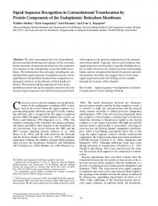
Fig. 6. Schematic of 7SL RNA indicating regions deduced for functional importance. Superimposed under this figure (modified from Siegel and Walter, 1988a; 7SL RNA secondary structure drawn according to Larsen and Zwieb, 1991) are regions known by a-sarcin footprinting to bind p68/72 and p19 (Siegel and Walter, 1988a) and deduced from the present work to bind p9/14. Areas of increased protection from chemical modification by binding to polysomes are shaded; single bases that also become protected by polysomes, such as nos 33 and 73, are not highlighted. Areas of increased protection by binding to rough endoplasmic reticulum membranes are indicated by brackets.
the bottom of stem III (130-134), spanning regions known to interact with p68/72 (Siegel and Walter, 1988a). The increased protection of these bases may be due either to (i) their direct protection by the SRP receptor or other components on the rough ER membrane, or (ii) protection by SRP proteins whose conformation could have been altered due to membrane binding.
Comparison between membrane bound SRP and soluble SRP
This comparison completes the SRP cycle and shows how the chemical accessibility (and therefore the exposure of 7SL RNA in SRP) decreases when SRP is no longer interacting with ribosomes or the SRP receptor (Figure 5B, open circles). A small number of bases become more exposed in soluble SRP (Figure 5B, filled circles).
Discussion A tertiary interaction in naked 7SL RNA The secondary structure of naked 7SL RNA that can be derived from our experimental results of chemical modification is in good agreement with the theoretical model derived by phylogenetic comparisons (Larsen and Zwieb, 1991). However, a few areas predicted to be base-paired have 'breathed' open, as indicated by brackets in Figure 2. One such area is near the tip of stem IV where bases
202-204 are chemically modified; therefore, they must not be base-paired with the '200' region opposite them as previously supposed. The '200' region is generally protected from modification, and we propose that it is involved in a long-range tertiary interaction with the '100' region, which is also protected from modification in naked 7SL RNA (boxed regions in Figure 2). Compensatory base changes support the base-pairing between regions '100' and '200' (Table II). In fact, Table II shows that this interaction can even be drawn for Escherichia coli 4.5S RNA which contains 774
region homologous to stem IV of eukaryotic 7SL RNAs (Poritz et al., 1988; Zwieb, 1988). The base-pairing we propose between the '100' and '200' regions would only occur in naked 7SL RNA, as the '100' region becomes sensitive to chemical modification in soluble SRP, polysome bound SRP and membrane bound SRP (Table I). The tertiary interaction suggested by our data could be a prerequisite for correct biogenesis of SRP. The tertiary interaction seen in naked 7SL RNA remains present even after binding by p19 (data not shown), but perhaps binding by other SRP proteins abolishes base-pairing between the '100' and '200' regions. For example, the subsequent binding of p54 might modulate the association of p19 with 7SL RNA. Similarly, the p54 homologue in E. coli (p48 in Romisch et al., 1989; equivalent to ffh protein in Bernstein et al., 1989), known to bind to E.coli 4.5S RNA (Poritz et al., 1990; Ribes et al., 1990), might destroy the comparable tertiary interaction that can be drawn for E. coli 4.5S RNA (Table II). The addition of SRP proteins might stabilize the close juxtaposition of stem II with stem IV (as indicated by cross-linking at other positions on stem II with stem IV; Zwieb and Schiiler, 1989) such that base-pairing between the '100' and '200' regions is now dispensable. 7SL RNA sites protected by SRP proteins Comparison of chemical modification of naked 7SL RNA a
and soluble SRP indicates sites of increased protection due protein binding (Figure 4A). Figure 6 portrays all the deduced protein binding sites on 7SL RNA. These regions coincide well with the a-sarcin footprints of p68/72 and p19 (Siegel and Walter, 1988a). No footprint data have yet been published for p9/14 known to associate with the Alu domain of 7SL RNA, but our results suggest p9/14 binds to stem I and the junction with stem II, both of which are in the Alu domain (Figure 4A). to
7SL RNA sites protected by polysomes SRP association with polysomes may be mediated by several interactions. First, p54 located at one end of SRP binds the
7SL RNA conformation
emerging signal sequence (Krieg et al., 1986; Kurzchalia et al., 1986; Wiedmann et al., 1987; Siegel and Walter, 1988b). Second, other parts of SRP such as the 7SL RNA component may bind directly to the ribosome. Bases that are more protected from chemical modification in polysome bound SRP than in soluble SRP (Figure 4B and shaded areas of Figure 6) are candidates for ribosome binding, though other explanations are also possible (see Results). It could be that 7SL RNA interacts directly with RNA of the ribosome to cause translational arrest. For example, an oligonucleotide for 97-110 of 7SL RNA can prime synthesis of 18S rRNA (Ullu and Weiner, 1985). Note that this 97-110 region includes nucleotides 98, 101 and 107 which we find more protected from chemical modification when SRP is bound to polysomes. In a second example, some species have sequence similarity between two regions in 7SL RNA and 5S RNA, so 7SL RNA could compete for 5S RNA binding sites on rRNA (Zwieb, 1985; Boehm, 1987). However, the similarity between 7SL RNA and 5S RNA does not hold up in some plants (Haas et al., 1988). Nonetheless, it is intriguing to note that polysome protection seen in our experiments for (i) nos 98, 101 and 107, and (ii) nos 233 and 239 are within or near the 5S-like regions of 7SL RNA of nos 104-109 and 222 -263, respectively (Zwieb, 1985; Boehm, 1987). Curiously, both regions are in the S domain of 7SL RNA, rather than the Alu domain that has been implicated in translational arrest (Siegel and Walter, 1985, 1986). Nucleotides 33 and 73 are the only ones in the Alu domain where we find increased protection from chemical modification after binding to polysomes. The area of the Alu domain containing nucleotide 33 mimics the shape of tRNA and could compete for tRNA binding sites on the ribosome (Zwieb, 1986). Site directed mutagenesis of each of the bases we find more protected in both the Alu and S domains in polysome bound SRP could reveal which bases might in fact be recognition points on 7SL RNA for ribosome binding. 7SL RNA sites protected by membranes The brackets in Figure 6 indicate regions in 7SL RNA that are more protected from chemical modification in membrane bound SRP than in polysome bound SRP (see also Figure SA). These regions in 7SL RNA could either be (i) protected directly by the SRP receptor or other components of the rough ER (e.g. mp3O binds SRP in the absence of SRP receptor, Tajima et al., 1986) or (ii) protected by SRP proteins whose conformation could have been altered due to membrane binding. Notice that most of the bracketed areas of 7SL RNA (Figure 6) coincide with p68/72 footprint regions (Siegel and Walter, 1988a), thus supporting possibility (ii).
Conformational changes in 7SL RNA during the SRP cycle The areas discussed above become increasingly protected from chemical modification during progression from one stage to the next of the SRP cycle. Other nucleotides show increased sensitivity moving through the SRP cycle, indicative of conformational changes in 7SL RNA. In fact, it has previously been demonstrated by gel electrophoresis that 7SL RNA can exist in several conformations (Zwieb, 1985; Zwieb and Ullu, 1986). One example of a conformational change that we found is the tertiary interaction between the '100' and '200' regions seen for naked 7SL
RNA but not for soluble SRP, discussed above. In addition, several sites of increased sensitivity going from soluble SRP to polysome bound SRP (Figure 4B, open symbols), or from polysome bound SRP to membrane bound SRP (Figure SA, open symbols), or from membrane bound SRP to soluble SRP (Figure SB, closed symbols) are scattered throughout the 7SL RNA molecule, though predominantly found in stem II and the no 170 junction between stems III and IV. These changes in chemical sensitivity indicate conformational changes in 7SL RNA between each of these stages of the SRP cycle. One result of these changes is that soluble SRP is in a more closed, protected conformation than polysome bound SRP or membrane bound SRP. In other words, when SRP is active during arrest of translation (polysome bound) or handing the signal complex over to the SRP receptor for translocation into the ER (membrane bound), the 7SL RNA moiety is more exposed than when SRP is inactive (soluble SRP). Most of the sites indicative of conformational changes were not protected by SRP proteins (i.e. not sensitive to chemicals in naked 7SL RNA and then protected in soluble SRP), making unlikely the alternative possibility that the nucleotides indicative of conformational changes only become more sensitive because of displacement of SRP
proteins. Do RNA -RNA interactions drive some steps of the SRP cycle? RNA-RNA interactions are important for translation (reviewed by Dahlberg, 1989). For example, rRNA interacts with mRNA during initiation (Shine -Dalgarno interaction) and during elongation (seen by ribosomal frame shifting experiments; Weiss et al., 1987). In another example, tRNA interacts with mRNA (anticodon -codon pairing) and with rRNA (seen by A, P and E site footprints; Moazed and Noller, 1986, 1989a). Intramolecular interactions within rRNA may also be important; the conformation of rRNA changes between inactive and active 30S ribosomal subunits in prokaryotes (Moazed et al., 1986) and between subunits and monosomes in eukaryotes (Stebbins-Boaz and Gerbi, 1990). Moreover, analysis of tRNA hybrid sites suggests that tRNA translocation is accompanied by changes in
interactions between ribosomal subunits (Moazed and Noller, 1989b). Just as rRNA conformational changes may be implied for the ribosome cycle, so too our data presented here demonstrate that 7SL RNA changes its conformation during the SRP cycle. There could be an interplay between the ribosome cycle and the SRP cycle via RNA-RNA interactions that alter RNA conformation. We speculate that changes in rRNA conformation during the elongation phase of the ribosome cycle could be responsible for tRNA translocation. Interaction of 7SL RNA with rRNA once SRP binds polysomes could block further conformational changes in rRNA, resulting in arrest of translation. Zwieb (1989) has hypothesized specific ways by which 7SL RNA may interact with the ribosomes, resulting in translational arrest. This translational arrest would be relieved when the 7SL RNA-rRNA interaction is discontinued upon docking on the rough ER. A major point to be made in our model is that SRP binding to polysomes is mediated both by p54-signal peptide and 7SL RNA-rRNA interactions. Similarly, SRP release upon docking on the rough ER requires that both sets of interactions be abolished. Our data presented here indicate that 7SL RNA changes 775
M.Andreazzoli and S.A.Gerbi
its conformation during the SRP cycle. It will be fascinating in future experiments to discover if there is a causal link between these conformational changes and progression through the SRP cycle as hypothesized here, as well as to substantiate the proposed overlap via RNA -RNA interactions between the SRP cycle and the ribosome cycle.
Materials and methods Preparation of microsomal membranes, SRP and polysomes Microsomal membranes were prepared as described by Walter and Blobel (1983c) and shown to be active by assaying translocation activity in wheat germ extract (Promega) programmed with bovine preprolactin mRNA obtained by in vitro transcription of pSPBP4 (Siegel and Walter, 1988b; a generous gift from V.Siegel). SRP was solubilized from microsomal membranes by salt extraction as described by Walter and Blobel (1983d). Both microsomal membranes and solubilized SRP were used for chemical modification experiments without further purification. Polysomes were prepared essentially as described by Gunning et al. (1981). Briefly, canine pancreas was homogenized in the usual buffer but in the presence of 100 mM potassium acetate, since at this concentration much SRP is ribosome bound (Walter and Blobel, 1983b). The postmitochondrial supernatant was layered on a discontinuous sucrose gradient consisting of 2.0 M sucrose and 0.8 M sucrose containing the same ionic conditions as the homogenization buffer. After centrifugation at 90 000 g for 18 h at 4°C in a SW-28 rotor, the pellets were collected and resuspended in 50 mM triethanolamine (TEA), pH 7.5, 100 mM potassium acetate, 250 mM sucrose and 1 mM DTT. Soluble SRP and membranes do not pellet under these conditions (Gunning et al., 1981). This pellet, which we refer to as the 'polysome' fraction, contains primarily polysomes (Falvey and Staehlin, 1970); in our conditions it has - 75 % polysomes and 25 % monosomes as assayed by respinning the resuspended pellet in a 10-30% sucrose gradient and checking fractions for the presence of 7SL RNA by primer extension. Samples were stored at -20'C and generally used soon thereafter for chemical modification.
Preparation of naked 7SL RNA To prepare naked 7SL RNA, soluble SRP was extracted twice with phenol, followed by chloroform, 0.1 % SDS. RNA was precipitated with 1/10 volume of 2.5 M sodium acetate (pH 5.1) and 2.5 volumes of 95% ethanol at -20°C overnight, pelleted and resuspended in 50 mM TEA (pH 7.5), 50 mM potassium acetate, 250 mM sucrose, 1 mM DTT. Samples were stored at -20'C and generally used soon thereafter for chemical modification and sequencing. Reconstitution of p19 with 7SL RNA Naked 7SL RNA prepared as described above was mixed with SRP protein p19 (a generous gift from P.Walter) under reconstitution conditions (Siegel and Walter, 1985). The molar ratio of 7SL RNA to p19 was 1:17.5; therefore, p19 was in excess. We used primer extension to confirm that p19 had indeed bound to 7SL RNA, and we obtained protection of 7SL RNA in regions predicted by the ca-sarcin footprint (Siegel and Walter, 1988a).
Chemical modification of RNA substrates Chemical modification of naked 7SL RNA, soluble SRP, membrane bound SRP and polysome bound SRP was carried out according to the procedure described by Stebbins-Boaz and Gerbi (1990). Primers Two 20-mer deoxyoligonucleotide primers, complementary to unique regions within the 7SL RNA, were synthesized using a Biosearch 8600 DNA synthesizer. The sequences of the primers were: (7SL 272-253) 5'-GGCTGGAGTGCAGTGGCTAT-3'; (7SL 187-168) 5'-CGGTTCACCCCTCCTTAGGC-3'. The primers were purified by gel electrophoresis through 20% polyacrylamide (19:1 bis-acrylamide), 7 M urea and 1 x TBE (0.8 M Tris-borate, 1 mM EDTA, pH 8.0). Primers were eluted from gel fragments by incubating with 1 ml of 0.1 M ammonium bicarbonate. Urea was removed from the eluted primer by repeated precipitations with 95 % ethanol. Purified primers were resuspended in H20 at a final concentration of 20 pmol/Ail and stored at -20'C.
End-labeling, sequencing and primer extension End-labeling of the primers and RNA sequencing were carried out as described by Stebbins-Boaz and Gerbi (1990), while primer extensions of 776
chemically modified RNAs were performed according to the procedure described by Lillie et al. (1986).
Gel electrophoresis Primer extension products were run on 0.4 mm thick sequencing gels (8% polyacrylamide, 7 M urea, 1 x TBE) using a sharkstooth comb. Gels were run at 20 mA, constant current, for 2.5-5 h and exposed to Kodak X-ray film at room temperature overnight (12-16 h). Adjustments in volume of samples loaded were made following a preliminary run in order to equalize the relative signals between sample lanes; when background bands were of equal intensity between lanes, samples were judged to be equivalent in loading. Modified bands can only be compared between different lanes when identical amounts of sample are loaded in each lane. Within a given lane the strength of modification signal was judged to be weak, moderate, strong or hypersensitive by comparison between bands in that one lane.
Acknowledgements We are grateful to V.Ware, V.Siegel and B.Dobberstein for teaching us how to isolate SRP, to B.Stebbins-Boaz for instruction in chemical modification, to C.Zwieb for sharing unpublished data on 7SL RNA conformation, and to P.Walter for a gift of pI9. We thank the following for their helpful comments on this paper: M.Firpo, P.Grabowski, B. Hoffman, H.Hoffmann, P.Milos, R.Rivera-Leon, R.Savino and B.StebbinsBoaz. The assistance of D.Angeloni with the figures was greatly appreciated. Our research is funded by NIH GM20261.
References Andrews,D.W., Walter,P. and Ottensmeyer,F.P. (1985) Proc. Natl. Acad. Sci. USA, 82, 785-789. Andrews,D.W., Walter,P. and Ottensmeyer,F.P. (1987) EMBO J., 6, 3471 -3477. Bernstein,H.D., Poritz,M.A., Strub,K., Hoben,P.J., Brenner,S. and Walter,P. (1989) Nature, 340, 482-486. Boehm,S. (1987) FEBS Lett., 212, 15-20. Campos,N., Palau,J., Torrent,M. and Ludevid,D. (1988) J. Biol. Chem., 263, 9646-9650. Dahlberg,A.E. (1989) Cell, 57, 525-529. Dobberstein,B. (1987) Mol. Biol. Rep., 12, 213-222. Falvey,A.K. and Staehlin,T. (1970) J. Mol. Biol., 52, 1-19. Gilmore,R. and Blobel,G. (1983) Cell, 35, 677-685. Gilmore,R. and Blobel,G. (1985) Cell, 42, 497-505. Gilmore,R., Blobel,G. and Walter,P. (1982a) J. Cell Biol., 95, 463 -469. Gilmore,R., Walter,P. and Blobel,G. (1982b) J. Cell Biol., 95, 470-477. Gundelfinger,E.D., Krause,E., Melli,M. and Dobberstein,B. (1983) Nucleic Acids Res., 11, 7363-7374. Gundelfinger,E.D., DiCarlo,M., Zopf,D. and Melli,M. (1984) EMBO J., 3, 2325-2332. Gunning,P.W., Beguin,P., Shooter,E.M., Lawrence,A. and Jeffrey,P.L. (1981) J. Biol. Chem., 256, 6670-6675. Haas,B., Klanner,A., Ramm,K. and Sanger,H.L. (1988) EMBO J., 7, 4063 -4074. Hagenbiichle,O., Santer,M., Steitz,J.A. and Mans,R.J. (1978) Cell, 13, 551 -563. Hortsch,M. and Meyer,D.I. (1986) Int. Rev. Cytol., 102, 215-242. Ibrahimi,I. (1987) J. Cell Biol., 104, 61 -66. Krieg,U.C., Walter,P. and Johnson,A.E. (1986) Proc. Natl. Acad. Sci. USA, 83, 8604-8608. Kurzchalia,T.V., Wiedmann,M., Girschovich,A.S., Bochkareva,E.S., Bielka,H. and Rapoport,T.A. (1986) Nature, 320, 634-636. Larsen,N. and Zwieb,C. (1991) Nucleic Acids Res., 19, 209-215. Lillie,J.W., Green,M. and Green,M.R. (1986) Cell, 46, 1043-1051. Lipp,J., Dobberstein,B. and Haeuptle,M.-T. (1987) J. Biol. Chem., 262, 1680-1684. Meyer,D.I. (1985) EMBO J., 4, 2031-2033. Meyer,D.I., Krause,E. and Dobberstein,B. (1982) Nature, 297, 647-650. Moazed,D. and Noller,H.F. (1986) Cell, 47, 985-994. Moazed,D. and Noller,H.F. (1989a) Cell, 57, 585-597. Moazed,D. and Noller,H.F. (1989b) Nature, 342, 142-148. Moazed,D., Van Stolk,B.J., Douthwaite,S. and Noller,H.F. (1986) J. Mol. Biol., 191, 483 -493. Mueller,M., Ibrahimi,I., Chang,C.N., Walter,P. and Blobel,G. (1982) J.
Biol. Chem., 257, 11860-11863. Portiz,M.A., Strub,K. and Walter,P. (1988) Cell, 55, 4-6.
7SL RNA conformation Poritz,M.A., Bernstein,H.D., Strub,K., Zopf,D., Wilhelm,H. and Walter,P. (1990) Science, 250, 1111-1117. Prehn,S., Wiedmann,M., Rapoport,T.A. and Zwieb,C. (1987) EMBO J., 6, 2093-2097. Rapoport,T.A., Heinrich,R., Walter,P. and Schulmeister,T. (1987) J. Mol. Biol., 195, 621-636. Ribes,V., Rornisch,K., Giner,A,. Dobberstein,B. and Tollervey,D. (1990) Cell, 63, 591-600. Romisch,K., Webb,J., Herz,J., Prehn,S., Frank,R., Vingron,M. and Dobberstein,B. (1989) Nature, 340, 478-482. Sanz,P. and Meyer,D.I. (1988) EMBO J., 7, 3553-3557. Siegel,V. and Walter,P. (1985) J. Cell Biol., 100, 1913-1921. Siegel,V. and Walter,P. (1986) Nature, 320, 81-84. Siegel,V. and Walter,P. (1988a) Proc. Natl. Acad. Sci. USA, 85, 1801-1805. Siegel,V. and Walter,P. (1988b) Cell, 52, 39-49. Stebbins-Boaz,B. and Gerbi,S.A. (1990) J. Mol. Biol., 217, 93-112. Tajima,S., Lauffer,L., Roth,V.L. and Walter,P. (1986) J. Cell Biol., 103, 1167-1178. Ullu,E. and Tschudi,C. (1984) Nature, 312, 171-172. Ul1u,E. and Weiner,A.M. (1985) Nature, 318, 371-374. Ullu,E., Murphy,S. and Melli,M. (1982) Cell, 29, 195-201. Walter,P. (1987) Nature, 328, 763-764. Walter,P. and Blobel,G. (1980) Proc. Natl. Acad. Sci. USA, 77, 7112-7116. Walter,P. and Blobel,G. (1981) J. Cell Biol., 91, 557-561. Walter,P. and Blobel,G. (1982) Nature, 299, 691 -698. Walter,P. and Blobel,G. (1983a) Cell, 34, 525-533. Walter,P. and Blobel,G. (1983b) J. Cell Biol., 97, 1693-1699. Walter,P. and Blobel,G. (1983c) Methods Enzymol., 96, 84-93. Walter,P. and Blobel,G. (1983d) Methods Enzymol., 96, 682-691. Walter,P. and Lingappa,V.R. (1986) Annu. Rev. Cell Biol., 2, 499-516. Walter,P., Ibrahimi,I. and Blobel,G. (1981) J. Cell Biol., 91, 545-550. Walter,P., Gilmore,R. and Blobel,G. (1984) Cell, 38, 5-8. Weiss,R.B., Dunn,D.M., Atkins,J.F and Gesteland,R.F. (1987) Cold Spring Harbor Symp. Quant. Biol., 52, 687-693. Welsh,M., Scherberg,N., Gilmore,R. and Steiner,D.F. (1986) Biochem. J., 235, 459-467. Wiedmann,M., Kurzchalia,T.V., Bielka,H. and Rapoport,T.A. (1987) J. Cell Biol., 104, 201-208. Wolin,S.L. and Walter,P. (1989) J. Cell Biol., 109, 2617-2622. Youvan,D.C. and Hearst,J.E. (1979) Proc. Natl. Acad. Sci. USA, 76, 3751 -3754. Zwieb,C. (1985) Nucleic Acids Res., 13, 6105-6124. Zwieb,C. (1986) Endocytobiosis Cell Res., 3, 167-177. Zwieb,C. (1988) Endocytobiosis Cell Res., 5, 327-336. Zwieb,C. (1989) Prog. Nucleic Acids Res. Mol. Biol., 37, 207-234. Zwieb,C. and Schuler,D. (1989) Biochem. Cell Biol., 67, 434-442. Zwieb,C. and Ullu,E. (1986) Nucleic Acids Res., 14, 4639-4657. Received on November 15, 1990; revised on Januarv 14, 1991
777