0013-7227/07/$15.00/0 Printed in U.S.A.
Endocrinology 148(12):5667–5679 Copyright © 2007 by The Endocrine Society doi: 10.1210/en.2007-0647
Changes in Adiponectin and Inflammatory Genes in Response to Hormonal Imbalances in Female Mice and Exacerbation of Depot Selective Visceral Adiposity by High-Fat Diet: Implications for Insulin Resistance Hui Zhang, Xinlei Chen, Jayaprakash Aravindakshan, and M. Ram Sairam Molecular Reproduction Research Laboratory, Clinical Research Institute of Montreal (affiliated with Universite´ de Montre´al), Montre´al, Que´bec, Canada H2W 1R7 Early obesity and late onset of insulin resistance associated with hormonal imbalances occur in FSH receptor-deficient follitropin receptor knockout female mice. This study tests the hypothesis that chronic high-fat diet aggravates obesogenic changes in a depot-specific manner and explores some molecular links of hormone imbalances with insulin resistance. In SV 129 mice, hormonal imbalances seem obligatory for exacerbation of diet-induced obesity. Visceral adiposity, glucose intolerance, and lipid disturbances in 9-month follitropin receptor knockout females were associated with decrease in adiponectin signaling. High-molecular-weight plasma adiponectin and adipose tissue adiponectin mRNA were decreased. Adiponectin receptors R1 and R2 mRNA was selectively altered in mesenteric fat but not periuterine fat. R2 decreased in the liver and R1 was higher in muscle. Whereas hepatic adenosine monophosphate T-activated protein kinase activity was down-regulated, both phosphoenolpyruvate car-
I
NCREASING PREVALENCE OF obesity and metabolic syndrome (MS) in postmenopausal women and association with sex hormone imbalance in endocrine disorders has attracted considerable interest because women in both these states have higher tendency for cardiovascular disease and type 2 diabetes (1). MS is a combination of visceral obesity, impaired glucose metabolism, atherogenic dyslipidemia, and blood pressure elevation, resulting from a primary disorder, insulin resistance (IR) (2). Although recent clinical intervention studies have shown that extended hormone therapy initiated after menopause does not confer cardiac protection and may actually increase the risk for coronary
First Published Online August 23, 2007 Abbreviations: AdipoR, Adiponectin receptor; AMPK, adenosine monophosphate T-activated protein kinase; AT, adipose tissue; CCL2, C-C motif chemokine ligand-2, also known as monocyte chemoattractant protein-1; CCR2, C-C motif chemokine receptor-2; DIO, diet-induced obesity; ER, estrogen receptor; FORKO, follitropin receptor knockout; G6Pase, glucose-6-phosphatase; HFD, high-fat diet; HMW, high molecular weight; IR, insulin resistance; ITT, insulin tolerance test; MAT, mesenteric adipose tissue; MS, metabolic syndrome; OGTT, oral glucose tolerance test; p-AMPK, phosphorylated AMPK; PAT, periuterine adipose tissue; PEPCK, phosphoenolpyruvate carboxykinase; QPCR, quantitative PCR; RD, regular diet; TC, total cholesterol; TG, triglyceride; WT, wild type. Endocrinology is published monthly by The Endocrine Society (http:// www.endo-society.org), the foremost professional society serving the endocrine community.
boxykinase and glucose-6-phosphatase enzymes were up-regulated. Longitudinally, diminishing sex hormone signaling in adipose tissue was associated with progressive down-regulation of adiponectin activity and gradual impaired glucose tolerance. Chronic high-fat diet in SV129 wild-type mice did not produce overt obesity but induced visceral fat depot changes accompanied by liver lipid accumulation, high cholesterol, and up-regulation of inflammation gene mRNAs. Thus, TNF-␣, C-C motif chemokine receptor-2, and C-C motif chemokine ligand-2 were selectively elevated in mesenteric fat without altering glucose tolerance and adiponectin signaling. Our study highlights adiponectin signaling and regulation to be involved in hormone imbalance-induced insulin resistance and demonstrates selective visceral adipose depot alterations by chronic high-fat diet and induction of inflammatory genes. (Endocrinology 148: 5667–5679, 2007)
heart disease in postmenopausal women (3), many underlying mechanistic issues remain to be explored and clarified. Thus, alternative interventions including appropriate timing might be necessary to improve the quality of life of aging women who live a third of their life in menopause. In addition, many women with polycystic ovarian syndrome, a hormonal imbalance disorder including hyperandrogenemia, also have obesity and IR at an early age. An understanding of the molecular basis that links sex hormone imbalances and IR could help prevent or overcome the occurrence of IR in aging women and polycystic ovarian syndrome subjects. Animal models that can mimic many, if not all, of the cluster of pathologies leading to IR in humans could also help define the molecular links between sex hormonal imbalances and IR. Two sex hormone imbalance (low estrogen to testosterone ratio) mouse models recapitulate different aspects of the MS phenotype. Studies from our group showed that follitropin receptor knockout (FORKO) female mice experiencing early obesity exhibit age-related mimicry of MS (4). Similarly aromatase-knockout mutants also show progressive development of IR in an age-dependent manner in male mice (5). Consumption of high-fat diet (HFD) is also known to be a major environmental factor in promoting obesity (6, 7) and reducing insulin sensitivity (8). Because obesity-related effects occur in a gene dosage-dependent manner in FORKO mice, we hypothesized that chronic high-fat diet might exacerbate adiposity effects in a
5667
5668
Endocrinology, December 2007, 148(12):5667–5679
depot-selective manner and explored mechanisms related to this phenomenon. Visceral obesity is a key etiological factor in inducing IR (9). In obese and related IR subjects, adipose tissue (AT) function is altered in several ways including changes in morphology, aberrant endocrine and metabolic functions as well as low-grade inflammation (10). AT responds to metabolic signals releasing many adipokines, and their aberration is critical for onset of obesity, glucose, and lipid disturbance (10). Among many alterations, up-regulations in TNF-␣ (11), the monocyte chemoattractant protein-1, also known as C-C motif chemokine ligand-2 (CCL2) (12) and C-C motif chemokine receptor-2 (CCR2) (13) couple adiposity with IR. Unlike many adipokines, adiponectin (also known as Acrp30, AdipoQ, and GBP28), an adipose tissue-specific secretory protein, is negatively correlated with body weight, fat content, and insulin levels and declines with progression toward the diabetic state (9). High-molecular weight (HMW) complexes of adiponectin consisting of 12–18 subunits constitute the active form of adiponectin determining insulin sensitivity (14). Adiponectin actions are mediated by two cell membrane receptors, adiponectin receptor (AdipoR)-1 and -2 (15), activating a protein kinase cascade, AMP kinase (AMPK), increasing fatty acid oxidation in muscle, and potentiating insulin inhibition of hepatic gluconeogenesis (16). Although adiponectin is causally related to IR and it has been noted as an antiinflammatory, antidiabetic, and antiatherogenic protein (9), little is known about the regulation of adiponectin signaling in response to sex hormone imbalances. In the present study, we addressed the regulation of adiponectin signaling and evaluated selected AT inflammation gene expressions in FORKO female mice with or without HFD for different periods. Our data revealed adiponectin signaling down-regulation and adipose tissue inflammation gene up-regulations could be part of mechanisms that link sex hormone imbalances with the onset of obesity and IR. Chronic HFD challenge in FORKO female mice in the SV129 background [a diet induced obesity resistance mouse strain (DIO)] could exacerbate the obesity phenotype. This is the first comprehensive study on adiponectin signaling aberration in sex hormone imbalance-induced obesity and related IR state. It is also the first report linking obesity-related phenotype and adipokine disturbance under the influence of chronic HFD in FORKO female mice. We also note differential impact on selective fat depots that lead to IR. Thus, a better understanding and dissection of adipokine alterations in specific fat depots could assist in developing targeting remedies that might preserve benefits of hormone replacement therapy and avoid adverse cardiovascular events (17). Materials and Methods This study was approved by an institutional ethics committee. Mice of all three genotypes were produced (18, 19) by breeding 129T2/SV EmsJ FSH-R ⫹/⫺ males and females. Wild-type (WT) (⫹/⫹) virgin, knockout (⫺/⫺) female mice (FORKO) from 3 to 9 months were used as indicated in the figures. Mice were housed (n ⫽ 6 or less per cage, 12 h light, 12 h dark) with free access to water and feed. At 1 month, mice were divided into four groups based on genotype and diet selection: group 1, WT, provided with regular chow (WT-RD; 4.5% kcal fat diet, rodent laboratory chow, 5001/Harlan Teklad 7004 diet 1:1 mixture; Harlan Teklad, Madison, WI); group 2, WT, provided with HFD (WT-HFD;
Zhang et al. • Adipokine Changes in FORKO Mice
rodent diet with 45% kcal fat from lard and soybean oil; Research Diet, Inc., New Brunswick, NJ); group 3, FORKO, provided with regular chow diet (FORKO-RD); group 4, FORKO, provided with HFD (FORKOHFD), except when overnight fasting was required. All comparisons were performed using littermates.
Oral glucose tolerance test (OGTT) Groups of age-matched females were fasted overnight (16 –18 h) and orally administered glucose (1.5 g/kg body weight). A blood sample was collected from the tail at t ⫽ 0, 30, 60, and 120 min. Glucose concentration was analyzed using a diagnostic glucometer from Abbott Laboratories (Montre´al, Que´bec, Canada).
Insulin tolerance test (ITT) Groups of age-matched females (8 months) were fed as usual and ip challenged with porcine insulin at 0.75 mU/g body weight (SigmaAldrich Canada, Oakville, Ontario, Canada). There was no food provided during the test, and blood samples were collected from the tail at t ⫽ 0, 15, 30, 60, and 90 min for determining glucose concentration, which was analyzed using a diagnostic glucometer (Abbott Laboratories).
Histological and morphometric analysis of adipose tissue Visceral adipose tissue from two sites were evaluated in this study. The visceral white adipose tissue surrounding mesenteric vessels (MAT) and adipose tissue surrounding the uterus-periuterine adipose tissue (PAT) were dehydrated and embedded in paraffin. Five-micrometerthick sections were cut and stained by standard protocols using hematoxylin and eosin for microscopic examination. For the quantification of the size of adipocytes in MAT and PAT, the area of 10 cells in random sectional areas from six fields of five to six animals per experimental group were analyzed under light microscopy using Image J system software (National Institutes of Health, Bethesda, MD). Results are expressed as mean ⫾ sem.
Liver oil red O stain and lipid quantification Frozen section from liver was used for oil red staining (20). Livers were sectioned (10 m), fixed in 10% formalin for 12 min, washed well in tap water, and rinsed in distilled water and stained with oil red O (Sigma-Aldrich Canada) for 10 min to identify neutral lipids, cholesterol, and fatty acids (red color). After rinsing with water, nuclei were counterstained (blue) with Mayer’s hematoxylin (Sigma-Aldrich Canada) for 1 min and washed in tap water and distilled water. Slides were mounted with Mount quick (Daido Sangyo Co. Ltd., Tokyo, Japan). Sections from livers in each experimental group were processed simultaneously. Finally, slides were covered in aqueous mount under a coverslip for viewing with a light microscope at equal light intensity. Six images from the stained slides of six mice per group were initially acquired using a 24-bit file format (8 bits per red-green-blue color) with a Micropublisher 3.3 RTV (Q-Imaging, Surrey, British Columbia, Canada) and the Northern Eclipse version 7.1 software (Empix Imaging Inc., Toronto, Ontario, Canada) under 10 ⫻ 1.6 magnification. Each image was processed using MatLab version 7.3 (The MathWorks, Inc., Natick, MA) by reducing the color numbers to a 64 gray tone per red-green-blue color (instead of 256). Representative images were used to select colors attributed to the stain to create a color file. Each image was then processed back to 64 gray tone. Staining percentage was given by the ratio of colors from the image having pixels that matches colors from the color file to total pixel number from the selected area (percent). Thus, the total size of lipid drops in proportion to total area from each image (percent) was quantified.
Plasma total cholesterol and total triglycerides Blood was collected by heart puncture. Plasma was separated and samples were kept at ⫺80 C until use. Total triglyceride (TG) was measured with a commercially available triglyceride reagent kit and total cholesterol (TC) was measured with a cholesterol reagent set (Pointe Scientific, Inc., Canton, MI). To confirm the results of plasma TG and TC, the same samples were also measured using an autoanalyzer (Cobas Miras; Roche, Montre´al, Que´bec, Canada).
Zhang et al. • Adipokine Changes in FORKO Mice
Endocrinology, December 2007, 148(12):5667–5679
5669
SDS-PAGE and immunoblotting
Statistical analysis
Liver and gastrocnemius muscle samples were collected on ice and stored at ⫺80 C for analysis of protein expression of AMPK, and phosphorylated AMPK (p-AMPK). Tissues were homogenized in ice-cold lysis buffer [1% Triton X-100, 125 mm NaCl, 10 mm Tris (pH 7.4), 1 mm EDTA, 1 mm EGTA, 2 mm Na3VO4, 10 mm sodium pyrophosphate, and 25 mm NaF] that contained a protease inhibitor cocktail (Roche Diagnostics). After insoluble components were removed by centrifugation (14,000 rpm, 5 min, 4 C), protein concentrations of supernatants were quantified using a commercial reagent (Bio-Rad Laboratories, Hercules, CA). Equal amounts of protein (50 g total lysates) were mixed with the sample buffer. Sample buffer for reducing conditions was 3% sodium dodecyl sulfate, 50 mm Tris-HCl (pH 6.8), 5% 2-mercaptoethanol, and 10% glycerol. Components were separated on a 7.5% SDS-PAGE gel. HMW adiponectin analysis in plasma was performed according to a published method with some modifications (21). Briefly, equal amount of plasma (1 l) was diluted to 20 l with lysis buffer to be separated by 6% SDS-PAGE under nonreducing conditions and without boiling. In addition, for nonreducing conditions, 2-mercaptoethanol was excluded from the sample buffer described above. The sample was mixed with 6⫻ buffer and incubated for 1 h at room temperature. Upon heating and under reducing conditions, adiponectin was converted to a 30-kDa monomer (22). Total adiponectin analysis in plasma was assessed by using reducing and heating conditions. Equal amounts of plasma (1 l) were diluted to 20 l with lysis buffer and mixed with 6⫻ sample buffer and heated for 10 min at 95 C. Samples were separated by 10% SDSPAGE. Proteins were transferred to nitrocellulose membrane, blocked with 5% dry nonfat milk and incubated with first antibody for 16 h at 4 C. The following rabbit polyclonal antibodies were used for Western blots: antiadiponectin (1:3000) (gift from Dr. P. Scherer, Albert Einstein College of Medicine, Bronx, NY), anti-AMPK (1:1000), and anti-p-AMPK (1:800) (Cell Signaling Technology, Danvers, MA). After three washes, membranes were incubated with the secondary antibody, goat antirabbit coupled to horseradish peroxidase (Sigma-Aldrich Canada), diluted 1:15,000 in milk followed by chemiluminescence reagent (GE Healthcare, Piscataway, NJ) and exposure to film (X-omat, XRP-5; Eastman Kodak, Rochester, NY). Band intensity was quantified using scanning densitometry (NIH Image software).
Statistical analyses were performed using SigmaStat3.1 (SYSTAT, Point Richmond, CA). Values are presented as means ⫾ sem. The significance of the results was determined by using the two-way ANOVA (two main effects: diet, genotype, and their interaction are assessed, except for those in Fig. 4C; genotype and age are two main effects) followed by Bonferroni t test for multiple comparison. The t test was used (for Fig. 3) when comparing genotype factor between two groups. Repeated-measures ANOVA was used for analyzing body weight, OGTT, and ITT experimental data. Three-way ANOVA (three main effects: diet, genotype, age, and their interaction) was used for adipocyte size quantification experiment in Fig. 1D. Statistical differences were considered significant at P ⬍ 0.05.
RNA extraction and real-time PCR Total RNA was extracted from PAT, MAT, liver, and muscle with Trizol reagent following the supplier’s protocol, and 1–2 g of RNA were reverse transcribed using the first-stand cDNA synthesis kit (GE Healthcare). Quantitative PCR (Q-PCR) was performed using Platinum SYBR Green qPCR SuperMix UDG kit (Invitrogen, Carlsbad, CA). Expressions of different transcripts were determined in relation to -actin with real-time PCR on Mx4000 (Stratagene, La Jolla, CA). All reactions were done in duplicate. Q-PCR parameters were 95 C for 10 min, followed by 40 cycles at 95 C for 30 sec, 57 C for 60 sec, and 72 C for 45 sec. No amplification of fragments occurred in control samples without reverse transcriptase. Quantity of mRNA was calculated using the ⌬⌬Ct method [⌬⌬Ct ⫽ ⌬Ct (treated) ⫺ ⌬Ct (control); ⌬Ct ⫽ Ct-actin ⫺ Cttarget; Ct ⫽ threshold cycle], as described in a previous report (23). The sequences of the specific PCR primers for Q-PCR are shown in Table 1.
Results Alteration of body weight, adipose tissue, and liver lipid accumulation in response to sex hormone imbalance and HFD
To characterize obesity phenotype in FORKO female mice challenged with chronic HFD, body weight was calculated monthly (Fig. 1A). When fed with regular diet (RD), FORKO females showed significant increase in body weight from 3 months old (P ⬍ 0.05), compared with age-matched WT mice. When fed with a HFD, body weight of FORKO females was higher from 2 months onward (P ⬍ 0.05), compared with WT mice under HFD. Body weight of FORKO-HFD group was also significantly higher than FORKO-RD group beginning at 4 months (P ⬍ 0.05) and continuously rose during the next 5–9 months. Interestingly provision of HFD including a long duration had no distinguishable effect on body weight of WT mice under identical conditions. This observation is consistent with previous reports that mice of the SV129 background strain are relatively resistant to an obesogenic diet (24). However, HFD augmentation of body weight gain in FORKO mice of the same strain suggests that hormone imbalances combined with dietary factors could exacerbate obesity. The adverse metabolic consequences of obesity are best predicted by changes (or accumulation of) visceral adipose mass. We have previously shown that deep visceral adiposity is readily evident in FORKO mice fed RD as indicated by the surrounding cover of adipose tissue around mesenteric vessels (4). Following this method, we measured the average width of the mesenteric vessel with fat at the widest point in the presence of HFD (supplemental Fig. 1, published as supplemental data on The Endocrine Society’s Journals Online Web site at http://endo.endojournals.org). As shown in Fig.
TABLE 1. Primers used for Q-PCR experiments Gene
Forward primer (5⬘–3⬘)
Reverse primer (5⬘–3⬘)
Reference
Length (bp)
Adiponectin AdipoR1 AdipoR2 TNF-␣ CCL2 CCR2 ER␣ ER G6Pase PEPCK Actin
GGAACTTGTGCAGGTTGGAT ATGCCATGGAGAAGATGGAG GCCCAGCTTAGAGACACCTG CAAACCACCAAGTGGAGGAG AGGTCCCTGTCATGCTTCTG TACGATGATGGTGAGCCTTG AATGAAATGGGTGCTTCAGG TGGCGACGACGGCACGGT AGGCATGCAGAGTCTTTGGT CTGGCACCTCAGTGAAGACA CGTGGGCCGCCCTAGGCACCA
GCTTCTCCAGGCTCTCCTT GTTGCCAGTCTCTGTGTGGA GCCTTCCCACACCTTACAAA GTGGGTGAGGAGCACGTAGT GCTGCTGGTGATCCTCTTGT AGCAGGAAGAGCAGGTCAGA AGGTGGACCTGATCATGGAG GCTGCTGGGAAGAGATTCCACTCTT ACCGCAAGAGCATTCTCAGT CTACGGCCACCAAAGATGAT TTGGCCTTAGGGTTCAGGGGGG
NM009605 NM028320 NM197985 U68414 BC055070 NM009915 NM007956 NM207707 NM008061 NM011044 NM007393)
221 175 289 179 198 203 292 203 302 301 243
5670
Endocrinology, December 2007, 148(12):5667–5679
Zhang et al. • Adipokine Changes in FORKO Mice
FIG. 1. Effect of chronic hormone imbalance and HFD on obesity phenotypes in SV129 female mice. Body weight gains of four groups (WT-RD, WT-HFD, FORKO-RD, and FORKO-HFD) were measured monthly. By repeat-measurement ANOVA, the main effect of groups factor (P ⬍ 0.01, F ⫽ 33.042, df ⫽ 3) and age factor (P ⬍ 0.001, F ⫽ 323.177, df ⫽ 8), and for the interactions (P ⬍ 0.01, F ⫽ 5.578, df ⫽ 24) were significant. Furthermore, pair-wise comparison by Bonferroni t test showed body weight of FORKO-RD was statistically different from WT-RD (**, P ⬍ 0.01,*, P ⬍ 0.05, n ⫽ 10). Body weight of FORKO-HFD was statistically different from WT-HFD (##, P ⬍ 0.01, #, P ⬍ 0.05, n ⫽ 10). High-fat feeding augmented the body weight gain in FORKO mice (ˆ, P ⬍ 0.05, n ⫽ 10). Provision of HFD had no effect on body weight in WT. B, Comparison of the width of mesenteric vessel with surrounding fat with and without HFD and combined effect of hormonal imbalances in mutants. By two-way ANOVA, the main effect of genotype (P ⬍ 0.01, F ⫽ 31.19, df ⫽ 1) and diet (P ⫽ 0.033, F ⫽ 5.78, df ⫽ 1) were significant. Furthermore, pair-wise comparison by Bonferroni t test were: *, P ⬍ 0.05 vs. WT-RD; #, P ⬍ 0.05 vs. FORKO-HFD (n ⫽ 4). C, Measure of visceral fat accumulation. Fat mass measured in the biggest visceral fat depot-PAT at 9 months in the four groups. PAT mass values normalized to body weight are shown. By two-way ANOVA, the main effect of genotype (P ⬍ 0.01, F ⫽ 22.29, df ⫽ 1) was significant. Furthermore, pair-wise comparison within each factor by Bonferroni t test showed that FORKO-RD was significantly higher than WT-RD (*, P ⬍ 0.05, n ⫽ 5). FORKO-HFD was significantly higher than WT-HFD (*, P ⬍ 0.05, n ⫽ 5). When only two groups were concerned by t test, FORKO-HFD was significantly higher than FORKO-RD (*, P ⬍ 0.05, n ⫽ 5). D, Analysis of PAT and MAT adipocyte area performed in four groups at 5 and 9 months. By three-way ANOVA, in PAT, the main effects of genotype (P ⬍ 0.001, F ⫽ 14.98, df ⫽ 1), diet (P ⬍ 0.001, F ⫽ 25.52, df ⫽ 1), and
Zhang et al. • Adipokine Changes in FORKO Mice
Endocrinology, December 2007, 148(12):5667–5679
5671
FIG. 2. Effect of chronic hormone imbalance and HFD on glucose homeostasis and lipid profile in SV129 female mice. A, Upper and lower graphs, Mice at 8 months were fasted overnight and orally fed glucose (1.5 g/kg body weight). Glucose levels in blood drawn from the tail at indicated time was determined by glucometer. By repeat-measurement ANOVA, the main effects of group factor (P ⬍ 0.01, F ⫽ 33.04, df ⫽ 3) and time factor (P ⬍ 0.001, F ⫽ 323.18, df ⫽ 3) for the interactions (P ⬍ 0.01, F ⫽ 5.58, df ⫽ 24) were significant. Furthermore, pair-wise comparison by Bonferroni t test showed impaired glucose tolerance in FORKO-RD (*, P ⬍ 0.05, n ⫽ 6), compared with WT-RD, and impaired glucose tolerance in FORKO-HFD (#, P ⬍ 0.05, n ⫽ 6), compared with WT-HFD, but no effect of HFD on glucose level, compared with RD. B, Upper and lower graphs, Mice at 8 months were fed freely and then given 0.75 mU of insulin per gram of body weight. Glucose levels were measured at the times indicated. Similarly, with repeat-measurement ANOVA, the main effects of group factor (P ⬍ 0.05, F ⫽ 3.85, df ⫽ 3) and time factor (P ⬍ 0.001, F ⫽ 46.79, df ⫽ 4) were significant. Following pair-wise comparison by Bonferroni t test insulin tolerance tests showed significant impaired insulin sensitivity in FORKO-RD (*, P ⬍ 0.05, n ⫽ 6), compared with WT, and in FORKO-HFD (#, P ⬍ 0.05, n ⫽ 6), compared with WT-HFD. C, Nonfasted plasma TC at 7 months was responsive to both genotype factor (P ⫽ 0.003, F ⫽ 13.47, df ⫽ 1) and diet (P ⫽ 0.007, F ⫽ 10.61, df ⫽ 1); pair-wise comparison indicated that TC is higher in WTHFD, compared with WT-RD, and was also higher in FORKO-RD, compared with WT-RD (*, P ⬍ 0.05, n ⫽ 4). D, Nonfasted plasma TG at 7 months showed no significant difference between groups.
1B, the width of mesenteric vessel and surrounded AT in WT-HFD was 50% (P ⬍ 0.05) wider than that in WT-RD. It was 90% (P ⬍ 0.05) wider in FORKO-RD than WT-RD. In the FORKO-HFD group, it was 82% (P ⬍ 0.05) wider than in WT-HFD and 40% (P ⬍ 0.05) wider than in FORKO-RD. To secure a quantitative estimate of AT weight changes, at 9 months we measured the weight of PAT, the biggest visceral adipose depot that can be dissected rapidly. As shown in Fig. 1C, due to chronic hormone imbalance, FORKO-RD PAT mass normalized to body weight was 52% higher (P ⬍ 0.05) than WT-RD. FORKO-HFD PAT was 110% higher (P ⬍ 0.01) than WT-HFD and 46% higher (P ⬍ 0.05) than FORKO-RD. Therefore, HFD also augmented PAT mass in FORKO females. Interestingly HFD had no distinguishable effect on PAT mass in WT siblings. It is known that the size of the adipocyte is inversely correlated with insulin sensitivity (4), and adipocyte morphology is also functionally altered in MS subjects (10). Therefore, morphometric analysis was performed in both MAT and PAT at 5 and 9 months of age. As shown in Fig.
1D, adipocyte size is bigger in FORKO mice than WT in MAT (P ⬍ 0.05, n ⫽ 6) and PAT (P ⬍ 0.01, n ⫽ 6) at both ages. Under the influence of HFD, MAT adipocyte size increased (P ⬍ 0.05, n ⫽ 6) in both WT and FORKO mice at 9 months; PAT adipocyte size increased (P ⬍ 0.01, n ⫽ 6) in FORKOs at both ages. As far as age factor is concerned, adipocyte size significantly increased at 9 months (P ⬍ 0.01, n ⫽ 6), compared with 5 months in PAT in both WT and FORKO mice. Thus, adipose size increased in response to hormone imbalance and HFD as well as aging. In addition, challenging FORKOs with chronic HFD further augmented changes in adipocyte size. Because liver fat accumulation results in hepatic insulin resistance (25), lipid accumulation was quantified and compared between the groups, and data for 9-month old mice are shown (Fig. 1E). There is significantly greater liver lipid accumulation in FORKO-RD (P ⬍ 0.05), compared with WTRD. It was also greater in FORKO-HFD (P ⬍ 0.01), compared with FORKO-RD and WT-HFD. Thus, chronic HF diet augments lipid accumulation in FORKO (P ⬍ 0.05), compared
age (P ⬍ 0.001, F ⫽ 16.54, df ⫽ 1) were significant. In MAT, the main effects of genotype (P ⫽ 0.02, F ⫽ 5.981, df ⫽ 1), diet (P ⫽ 0.005, F ⫽ 8.90, df ⫽ 1), and age (P ⬍ 0.001, F ⫽ 13.13, df ⫽ 1) were significant. When compared within genotype factor, adipocyte size is bigger in FORKO mice than WT in MAT (P ⬍ 0.05, n ⫽ 6) and PAT (P ⬍ 0.01, n ⫽ 6) in both ages. Within diet factor, under HFD, MAT adipocyte size increased (P ⬍ 0.05, n ⫽ 6) in both WT and FORKO; PAT adipocyte size increased (P ⬍ 0.01, n ⫽ 6) in FORKO. Within age factor, adipocyte size significantly increased in 9 months (P ⬍ 0.05, n ⫽ 6), compared with 5 months in PAT in both WT and FORKO mice. Note changes MAT also occur in WT-HFD mice. E, Representative images and quantification analysis of lipid accumulation in liver by oil red O staining in liver at 9 months of four groups. Both the main factors of genotype (P ⬍ 0.001, F ⫽ 76.47, df ⫽ 1), diet (P ⬍ 0.001, F ⫽ 88.80, df ⫽ 1), and their interaction (P ⬍ 0.01, F ⫽ 33.34, df ⫽ 1) were significant. Bars, 50 m. Furthermore, pair-wise comparison was done by Bonferroni t test (*, P ⬍ 0.01 vs. WT-RD; ##, P ⬍ 0.05 vs. FORKO-HFD, n ⫽ 5– 6).
5672
Endocrinology, December 2007, 148(12):5667–5679
with FORKO-RD (Fig. 1E). Interestingly, in the WT, the addition of HFD also caused an increase in liver lipid accumulation despite no significant increase in body weight (Fig. 1A). From these data, we note that hormonal imbalance and HFD are two modifiable factors, which aggravate the obesity phenotype. Alteration of glucose homeostasis and lipid profile in response to hormone imbalance and HFD
We next questioned whether hormone imbalance and HFD-induced obesity could also lead to additional glucose and lipid metabolic disturbances. To assess this, OGTT was first performed at 4 months because significant body weight gain was already apparent at this age. However, there was no distinguishable difference in OGTT between groups at this age (data not shown). When OGTT was performed at 8 months, the glucose level in FORKO-RD and FORKO-HFD were significantly higher (P ⬍ 0.05) than WT-RD and WTHFD, respectively (Fig. 2A). However, HFD did not exacerbate glucose intolerance in FORKOs at this age. HFD also had no distinguishable effect on glucose tolerance in WT mice. To confirm the impaired glucose response in FORKO, an ITT was also performed at 8 months (Fig. 2B). The glucose-lowering effect of insulin was significantly blunted (impaired) in FORKO-RD and FORKO-HFD (P ⬍ 0.05), compared with WT-RD and WT-HFD, respectively. These results demonstrate the onset of impaired glucose/insulin response in response to sustained and chronic hormone imbalance. Although HFD had no apparent additional effect in disturbing glucose metabolism at this age, deleterious effects could appear at a much later age. Nonfasted plasma lipid profile was also measured at 7 months. TC increased by 20% (P ⬍ 0.05) in WT-HFD and increased by 22% in FORKO-RD (P ⬍ 0.05), compared with WT-RD. TC level did not exacerbate in FORKO-HFD, compared with FORKO-RD (Fig. 2C). Nonfasted plasma TG was not significantly altered in WT-HFD, FORKO-RD, and FORKO-HFD (Fig. 2D). These results derived from using reagent kits were independently confirmed by measurements on the autoanalyzer and similar patterns were observed (data not included). Therefore, alterations in TC occur in response to both HFD and hormone imbalances, but no synergistic effects are evident at this age.
Zhang et al. • Adipokine Changes in FORKO Mice
depot is important in understanding potential adipose tissue steroid responses. We thus questioned whether alterations in ER␣ and ER levels mediate adipose tissue hormone signaling in FORKO females and whether there is selectivity in MAT and PAT depots at different ages. At 5 months, both ER␣ and ER mRNA did not change in either MAT or PAT (data not shown) in all groups. However, at 9 months, ER␣ was reduced by 46% (P ⬍ 0.05) in MAT and 55% (P ⬍ 0.05) in PAT, compared with WT mice in respective fat depots (Fig. 3A). ER did not change significantly in both fat depots (Fig. 3B), although there was a decreasing tendency only in MAT of FORKO females. Therefore, during sustained estrogen deficiency in FORKO females (3–12 months) (4), ER␣ was down-regulated only at a later age. There was no significant change for both ER␣ and ER mRNA level in liver tissue in FORKO female at 9 months (data not shown). These data suggest that progressive down-regulation specifically in adipose tissue estrogen signaling could be causally related to gradual occurrence of glucose intolerance and IR in FORKO female mice. However, this does not preclude changes related to other partners or players involved in estrogen actions. Adiponectin expression regulation in response to hormone imbalance and HFD
Aberration of adipokines is now recognized as a major aspect of adipose dysfunction leading to IR. Among many adipokines impacting metabolic homeostasis, adiponectin appears to play a dominant role. Although the expression of AT adiponectin mRNA remained constant in 5-month FORKO-RD (data not included), we thought that diet-in-
Modulation of estrogen receptors in adipose tissue in FORKO female mice
The low estrogen to androgen ratio that becomes apparent at an early age is an extraordinary feature of FORKO female mice. Sex hormone levels described in recently published data from our group (4) indicate that, as early as 3 months, plasma estradiol-17 level in FORKO is 92% lower than WT, and estrogen deficiency in mutants is also accompanied by hyperandrogenemia. Plasma testosterone level in FORKO was 8.2-fold higher than WT. The ratio of estrogen to androgen remained unaltered at 12 months. Estrogen receptors (ERs)-␣ and ER are both present in mouse adipose tissue with the predominance of ER␣ that modulates gene expressions involved in insulin sensitivity and glucose homeostasis (26). Thus, modulation in the mRNAs of ERs in adipose
FIG. 3. Modulation of ERs in MAT and PAT at 9-month FORKO under a RD. ER␣ (A), ER (B) mRNA in MAT and PAT in four groups at 9 months were measured by Q-PCR (*, P ⬍ 0.05 vs. WT-RD, n ⫽ 4).
Zhang et al. • Adipokine Changes in FORKO Mice
duced exacerbation of obesity might produce different effects. A state of sustained hormonal imbalance could also have additional effects. Coincident with impaired glucose response at later age, adiponectin mRNA expression at 9 months declined by 48% (P ⬍ 0.05) in MAT and 38% (P ⬍ 0.05) in PAT when comparing FORKO-RD with WT-RD (Fig. 4A). It is important to note that even in WT females in which there was no outward appearance of obesity, HFD induced differential effects on deep visceral adipose depots. In these mice adiponectin mRNA was reduced significantly by 72% (P ⬍ 0.05) only in MAT but not in PAT. HFD did not contribute further to the already significant decline in adiponectin mRNA in FORKOs in both MAT and PAT depots. (Fig. 4A). Therefore, adiponectin mRNA down-regulation occurred in response to both chronic hormone imbalance and chronic HFD, but additional effects were not apparent in FORKOs. It is important to highlight that adiponectin mRNA alteration was relatively more severe in MAT, a fact also highlighted by differential effects of HFD on fat depots (Fig. 1). Regulation of plasma HMW adiponectin in response to sex hormone imbalance, HFD, and age
Next, we questioned whether dysregulation of adiponectin mRNA in FORKOs could be functionally associated with IR via its state of oligomerization. Plasma adiponectin protein level was thus examined. After heating and reducing SDS-PAGE analysis, total adiponectin in plasma was un-
Endocrinology, December 2007, 148(12):5667–5679
5673
changed between groups (supplemental Fig. 2, A and B, published as supplemental data on The Endocrine Society’s Journals Online Web site at http://endo.endojournals.org). To analyze HMW adiponectin complexes, which are critical in determining insulin sensitivity (14, 21), we used SDSPAGE in nonreducing and nonheating conditions and estimated the ratio relative to total adiponectin (Fig. 4B). At 9 months, HMW adiponectin in FORKO-RD was reduced by 33.1% (P ⬍ 0.05), compared with WT-RD, which is also consistent with adiponectin mRNA level at this age. In WT mice fed HFD, there was a downward trend (although not significant; 18% reduction) in HMW adiponectin. HFD did not exacerbate this change in FORKO (Fig. 4B). Therefore, at 9 months, alteration of HMW adiponectin was correlated with glucose intolerance in response to hormone imbalance. Unchanged HMW adiponectin under HFD was also correlated with normal glucose handling at this age. Therefore, these data indicated that adiponectin might be functionally involved in hormone imbalance-induced IR in an age-dependent manner. To further confirm the link between adiponectin regulation and chronic hormone imbalance in an age-dependent manner, plasma HMW adiponectin protein was measured at three different ages (3, 5, and 9 months) for both WT and FORKO mice on a regular chow and normalized to total adiponectin at each age. Total adiponectin in plasma was unchanged between groups (supplemental Fig. 2B and Fig. 4C). In WT, HMW adiponectin showed a trend to increase
FIG. 4. Expression of adiponectin mRNA and alteration in the regulation of plasma HMW adiponectin in response to hormone imbalance, HFD, and age. A, Adiponectin mRNA in MAT and PAT in four groups at 9 month were measured by Q-PCR. In MAT, both the main factor of genotype (P ⬍ 0.05, F ⫽ 5.94, df ⫽ 1) and the main factor of diet (P ⬍ 0.01, F ⫽ 13.11, df ⫽ 1) were significant. In PAT, only genotype effect (P ⬍ 0.05, F ⫽ 7.45, df ⫽ 1) was significant. Pair-wise comparison indicated as in panel A (**, P ⬍ 0.01, *, P ⬍ 0.05 vs. WT-RD, n ⫽ 4). B, Differences in HMW forms of plasma adiponectin in mutants. Western blots were performed on samples separated by SDS-PAGE under nonreducing and reducing conditions (representative gels used for quantitation are shown in supplemental Fig. 2). Densitometry analysis of HMW adiponectin normalized to total adiponectin found in the plasma samples showed that only genotype effect (P ⬍ 0.01, F ⫽ 11.41, df ⫽ 1) was significant. Pair-wise comparison were shown in panel B (*, P ⬍ 0.05, n ⫽ 4). C, Age-related changes in HMW adiponectin forms. Western blots performed as in B on plasma samples from mice at 3, 5, and 9 months. Densitometry analysis of HMW adiponectin by normalized to total adiponectin are shown. Two-way ANOVA indicated interaction of genotype with age (P ⬍ 0.001, F ⫽ 11.33, df ⫽ 2) was significantly related to expression level of HMW adiponectin. Pair-wise comparison indicated in panel C (*, P ⬍ 0.05, n ⫽ 4).
5674
Endocrinology, December 2007, 148(12):5667–5679
progressively with age. There was an opposite trend in FORKOs; in these mutants, HMW adiponectin that rose in the early ages declined later. Thus in 9-month FORKOs, plasma HMW adiponectin decreased by about 35% (P ⬍ 0.05), compared with 3- and 5-month FORKOs or the 9-month WT (Fig. 4C). Therefore, it appears that chronic hormone imbalance contributed to gradual decrease of adiponectin functionality along with age. Regulation of AdipoR1 and AdipoR2 in response to hormone imbalance and HFD
Suspecting that adiponectin signaling could also be regulated at the receptor level, we questioned how adiponectin receptors are modulated in response to hormone imbalance and HFD. Two types of adiponectin receptors mediate most effects of the adiponectin on target tissues (15). AdipoR1 is predominantly expressed in skeletal muscle, whereas AdipoR2 is abundant in liver. Both receptor types are present in adipocytes, which suggest that adiponectin may also act on these cells in an autocrine or paracrine manner (27). Indeed recent studies (28) have shown that physiological variations in adiponectin receptor mRNA and protein levels are directly related. Therefore, we measured AdipoR1 and AdipoR2 mRNA expression in AT, muscle, and liver. Interestingly, both AdipoR1 (Fig. 5A) and AdipoR2 mRNA (Fig. 5B) in MAT and PAT were altered differentially and in a site-specific manner. At 9 months, in MAT, AdipoR1 fell by 59% (P ⬍ 0.05) in WT-HFD, reduced by 25% (P ⬍ 0.05) in FORKO-RD, compared with WT-RD. In MAT, AdipoR2 was reduced by 60% (P ⬍ 0.05) in WT-HFD, compared with WT-RD and
Zhang et al. • Adipokine Changes in FORKO Mice
reduced by 55% (P ⬍ 0.05) in FORKO-HFD, compared with FORKO-RD. In PAT, AdipoR1 and AdipoR2 were unaffected (Fig. 5, A and B). At 9 months, liver AdipoR2 decreased by 25% (P ⬍ 0.05) only in FORKO-HFD, compared with WTHFD. Liver AdipoR1 was unchanged in response to hormone imbalance and HFD (Fig. 5C). In contrast to the above effects, muscle AdipoR1 was 35.9% (P ⬍ 0.05) higher in WT-HFD and 31.7% (P ⬍ 0.05) higher in FORKO-RD as compared with WT-RD. Muscle AdipoR2 remained unchanged in response to hormone imbalance and HFD (Fig. 5D). Overall, among the tissues studied, these results indicated that during hormone imbalances and HFD, AdipoR1 and -R2 are regulated in a tissue-specific and fat depot-specific manner. Regulation of adiponectin downstream signaling in target tissue in response to sex hormone imbalance and HFD
To further examine whether adiponectin is functionally involved in the onset of IR induced by hormone imbalance and HFD, aspects of adiponectin signaling were studied. AMPK is one of the major downstream targets of adiponectin, through which it regulates glucose metabolism and insulin sensitivity (16). To get an indication of adiponectin function at the target level, we measured phosphorylation status of AMPK in liver and muscle at 9 months. Although there were no changes in AdipoR1 mRNA, the enzyme active form p-AMPK, was reduced in liver by 44% (P ⬍ 0.05) in FORKO-RD as compared with WT-RD and reduced by 40% (P ⬍ 0.05) in FORKO-HFD as compared with FORKO-RD. HFD did not exacerbate down-regulation of p-AMPK in FORKO. p-AMPK remained unchanged in WT-HFD, com-
FIG. 5. Alteration in adiponectin receptors in response to hormone imbalance and HFD in adipose tissue and other targets. AdipoR1 (A) and AdipoR2 (B) mRNA in MAT and PAT in four groups at 9 months were measured by Q-PCR. In MAT, AdipoR1 was responsive to the main factor diet (P ⫽ 0.003, F ⫽ 13.69, df ⫽ 1). Within a RD, AdipoR1 was higher in FORKO than WT and within WT, and AdipoR1 was higher in HFD than in RD (*, P ⬍ 0.05, n ⫽ 4). AdipoR2 was responsive to the main factor diet (P ⫽ 0.005, F ⫽ 11.92, df ⫽ 1) only. Pair-wise comparison is indicated in figure (*, P ⬍ 0.05, n ⫽ 4). When AdipoR1 and AdipoR2 mRNA in liver (C) were measured in four groups at 9 months by Q-PCR. Only AdiopoR2 was responsive to diet (P ⫽ 0.019, F ⫽ 7.39, df ⫽ 1). Pair-wise comparison is shown in figure. In muscle (D) AdiopoR1 responded significantly to both factors: genotype (P ⬍ 0.05, F ⫽ 4.39, df ⫽ 1) and diet (P ⬍ 0.05, F ⫽ 6.052, df ⫽ 1). Pair-wise comparison is shown in figure (*, P ⬍ 0.05, n ⫽ 4).
Zhang et al. • Adipokine Changes in FORKO Mice
pared with WT-RD (Fig. 6A). p-AMPK level also remained unchanged in muscle (data not shown). Phosphoenolpyruvate carboxykinase (PEPCK) and glucose-6-phosphatase (G6Pase), two downstream targets for AMPK, are involved in gluconeogenesis. Normally activation of p-AMPK reduces PEPCK and G6Pase expression in hepatocytes (16). As shown in Fig. 6B, PEPCK mRNA expression in liver tissue increased by 2.6-fold (P ⬍ 0.05) in FORKO-RD, an increase that was also sustained in FORKO-HFD as compared with WT-HFD (P ⬍ 0.05). G6Pase mRNA expression in liver (Fig. 6C) increased in FORKO-RD by 3.3-fold (P ⬍ 0.05), as compared with WT-RD. Therefore, chronic hormone imbalance decreased events associated with adiponectin signaling: down-regulation of liver p-AMPK and up-regulation of liver PEPCK and G6Pase contributed to the disturbance of glucose metabolism and IR in FORKO. Altered inflammation genes in adipose tissue sites in response to hormone imbalance and HFD
Inflammation and macrophage accumulation in AT are also among major alterations of adipose tissue dysfunction, which lead to IR (10). In addition, there is interaction between adiponectin and inflammation genes, such as TNF-␣ (29). We therefore questioned whether inflammation genes are misregulated in response to hormone imbalance and HFD. To address this issue, mRNA levels of a classic inflammation marker TNF-␣ and a recently reported inflammation pathway CCL2/CCR2 (12, 13) were examined in MAT and PAT at 9 months. In this regard dramatic changes became apparent in MAT. In this deep visceral depot, TNF-␣ increased by 1.5-fold in FORKO-RD (P ⬍ 0.05, t test, when only genotype is concerned) in comparison with WT-RD. Feeding HFD produced remarkable changes in both WT and FORKO fe-
FIG. 6. Patterns of change in adiponectin downstream signaling events in response to hormone imbalance and HFD. A, AMPK activation was measured in liver at 9 months in 4 groups. A representative Western blot pattern is shown. A 50-g liver homogenate was subjected to immunoblotting for p-AMPK (top band) and total AMPK (bottom band). Densitometry analysis of p-AMPK by normalized to total AMPK. By two-way ANOVA, only the main effect of genotype (P ⬍ 0.01, F ⫽ 9.99, df ⫽ 1) was significant. Comparison within the factor is indicated in figure (*, P ⬍ 0.05, n ⫽ 4). B, Liver PEPCK mRNA in four groups at 9 months were measured by Q-PCR. By two-way ANOVA, only the main effect of genotype (P ⬍ 0.001, F ⫽ 32.81, df ⫽ 1) was significant. Comparison within the factor is indicated in figure (*, P ⬍ 0.05, n ⫽ 4). C, Liver G6Pase mRNA in four groups at 9 months were measured by Q-PCR. By two-way ANOVA, only the main effect of genotype (P ⬍ 0.05, F ⫽ 5.80, df ⫽ 1) was significant. Comparison within the factor is indicated in figure (*, P ⬍ 0.05, n ⫽ 4).
Endocrinology, December 2007, 148(12):5667–5679
5675
males. In WT mice HFD induced a 6.5-fold (P ⬍ 0.01) increase, and in FORKO-HFD, TNF-␣ was 5.3-fold higher (P ⬍ 0.05) than WT-HFD. In PAT, TNF-␣ increased by 3.5-fold (P ⬍ 0.05) in FORKO-RD, compared with WT-RD (Fig. 7A). Interestingly, there was no change in TNF-␣ on feeding HFD to FORKOs. CCL2 in MAT did not change between WT and FORKO but increased by 1.9-fold in WT-HFD (P ⬍ 0.05), compared with WT-RD, and 4.4-fold in FORKO-HFD (P ⬍ 0.01) in MAT, compared with WT-HFD; levels remained unaltered in PAT (Fig. 7B). CCR2, the receptor for CCL2, increased in MAT in WT-HFD by 2.5-fold (P ⬍ 0.05); no changes were perceptible in any group for PAT (Fig. 7C). Therefore, these results indicated that whereas TNF-␣ changed dramatically in response to both HFD and hormone imbalances, other components such as CCL2/CCR2 were regulated only in response to HFD at the ages examined. In particular, these inflammation gene marker changes appear to be more severe in MAT than PAT, suggesting strong and differential impact within sites of the deep visceral fat depots. Discussion
FORKO female mice, with low estrogen and hyperandrogenemia, mimicking hormone imbalances in postmenopausal women or states of endocrine dysfunction, display early visceral obesity and age-related MS (4). In this study we found that chronic HFD aggravates AT dysfunction in many ways. These include higher fat mass with larger adipocytes; adiponectin signaling down-regulation, hepatic lipid accumulation and up-regulation of TNF-␣, CCL2, and CCR2. To our knowledge this is also the first study demonstrating altered adiponectin signaling and regulation in a sex hormone imbalance model because it establishes an essential link between hormone imbalance and appearance of glucose
5676
Endocrinology, December 2007, 148(12):5667–5679
FIG. 7. Alteration in selected inflammation genes in MAT and PAT at 9 months. TNF-␣ (A), CCL2 (B), and CCR2 (C) mRNA in MAT and PAT in four groups at 9 months were measured by Q-PCR. Using two-way ANOVA, in TNF-␣, in MAT, the main effect of diet (P ⬍ 0.001, F ⫽ 111.368, df ⫽ 1) was significant. In PAT, only the main effect of genotype (P ⬍ 0.01, F ⫽ 10.07, df ⫽ 1) was significant. CCL2, the main effect of diet (P ⬍ 0.001, F ⫽ 21.227, df ⫽ 1) was significant in MAT. CCR2, the main effect of diet (P ⬍ 0.01, F ⫽ 9.89, df ⫽ 1) was significant. Comparison within the factor is indicated in figure (*, P ⬍ 0.05; **, P ⬍ 0.01, n ⫽ 4).
intolerance. Importantly, even though overt obesity as reflected in body weight did not occur after chronic HFD in WT of the SV129 background, some of the effects noted above for mutants were evident. Thus, the potential exists for even more adverse health consequences by a combination of hormonal imbalances and chronic HFD. Adiponectin present in high concentration in circulation is implicated in determining glucose homeostasis. Decreases in adiponectin can be caused by interactions of genetic (30) and environmental factors such as HFD or sedentary lifestyle (31). In addition, sexual dimorphism in adiponectin levels suggest sex hormonal regulation of adiponectin production (32). However, adiponectin signaling and regulation under sex hormone imbalance-induced IR (pathophysiological condition) is unclear. We demonstrate that chronic sex hormone
Zhang et al. • Adipokine Changes in FORKO Mice
imbalance in FORKO female mice caused a decrease adiponectin mRNA expression in AT and plasma HMW adiponectin (protein level). The effects becoming dominant only at 9 months but not 5 months suggest an age-dependent phenomenon for the induction of changes. Longitudinally in WT mice, plasma HMW adiponectin levels show an increasing trend, which is in line with another study that found adiponectin increase in aging (32). However, alteration in sex hormones (or ratios as in mutants) dramatically changes adiponectin profile, decreasing plasma HMW forms progressively with age. In male mice, testosterone administration decreases plasma adiponectin level as well as HMW adiponectin secretion from adipocyte (33). Our data are generally consistent with these reports. It differs from the effects of total ovariectomy in adult mice, a procedure that eliminates sex steroids including majority of circulating androgens, increases plasma adiponectin 10 d after surgery. Sustained exposure to high estrogen then suppress adiponectin in female mice (32). This apparent contradiction of estrogen effect in ovariectomized mice might be related to the experimental design and time point of measurement of adiponectin. In fact, in 3-month-old FORKO, HMW adiponectin is higher than WT. At this age, FORKO has low estrogen to androgen ratio, increased adiposity, but no glucose intolerance (4). Thus, an early up-regulation of HMW adiponectin in FORKOs could be a compensatory mechanism that is overcome with aging. Decreases in plasma HMW adiponectin levels with age at 9 months coincided with emergence of impaired glucose tolerance. These data are consistent with our previous findings of apparent insulin resistance in 9- to 12-month-old FORKO females (4). It is increasingly evident that adiponectin is causally related to insulin sensitivity. Administration of adiponectin reverses insulin resistance and diabetes in db/db and KKA␥ mice, two different mouse models of type 2 diabetes characterized by obesity, hyperlipidemia, insulin resistance, and hyperglycemia (34). Furthermore, adiponectin-deleted mice develop diet-induced insulin resistance (29). These results confirm that adiponectin regulation is an important factor influencing insulin sensitivity. AdipoR1 and AdipoR2 are among other potential mediators thought to transmit the insulin-sensitizing effects of adiponectin. Modifications of their expression in insulinsensitive tissues (skeletal muscle, liver, and AT) could therefore play a role in the control of insulin sensitivity (15). Our studies demonstrate that only at 9 months but not earlier, both AdipoR1 and AdioR2 are down-regulated in AT in a depot-specific manner, suggesting a self-regulation mechanism of adiponectin by down-regulation of its own receptors in AT in response to chronic hormone imbalance and HFD. Besides, liver AdipoR2 is down-regulated in response to hormone imbalance and HFD in combination. Muscle AdipoR1, but not AdipoR2, is increased in response to both HFD and hormone imbalance, suggesting that receptor level regulations in target tissues also contribute to alterations in adiponectin signaling. Several reports have found that AdipoR1 and AdipoR2 levels are regulated differentially under states of IR and may partially depend on the site and/or oxidative status of the tissue being sampled (28, 31, 35, 36). Studying the adiponectin downstream target AMPK sheds
Zhang et al. • Adipokine Changes in FORKO Mice
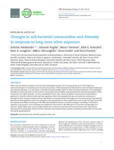
light on its functional involvement in hormone imbalanceinduced IR. Adiponectin stimulates AMPK phosphorylation and activation in the liver and muscle, thereby directly regulating glucose metabolism and insulin sensitivity in vitro and in vivo. Blocking AMPK activation by a dominant-negative mutant inhibited the effects of adiponectin, indicating that stimulation of glucose use and fatty acid combustion by adiponectin occurs through activation of AMPK (16). Reduced expression of gluconeogenic enzymes such as PEPCK and G6Pase is associated with elevated phosphorylation of hepatic AMPK (37), accounting for inhibition of endogenous glucose production by adiponectin (38). Our results extend these findings to sex hormone imbalance in FORKO females in which adiponectin signaling down-regulation also led to selective hepatic deactivation of AMPK and up-regulation of gluconeogenic enzymes PEPCK and G6Pase. Whereas we examined AMPK as one major known downstream pathway, the possible contribution of other pathways, such as peroxisomal proliferator-activated receptor-␣ activation, remain to be studied. This could become relevant because other studies have shown adiponectin to increase fatty acid combustion and energy consumption, in part via peroxisomal proliferator-activated receptor-␣ activation, coordinating increased insulin sensitivity (39). An important objective of our study was to test chronic HFD effect on hormone imbalance-induced obesity and IR in females. The response of DIO and IR in mice is strain dependent (40). C57BL/6 mice subjected to HFD develop severe obesity, hyperglycemia, and insulin resistance (31), whereas SV129 mice that are used in developing and propagating many gene knockouts are resistant to DIO and glucose intolerance (24). In this context, our observations in WT SV 129 strain are noteworthy. Despite the absence of body FIG. 8. Potential mechanisms linking visceral obesity and insulin resistance in FORKO mice. Chronic hormone imbalance causes visceral obesity to produce age-related IR in mutants. At the molecular level, this is preceded with decreases in components of adiponectin signaling pathway, including decrease in adiponectin mRNA and protein level in its active form; aberration of adiponectin receptors in AT, muscle, and liver and decrease of adiponectin downstream target AMPK activity in liver cause up-regulation of gluconeogenesis genes PEPCK and G6Pase in the liver. Therefore adiponectin appears to be an important mediator of hormone imbalances and eventual IR. Chronic HFD in FORKO mice causes dyslipidemia and lipid accumulation in tissues (e.g. hepatic and deep visceral fat depot) and associated with up-regulation of inflammation genes. Chronic HFD also conspires with chronic hormone imbalance exacerbating visceral obesity in FORKO, with additional potential to induce damage at later ages. Although not shown in this diagram chronic HFD selectively alters MAT depot in WT mice that are perceived to be resistant to DIO. Eventually such changes could independently lead to gradual and age-related metabolic disturbances.
Endocrinology, December 2007, 148(12):5667–5679
5677
weight gain and impaired glucose tolerance at 8 months in WT females challenged by chronic HFD, other adverse effects of potential metabolic significance are clearly evident. Thus, changes such as increased hepatic lipid accumulation and plasma TC as well as higher fat around mesenteric vessels and larger adipocyte size are notable and also accompanied by other changes at the molecular level. Up-regulation of components of inflammation pathway TNF-␣, CCL2, CCR2 and adipose depot-specific alteration of adiponectin mRNA and receptors are part of these alterations. It is also of interest to note that provision of HFD greatly exaggerated body weight, PAT mass, PAT adipose size, and liver lipid accumulation when conditions of chronic hormone imbalance prevailed. Inflammatory changes such as elevated TNF-␣ are associated with glucose and lipid disturbances (10). Our findings of TNF-␣ mRNA increasing in response to HFD, specifically in MAT depot in WT, and hormone imbalance in both MAT and PAT depot are of added significance. These are in general agreement with data in adiponectin knockout mice in which HFD-induced obesity and IR is accompanied by high TNF-␣ level (29). CCL2, a chemokine up-regulated in human obesity, modulates macrophage trafficking and activation (12). The CCL2/CCR2 pathway is necessary for macrophage recruitment, influencing the development of obesity and insulin resistance via adipose macrophage accumulation and inflammation (13). Our finding of up-regulation of CCL2/ CCR2 in response to HFD exclusively in MAT depot but not PAT suggests an important role for differential endocrine influence among sites within the AT depots because marked differences in their functional capacities and responsiveness to nutritional manipulations could exist. Thus, we highlight distinction within the visceral WAT, namely MAT with PAT,
5678
Endocrinology, December 2007, 148(12):5667–5679
assigning particular significance for MAT (a site in deep visceral fat). Because adipokines secreted from MAT go directly to portal circulation exerting relatively greater effects on hepatic function (41, 42), the possibility for enhancing metabolic alteration and damage appears greater. Higher CCL2 in MAT, compared with epididymal, perenephric, and sc fat depot in high-fat-fed obese mice, has also been reported by others (43). Taken together, our findings suggest that the increase in CCL2/CCR2 inflammatory signal from the MAT site might play a critical role in aggravating visceral obesityrelated conditions. The type of dietary fat (e.g. trans fat) could also become important in inducing depot-specific changes. Considering that low estrogen to androgen ratio regulates energy metabolism and induces obesity and MS (4) and findings of ER␣ as a main mediator of estrogen on energy homeostasis (44), the pattern of ER expression in FORKO female mice during aging becomes important. Although estrogen level is very low as early as 3 months and remains so at 12 months (4), ER␣ undergoes selective down-regulation only at a later age (9 months) but not earlier in both MAT and PAT depots. This suggests that loss of estrogen signaling in aging FORKOs is partly due to AT ER␣ down-regulation. Whether other components involved in estrogen signaling in AT are affected remains to be assessed. Nevertheless, the present results support the idea of drugs that can selectively modulate the activity of ERs could represent a frontier in treatment of IR (26); thus, our model might have implications for understanding the potential effects of treatments after long-term deprivation. In conclusion, we have constructed a probable scenario (Fig. 8) to explain the effects of hormonal imbalances on AT and IR in female mice. Chronic hormone imbalance-induced obesity and IR in 9-month FORKO female mice is associated with major changes in adiponectin and inflammation. In particular, we found down-regulation of adiponectin expression in AT and reduction of adiponectin active forms in plasma decrease p-AMPK and increase in PEPCK and G-6Pase in liver. Both AdipoR1 and AdipoR2 in AT are down-regulated; AdipoR1 in muscle is up-regulated and AdipoR2 in liver is down-regulated after HFD. ER␣ downregulation at later ages further diminish sex hormone signaling in AT, associated with gradual down-regulation of adiponectin activity, causing in parallel impaired glucose tolerance. Thus, adiponectin appears to be an important mediator of hormone imbalance-induced IR. Hence, interventions designed to restore adiponectin signaling could be useful in preventing hormone imbalance-induced IR. HFD alters visceral AT without necessarily inducing obesity (as in WT), and this effect is also associated with up-regulated inflammation genes. Chronic HFD conspires with hormone imbalance, exacerbating obesity phenotype inducing metabolic injury at later ages. Acknowledgments We thank Dr. P. E. Scherer (Albert Einstein College of Medicine, Bronx, NY) for the supply of antibody reagents used in this work. Analysis of lipids performed on the autoanalyzer with the cooperation of Dr. Lise Bernier in the hyperlipidemia and atherosclerosis laboratories of the Clinical Research Institute of Montreal is gratefully acknowledged.
Zhang et al. • Adipokine Changes in FORKO Mice
Received May 15, 2007. Accepted August 14, 2007. Address all correspondence and requests for reprints to: M. Ram Sairam, Ph.D., Director, Molecular Reproduction Research Laboratory, Clinical Research Institute of Montre´al, 110 Pine Avenue West, Montre´al, Que´bec, Canada H2W 1R7. E-mail:
[email protected]. This work was supported by a pilot grant from the Canadian Institutes for Health Research and focused giving program of Johnson & Johnson, USA. Disclosure statement: H.Z., X.C., and J.A. have nothing to declare. M.R.S. has received grant support from the focused giving program of Johnson & Johnson, USA.
References 1. Crawford SL, Johannes CB 1999 The epidemiology of cardiovascular disease in postmenopausal women. J Clin Endocrinol Metab 84:1803–1806 2. Reusch JE 2002 Current concepts in insulin resistance, type 2 diabetes mellitus, and the metabolic syndrome. Am J Cardiol 90:19G–26G 3. Manson JE, Hsia J, Johnson KC, Rossouw JE, Assaf AR, Lasser NL, Trevisan M, Black HR, Heckbert SR, Detrano R, Strickland OL, Wong ND, Crouse JR, Stein E, Cushman M, the Women’s Health Initiative Investigators 2003 Estrogen plus progestin and the risk of coronary heart disease. N Engl J Med 349:523–534 4. Sairam MR, Wang M, Danilovich N, Javeshghani D, Maysinger D 2006 Early obesity and age-related mimicry of metabolic syndrome in female mice with sex hormonal imbalances. Obesity 14:1142–1154 5. Takeda K, Toda K, Saibara T, Nakagawa M, Saika K, Onishi T, Sugiura T, Shizuta Y 2003 Progressive development of insulin resistance phenotype in male mice with complete aromatase (CYP19) deficiency. J Endocrinol 176: 237–246 6. Bray GA, Popkin BM 1998 Dietary fat intake does affect obesity! Am J Clin Nutr 68:1157–1173 7. Hill JO, Melanson EL, Wyatt HT 2000 Dietary fat intake and regulation of energy balance: implications for obesity. J Nutr 130:284 8. Chalkley SM, Hettiarachchi M, Chisholm DJ, Kraegen EW 2002 Long-term high-fat feeding leads to severe insulin resistance but not diabetes in Wistar rats. Am J Physiol Endocrinol Metab 282:E1231–E1238 9. Matsuzawa Y 2006 The metabolic syndrome and adipocytokines. FEBS Lett 580:2917–2921 10. Sharma AM, Staels B 2007 Peroxisome proliferator-activated receptor ␥ and adipose tissue— understanding obesity-related changes in regulation of lipid and glucose metabolism. J Clin Endocrinol Metab 92:386 –395 11. Hotamisligil G, Spiegelman B 1994 Tumor necrosis factor ␣: a key component of the obesity-diabetes link. Diabetes 11:1271–1278 12. Sartipy P, Loskutoff DJ 2003 Monocyte chemoattractant protein 1 in obesity and insulin resistance. Proc Natl Acad Sci USA 100:7265–7270 13. Weisberg S, Hunter D, Huber R, Lemieux J, Slaymaker S, Vaddi K, Charo I, Leibel R, Ferrante Jr AW 2006 CCR2 modulates inflammatory and metabolic effects of high-fat feeding. J Clin Invest 116:115–124 14. Pajvani UB, Hawkins M, Combs TP, Rajala MW, Doebber T, Berger JP, Wagner JA, Wu M, Knopps A, Xiang AH, Utzschneider KM, Kahn SE, Olefsky JM, Buchanan TA, Scherer PE 2004 Complex distribution, not absolute amount of adiponectin, correlates with thiazolidinedione-mediated improvement in insulin sensitivity. J Biol Chem 279:12152–12162 15. Yamauchi T, Kamon J, Ito Y, Tsuchida A, Yokomizo T, Kita S, Sugiyama T, Miyagishi M, Hara K, Tsunoda M, Murakami K, Ohteki T, Uchida S, Takekawa S, Waki H, Tsuno NH, Shibata Y, Terauchi Y, Froguel P, Tobe K, Koyasu S, Taira K, Kitamura T, Shimizu T, Nagai R, Kadowaki T 2003 Cloning of adiponectin receptors that mediate antidiabetic metabolic effects. Nature 423:762–769 16. Yamauchi T, Kamon J, Minokoshi Y, Ito Y, Waki H, Uchida S, Yamashita S, Noda M, Kita S, Ueki K, Eto K, Akanuma Y, Froguel P, Foufelle F, Ferre P, Carling D, Kimura S, Nagai R, Kahn BB, Kadowaki T 2002 Adiponectin stimulates glucose utilization and fatty-acid oxidation by activating AMPactivated protein kinase. Nat Med 8:1288 –1295 17. Turgeon JL, McDonnell DP, Martin KA, Wise PM 2004 Hormone therapy: physiological complexity belies therapeutic simplicity. Science 304:1269 –1273 18. Danilovich N, Babu PS, Xing W, Gerdes M, Krishnamurthy H, Sairam MR 2000 Estrogen deficiency, obesity, and skeletal abnormalities in follicle-stimulating hormone receptor knockout (FORKO) female mice. Endocrinology 141:4295– 4308 19. Dierich A, Sairam MR, Monaco L, Fimia GM, Gansmuller A, LeMeur M, Sassone-Corsi P 1998 Impairing follicle-stimulating hormone (FSH) signaling in vivo: targeted disruption of the FSH receptor leads to aberrant gametogenesis and hormonal imbalance. Proc Natl Acad Sci USA 95:13612–13617 20. Morton NM, Densmore V, Wamil M, Ramage L, Nichol K, Bunger L, Seckl JR, Kenyon CJ 2005 A polygenic model of the metabolic syndrome with reduced circulating and intra-adipose glucocorticoid action. Diabetes 54:3371– 3378 21. Waki H, Yamauchi T, Kamon J, Ito Y, Uchida S, Kita S, Hara K, Hada Y,
Zhang et al. • Adipokine Changes in FORKO Mice
22.
23. 24.
25. 26. 27.
28. 29.
30.
31. 32.
Vasseur F, Froguel P, Kimura S, Nagai R, Kadowaki T 2003 Impaired multimerization of human adiponectin mutants associated with diabetes: molecular structure and multimer formation of adiponectin. J Biol Chem 278:40352– 40363 Kim CH, Pennisi P, Zhao H, Yakar S, Kaufman JB, Iganaki K, Shiloach J, Scherer PE, Quon MJ, LeRoith D 2006 MKR mice are resistant to the metabolic actions of both insulin and adiponectin: discordance between insulin resistance and adiponectin responsiveness. Am J Physiol Endocrinol Metab 291:E298 – E305 Chen X, Aravindakshan J, Yang Y, Tiwari-Pandey R, Sairam MR 2006 Aberrant expression of PDGF ligands and receptors in the tumor prone ovary of follitropin receptor knockout (FORKO) mouse. Carcinogenesis 27:903–915 Challis BG, Coll AP, Yeo GSH, Pinnock SB, Dickson SL, Thresher RR, Dixon J, Zahn D, Rochford JJ, White A, Oliver RL, Millington G, Aparicio SA, Colledge WH, Russ AP, Carlton MB, O’Rahilly S 2004 Mice lacking proopiomelanocortin are sensitive to high-fat feeding but respond normally to the acute anorectic effects of peptide-YY3–36. Proc Natl Acad Sci USA 101:4695– 4700 Lavoie M, Gauthier S 2006 Regulation of fat metabolism in the liver: link to non-alcoholic hepatic steatosis and impact of physical exercise. Cell Mol Life Sci 63:1393–1409 Barros RPA, Machado UF, Gustafsson JA 2006 Estrogen receptors: new players in diabetes mellitus. Trends Mol Med 12:425– 431 Bauche IB, Ait El Mkadem S, Rezsohazy R, Funahashi T, Maeda N, Miranda LM, Brichard SM 2006 Adiponectin downregulates its own production and the expression of its AdipoR2 receptor in transgenic mice. Biochem Biophys Res Commun 345:1414 –1424 Inukai K, Nakashima Y, Watanabe M, Takata N, Sawa T, Kurihara S, Awata T, Katayama S 2005 Regulation of adiponectin receptor gene expression in diabetic mice. Am J Physiol Endocrinol Metab 288:E876 –E882 Maeda N, Shimomura I, Kishida K, Nishizawa H, Matsuda M, Nagaretani H, Furuyama N, Kondo H, Takahashi M, Arita Y, Komuro R, Ouchi N, Kihara S, Tochino Y, Okutomi K, Horie M, Takeda S, Aoyama T, Funahashi T, Matsuzawa Y 2002 Diet-induced insulin resistance in mice lacking adiponectin/ACRP30. Nat Med 8:731–737 Hara K, Boutin P, Mori Y, Tobe K, Dina C, Yasuda K, Yamauchi T, Otabe S, Okada T, Eto K, Kadowaki H, Hagura R, Akanuma Y, Yazaki Y, Nagai R, Taniyama M, Matsubara K, Yoda M, Nakano Y, Kimura S, Tomita M, Kimura S, Ito C, Froguel P, Kadowaki T 2002 Genetic variation in the gene encoding adiponectin is associated with an increased risk of type 2 diabetes in the Japanese population. Diabetes 51:536 –540 Bullen J, Bluher S, Kelesidis T, Mantzoros CS 2007 Regulation of adiponectin and its receptors in response to development of diet-induced obesity in mice. Am J Physiol Endocrinol Metab 292:E1079 –E1086 Combs TP, Berg AH, Rajala MW, Klebanov S, Iyengar P, Jimenez-Chillaron JC, Patti ME, Klein SL, Weinstein RS, Scherer PE 2003 Sexual differentiation,
Endocrinology, December 2007, 148(12):5667–5679
33.
34.
35. 36. 37.
38. 39.
40.
41. 42. 43. 44.
5679
pregnancy, calorie restriction, and aging affect the adipocyte-specific secretory protein adiponectin. Diabetes 52:268 –276 Xu A, Chan KW, Hoo RLC, Wang Y, Tan KCB, Zhang J, Chen B, Lam MC, Tse C, Cooper GJS, Lam KSL 2005 Testosterone selectively reduces the high molecular weight form of adiponectin by inhibiting its secretion from adipocytes. J Biol Chem 280:18073–18080 Yamauchi T, Kamon J, Waki H, Terauchi Y, Kubota N, Hara K, Mori Y, Ide T, Murakami K, Tsuboyama-Kasaoka N, Ezaki O, Akanuma Y, Gavrilova O, Vinson C, Reitman M, Kagechika H, Shudo K, Yoda M, Nakano Y, Tobe K, Nagai R, Kimura S, Kimura, Tomita M, Froguel P, Kadowaki T 2001 The fat-derived hormone adiponectin reverses insulin resistance associated with both lipoatrophy and obesity. Nat Med 7:941–948 Oana F, Takeda H, Matsuzawa A, Akahane S, Isaji M, Akahane M 2005 Adiponectin receptor 2 expression in liver and insulin resistance in db/db mice given a 3-adrenoceptor agonist. Eur J Pharmacol 518:71–76 Barnea M, Shamay A, Stark AH, Madar Z 2006 A high-fat diet has a tissuespecific effect on adiponectin and related enzyme expression. Obesity 14:2145– 2153 Lochhead PA, Salt IP, Walker KS, Hardie DG, Sutherland C 2000 5-Aminoimidazole-4-carboxamide riboside mimics the effects of insulin on the expression of the 2 key gluconeogenic genes PEPCK and glucose-6-phosphatase. Diabetes 49:896 –903 Combs TP, Berg AH, Obici S, Scherer PE, Rossetti L 2001 Endogenous glucose production is inhibited by the adipose-derived protein Acrp30. J Clin Invest 108:1875–1881 Yamauchi T, Kamon J, Waki H, Imai Y, Shimozawa N, Hioki K, Uchida S, Ito Y, Takakuwa K, Matsui J, Takata M, Eto K, Terauchi Y, Komeda K, Tsunoda M, Murakami K, Ohnishi Y, Naitoh T, Yamamura K, Ueyama Y, Froguel P, Kimura S, Nagai R, Kadowaki T 2003 Globular adiponectin protected ob/ob mice from diabetes and ApoE-deficient mice from atherosclerosis. J Biol Chem 278:2461–2468 Svenson KL, Von Smith R, Magnani PA, Suetin HR, Paigen B, Naggert JK, Li R, Churchill GA, Peters LL 2007 Multiple trait measurements in 43 inbred mouse strains capture the phenotypic diversity characteristic of human populations. J Appl Physiol 102:2369 –2378 Bjorntorp P 1990 “Portal” adipose tissue as a generator of risk factors for cardiovascular disease and diabetes. Arteriosclerosis 10:493– 496 Kershaw EE, Flier JS 2004 Adipose tissue as an endocrine organ. J Clin Endocrinol Metab 89:2548 –2556 Yu R, Kim CS, Kwon BS, Kawada T 2006 Mesenteric adipose tissue-derived monocyte chemoattractant protein-1 plays a crucial role in adipose tissue macrophage migration and activation in obese mice. Obesity 14:1353–1362 Musatov S, Chen W, Pfaff DW, Mobbs CV, Yang XJ, Clegg DJ, Kaplitt MG, Ogawa S 2007 Silencing of estrogen receptor ␣ in the ventromedial nucleus of hypothalamus leads to metabolic syndrome. Proc Natl Acad Sci USA 104:2501–2506
Endocrinology is published monthly by The Endocrine Society (http://www.endo-society.org), the foremost professional society serving the endocrine community.