We measured bone remodeling of the proximal tibia prospectively for 3 years after uncemented total knee arthroplasty (TKA) in 25 knees with primary arthrosis.
Acta Orthop Scand 1995;66 (6): 513-51 6
513
Changes in bone mineral density of the proximal tibia after uncemented total knee arthroplasty A 3-year follow-up of 25 knees
Acta Orthop Downloaded from informahealthcare.com by 186.93.205.60 on 05/20/14 For personal use only.
Michael M Petersen, Poul T Nielsen, Jes B Lauritzen and Bjarne Lund We measured bone remodeling of the proximal tibia prospectively for 3 years after uncemented total knee arthroplasty (TKA) in 25 knees with primary arthrosis. In the trabecular bone below the tibial component, bone mineral density (BMD) was measured in 6 different regions of interest (ROI), using dual photon absorptiometry (DPA). In the tibial condyles, where the change in knee alignment indicated
that the load was reduced postoperatively, a fast bone loss of 7-20% was seen during the first 6 months after surgery. A small, but significant increase in BMD of 2-7% was seen in the tibial condyles, where the load was increased. On average, the density for all ROI below the tibial component showed a significant and progressive decrease in BMD, reaching 22% at 3 years follow-up.
Department of Orthopedics U 2161, Rigshospitalet, University of Copenhagen, Blegdamsvej 9, DK-2100 Copenhagen 0, Denmark. Tel+45 35-452781. Fax -456733 Submitted 95-05-02. Accepted 95-09-17
Bone strength and bone mineral in the trabecular bone of the proximal tibia are a critical determinant of the failure rate in total knee arthroplasty (TKA) (Christensen et al. 1982, Hvid and Hansen 1986, Harada et al. 1988, Hvid 1988, Zysset et al. 1994). The failure rate following TKA has decreased during the past years (Knutson et al. 1994), but one problem is still the fixation of the tibial component (Windsor et al. 1989, Moran et al. 1991). Only a few investigators have measured prospectively the changes in bone mineral density (BMD) of the proximal tibia, below the tibial component, following TKA (Bohr and Lund 1987, Hvid et al. 1988). We measured the changes in BMD in the proximal tibia following uncemented TKA and evaluated the influence of pre- and postoperative knee alignment on bone remodeling.
the operations was evaluated by The Hospital for Special Surgery Knee-rating score (HSS knee-score) (Insall et al. 1976) (Table I). All knees had pre- and postoperative measurements of the mechanical axis, measured as the hipknee-ankle angle on long-leg standing radiographs (Table 1). 21 patients had preoperative varus alignment corrected to either valgus or neutral alignment (n 12), decreased varus alignment (n 7) or increased varus alignment (n 2). 2 patients had an unchanged alignment after the operation and 2 patients had valgus alignment corrected to varus and decreased valgus alignment, respectively. Measurements of BMD in the proximal tibia were performed by dual photon absorptiometry (DPA) using the radiation peaks of 44 and 100 KeV from a
Patients and methods
Table 1. Clinical data in 25 patients
28 consecutive patients with primary arthrosis of the knee were included in the study. All were given an uncemented TKA (PCA Modular@).3 patients were excluded during the study period; 1 patient died within the first postoperative year and 2 patients were unable to keep the knee still during examination, causing poor quality in the BMD scannings, thus leaving 25 patients with 25 TKAs for the study (Table 1). The operations were performed as decribed by Hungerford et al. (1982). and the clinical outcome of
Patients Age Femalehnale
70 (52-81) 19 I 6
HSS-score Preop. 1 year 3 years
51 (32-65) 84 (56-96) 85 (52-97)
Knee alignment (HKA) Preop. Postop. ~-
172" (160°-1860) 180" (172'-196") -_ --
Copyri%qht 0 Scandinavian University Press 1YY.5'. ISSN 0001-M70. Primed in Swerlerr - ull rights reserw(l.
___
-
-
Acta Orthop Downloaded from informahealthcare.com by 186.93.205.60 on 05/20/14 For personal use only.
514
Acfa Orthop Scand 1995;66 (6):51 3-51 6
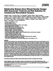
Figure 1 . Scan plot from a DPA-scanning performed in the proximal tibia after implantation of an uncemented total knee arthroplasty (PCA ModulaF)).The location of the ROI used for the measurements of BMD medially and laterally below the tibial component is shown. ROI 1 is the area located proximally between the fixation peg and the medialllateral peripheral rim of the bone, ROI 2 is the area located distally just below the fixation peg in the medialllateral tibial condyle, and ROI 3 is the area located proximally in the central part of the bone between the fixation screw and the medialllateralfixation peg.
153gadoliniumsource (200 mCi) in a custom-made knee scanner (GT-50, Tibia-la, Gammatec A/S, Stormly 16, Vrerl~se,Denmark). The scanner was specially designed for bone mineral measurements in the proximal tibia. Scanning was performed with a spatial resolution (pixel size) of 2 by 2 mm. To obtain reproducible scanning statistics, the scan speed (3-6 mm/s) was automatically adjusted according to source decay. The scannings were performed in the coronal plane, with the knee extended and the foot in an upright position, and placed in a device designed to secure the same rotation of the limb at follow-up. On the computerized scan plot 6 regions of interest
(ROI) in the trabecular bone below the tibial component were selected for BMD measurements; 3 ROI around the medial fixation peg and 3 ROI around the lateral peg (Figure 1). The ROIs in the majority of the patients were of 0.8 x 0.8 cm (16 pixels). In some of the patients one or more of the ROIs were smaller because of the anatomical dimensions of the proximal tibia; the minimal acceptable size was 8 pixels, which was very seldom used. The initial size of a ROI in a patient was kept at the same size throughout the study. In all patients BMD measurements were performed within 2 weeks after the operation and at follow-up after 6 months, I,2 and, 3 years. The precision of BMD measurements was evaluated in 8 patients measured twice on the same day, with full reposition between each scanning. Statistics The precision expressed as the coefficient of variation (CV) was calculated as CV = SD/mean. The nonparametric test a.m. Wilcoxon for paired data. and nonparametric two way analysis of variance (Friedman test) for repeated measurements were used. Results are given as mean and total range.
Results (Table 2) The mean precision of measurements of BMD in the trabecular bone of the proximal tibia below the tibial component was 3.8 (2.8-4.7)%. On the basis of the
Table 2. Changes in mean BMD (gkm2) at different locations in the proximal tibia in patients who had a change in knee allgnment (leading to altered load in the tibial condyles) following TKA (n 23),and the mean changes in BMD in all areas (ROI numbers 1-3 in both tibial condyles) (n 25). Mean (range) and percent change of mean compared to initial postop value -__ 6 months -
ROI number (load)
Postop
ROI 1 (decreased)
0.85 (0.52-1.33)
ROI 2 (decreased)
0.80 (0.561.24) 0.74 (0.45-1.08) 0.69 (0.39-1.04) 0.67 (0.40-1.08) 0.62 (0.49-0.93) 0.73 (0.51-0.99)
ROI 3 (decreased)
ROI 1 (increased) ROI 2 (increased)
ROI 3 (increased) All ROI (11-25) a
-.-
'
0.68 -20% (0.26-0.99) 0.70' -13% (0.40-1.04) 0.693 -7% (0.28-1 -03) 0.73 6% (0.35-1.21) 0.728 7% (0.42-1 .lo) 0.63 2% (0.18-1.23) 0.69 -5% (0.41-1.00)
Significantly different (p < 0.05) from initial value (Wlkxxon test). Friedman test p < 0.00005 (repeated measurementsover time).
1 year 0.62 ' -27% (0.19-0.86) 0.67' -16% (0.37-0.98) 0.65 -12% (0.23-0.99) 0.68 -1% (0.30-1.15) 0.69 3% (0.39-1.16) 0.61 -2% (0.16-1.20)
'
'
0.67 -8Oh (0.39-0.92)
2 years
3 years
0.60a -29% (0.17495) 0.64' -20% (0.31-1.06) 0.63' -15% (0.21-0.96)
0.51 a -40% (0.04-0.88) 0.61 ' -24% (0.32-1.05) 0.56' -24% (0.27-1 .OO)
0.63' -9% (0.31-1.02)
0.54 a -22% (0.14-1.00) 0.59 a -12% (0.24-1.12) 0.49" -21% (0.16-1 .lo) 0.57' -22% (0.30-0.91)
0% 0.67 (0.33-1.04)
0.55 -11% (0.23-1.17)
'
0.63 -14% (0.37-0.91)
Acta Orthop Scand 1995; 66 (6): 513-51 6
Acta Orthop Downloaded from informahealthcare.com by 186.93.205.60 on 05/20/14 For personal use only.
change in alignment of each knee, we decided whether the load in the medial and lateral tibial condyles had decreased or increased as a consequence of the operation. In the condyles with decreased load (n 23). a significant decrease in BMD, starting at 6 months and continuing throughout the study period, was seen in all ROIs. In the tibial condyles with increased load (n 23), a temporary increase in BMD was seen, followed by a slow decrease. The average density of all ROIs below the tibial component (n 25) showed a significant and progressive decrease in BMD, reaching 22% at 3 years of follow-up (Table 2).
Discussion The average precision of measurements of BMD in ROI in the trabecular bone below the tibial component was 3.8%. This precision is not essentially different from that obtained when BMD of the proximal tibia is measured in patients without TKA or other orthopedic implants (Madsen et al. 1994, Petersen et al. 1995). It is also on the same level of precision obtainable using DEXA around the femoral component in uncemented total hip or knee arthroplasty (Kearns McCarthy et al. 1991, Kiratli et al. 1992, Kilgus et al. 1993, Trevisan et al. 1993, Robertson et al. 1994). In the tibial condyles, with decreased load, a fast bone loss of 7-20% was seen. A small increase in BMD of 2-7% was seen in the tibial condyles, with increased load. These early changes in BMD are in agreement with Wolff’s Law and recent studies on bone remodeling as a response to altered mechanical load (Andersson and Nilsson 1979, Lanyon 1984, Margulies et al. 1986). Increased load proved to be a stimulus so strong that it could induce a local increase in BMD, despite the fact that the patients evaluated had a mean age of 70 years. In a study using quantitative computed tomography, Hvid et al. (1988) measured trabecular bone remodeling of the proximal tibia following TKA with a cemented non-metal-backed tibial component in 18 patients (9 with arthrosis and 9 with rheumatoid arthritis) within the first postoperative week and 2 years postoperatively. During the observation period, the mean bone density had decreased significantly in the preoperatively more loaded tibial condyle, while the density in the preoperatively less loaded condyles was unchanged. We consider the adaptive bone remodeling in this study to be in agreement with our results. Posttraumatic loss of bone mineral following fractures is well known (Andersson and Nilsson 1979, Petersen et al. 1992). but it is uncertain to which
515
degree the bone loss is caused by immobilization or by the trauma of the bone itself. Operation with insertion of a knee or a hip arthroplasty represents a substantial trauma to the bone. Several studies have shown, that even though the level of activity of these patients is increased postoperatively, no permanent increase in bone mineral occurs (Lindberg and Nilsson 1984, Ruegsegger et al. 1986, Bohr and Lund 1987, Hvid et al. 1988, Adolphson et al. 1993). In our study, the long-term change in average BMD of the proximal tibia following uncemented TKA was an overall loss of bone mineral of 22% during the first 3 postoperative years. In the study by Hvid et al. (1988). the average decrease in bone density after 2 years was 32% in rheumatoid arthritis and 11% in arthrosis. In a study by Bohr and Lund ( I 987). a temporary increase in average BMD in the proximal tibia was found 6 months after uncemented arthroplasty. During the next year, BMD decreased to the initial level. However, because of the study design, the results of this study were strongly influenced by ageand sex-related inter-individual differences in bone mineral of the proximal tibia (Bohr and Schaadt 1987, Petersen et al. 1993). An average bone loss of more than 20% in the proximal tibia 3 years after TKA must be considered to be of clinical importance in relation to the normal annual loss of 1% (Bohr and Schaadt 1987, Checovich et al. 1989, Petersen et al. 1993). It is well known that there is a close relation between BMD and the strength of trabecular bone (Hansson et al. 1980, Hvid et al. 1985). and theoretically an increased risk of fractures or loosening of the tibial component might be expected in patients with a high postoperative bone loss. However, the long-term results following TKA are satisfactory with a low revision rate and fractures of the proximal tibia are rare. Preoperatively, most patients with arthrosis of the knee have bone mineral values of the proximal tibia above average in normals (Petersen et al. 1993) and a part of the bone loss after TKA might represent a return to normal of the relatively high-density arthrotic bone.
Acknowledgements Financial support for this study was provided by the Velux Foundation of 1981 and Danish Hospital Foundation for Medical Research, Region of Copenhagen. The Faroe Islands and Greenland. The authors wish to thank lab. tech. Merete Tardrup for performing some of the BMD measurements.
51 6
Acta Orthop Downloaded from informahealthcare.com by 186.93.205.60 on 05/20/14 For personal use only.
References Adolphson P, von Sivers K, DalCn N, Jonsson U, Dahlborn fter hip arthroplasty. Acta M. Bone and muscle ma Orthop Stand 1993: 64:181-4. Antlersson S M, Nilsson B E. Changes in bone mineral content following tibia shaft fractures. Clin Orthop 1979; 144: 226-9. Bohr H H. Lund B. Bone mineral density of the proximal tibia following uncemented arthroplasty. J Arthroplasty 1987; 2: 309-12. Bohr H H, Schaadt 0. Mineral content of the upper tibia assessed by dual photon densitometry. Acta Orthop Stand 1987; 58: 557-9. Checovich M M, Kiratli B J, Smith E L. Dual photon absorptiometry of the proximal tibia. Calcif Tissue Int 1989; 45: 281-4. Christensen P, Kjaer J, Melsen F, Nielsen H E, Sneppen 0, Vang P-S. The subchondral bone of the proximal tibial epiphysis in osteoathritis of the knee. Acta Orthop Scand 1982; 53: 889-95. Hansson T, Roos B, Nachemson A. The bone mineral content and ultimate compressive strength of lumbar vertebrae. Spine 1980; 5 : 46-55. Harada Y. Wevers H W, Cooke T D V. Distribution of bone strength in the proximal tibia. J Arthroplasty 1988; 3: 167-75. Hungerford D S, Kenna R V, Krackow K A. The porouscoated anatomic total knee. Orthop Clin North Am 1982; 13: 103-22. Hvid 1. Trabecular bone strength at the knee. Clin Orthop 1988; 227: 210-21. Hvid I, Hansen S L. Subchondral bone strength in arthrosis. Acta Orthop Scand 1986; 57: 47-51. Hvid I, Jensen N C, Biinger C, S l u n d K, Djurhuus J C. Bone mineral assay: its relation to the mechanical strength of cancellous bone. Eng Med 1985; 1 4 79-83. Hvid I, Bentzen S M, Jorgensen J. Remodeling of the tibial plateau after knee replacement. Acta Orthop Scand 1988; 59: 567-73. Insall J N, Ranawat C S, Aglietti P, Shine J. A comparison of four models of total knee-replacement prostheses. J Bone Joint Surg (Am) 1976 58: 754-65. Keams McCarthy C, Steinberg G G, Agren M,Leahey D, Wyman E, Baran D T. Quantifying bone loss from the proximal femur after total hip arthroplasty. J Bone Joint Surg (Br) 1991; 73: 774-8. Kilgus D J, Shimaoka E E, Tipton J S, Eberle R W. Dualenergy X-ray absorptiometry measurement of bone mineral density around porous-coated cementless femoral implants. J Bone Joint Surg (Br) 1993; 75: 279-87. Kiratli B J. Heiner J P, McBeat A A, Wilson M A. Determination of bone mineral density by dual X-ray absorptiometry in patients with uncemented total hip arthroplasty. J M o p Res 1992; 1 0 836-44.
Acta Orthop Scand 1995;66 (6):51 3-51 6
Knutson K, Lewold S. Robertsson 0, Lidgren L. The Swedish knee arthroplasty register. A nation-wide study of 30,003 knees 19761992. Acta Orthop Scand 1994; 65: 375-86. Lanyon L E. Functional strain as a determinant for bone remodeling. CalcifTissue lnt (Suppl I ) 19x4; 36: 56-61. Lindberg H, Nilsson B. Changes in bone mineral content in the femur following total hip arthroplasty. Clin Orthop 1984; 183: 276-9. Madsen 0 R. Schaadt 0. Bliddal H, Egsmose C, Sylvest J. Bone mineral distribution of the proximal tibia in gonarthrosis assessed in vivo by photon absorption. Osteoarthritis Cartilage 1994; 2: 141-7. Margulies J Y,Simkin A, Leichter I, Bivas A, Steinberg R, Giladi M, Stein M, Kashtan H, Milgrom C. Effect of intense physical activity on the bone-mineral content in the lower limbs of young adults. J Bone Joint Surg (Am) 1986; 68: 1090-3. Moran C G, Pinder I M, Lees T A, Midwinter M J. Survivorship analysis of the uncemented porous-coated anatomic knee replacement. J Bone Joint Surg (Am) 1991; 73: 848-57. Petersen M M, Olsen C, Lauritzen J B, Lund B. Changes in bone mineral content in the proximal tibia following ankle fracture. Eur J Exp Musculoskel Res 1992; I: 7780. Petersen M M, Olsen C, Lauritzen J B, Lund B. Bone mineral content assessed by dual photon absorptiometry in the proximal tibia: normative data and measurements in orthopedic conditions. Eur J Exp Musculoskel Res 1993; 2: 121-6. Petersen M M, Olsen C, Lauritzen J B, Lund B, Hede A. Late changes in bone mineral density in the proximal tibia following total versus partial medial meniscectomy. A randomised study. J Orthop Res 1995 (in press). Robertson D D, Mintzer C M, Weisman B N, Ewald F C, LeBoff M, Spector M. Distal loss of femoral bone following total knee arthroplasty. J Bone Joint Surg (Am) 1994; 76: 66-76. Riiegsegger P, Seitz P, Gschwend N, Dubs L. Disuse osteoporosis in patients with total hip prostheses. Arch Orthop Trauma Surg 1986; 105: 268-73. Trevisan C. Bigoni M, Cherubini R, Steiger P, Randelli G, Ortolani S. Dual X-ray absorptiometry for the evaluation of bone density from the proximal femur after total hip arthroplasty: analysis protocols and reproducibility. Calcif Tissue Int 1993; 53: 158-61. Windsor R E, Scuderi G R, Moran M C, Insall J N. Mechanisms of failure of the femoral and tibial components in total knee arthroplasty. Clin Orthop 1989; 248: 15-20. Zysset P K, Sonny M, Hayes W C. Morphology-mechanical property relations in trabecular bone of the osteoarthritic proximal tibia. J Arthroplasty 1994; 9: 203-16.