Changes in Postural Control Parameters after Vestibular ...
Recommend Documents
Hamlet Suarez. Laboratory of Audiology and Vestibular Pathophysiology. School of Medicine. C. de Guayaquil 1332. Montevideo 11400. Uruguay. Tel.: +598 2 ...
Apr 26, 2012 - Training Using a Video Game Balance Board Program and 2. Standard ...... AccuSway System; Advanced Mechanical Technology Inc, 176.
The authors declare no conflicts of interest. Address correspondence to: Fabiano Politti, PT, PhD, Rua. Vergueiro, 2355 â Liberdade, São Paulo 01504-001, SP,.
Apr 22, 2014 - Background: Women undergoing mastectomy may have postural asymmetries due to mutilation, culminating in changes in postural balance.
Goldfish (Carassius auratus) of the comet variety were used, ranging in length ... An experimental tank, in which postural behaviour could be recorded under.
of the slovak Ministry of education and the slovak academy of sciences (project. 1/0070/11). Tento clanek podleha autorskemu zakonu a jeho vyuziti je mozne v ...
Vestibular dysfunction and postural balance in ... - SciELO › publication › fulltext › Vestibular... › publication › fulltext › Vestibular...by JJ Melo · 2018 · Cited by 1 · Related articlesTratado de Audiologia. São Paulo: Santos; 2011. p.404-25.
AbstractâThe objective for this study was to investigate whether the adaptation of postural control was similar during galvanic vestibular stimulation and during ...
42nd Department of Obstetrics and Gynecology, Aretaieio Hospital, School of Medicine, University of Athens, Athens, .... standard physiotherapy protocol.
children that causes poor cosmesis and function [1â3]. .... FDL, flexor digitorum longus; FDO, femoral derotational osteotomy; FHL, flexor hallucis longus; psoas,.
Contraversive pushing, called. ''Pusher syndrome,'' is the most .... assessed by the Berg Balance Scale [23], and the acquisition of APAs associated with a newly ...
our algorithms in assessing balance, we investigated the two-dimensional sway of COM and RCI in 21 healthy subjects and 17 patients with diabetic peripheral ...
Neurodegenerative diseases such as diabetic neuropathy, Parkinson's, and ...... healthy subjects to compensate movement of the proximal segment via ...
Page 01 of 15 ... delimitação de publicação dos últimos 10 anos, em lÃngua inglesa, com as ... com a perda do equilibrio e aumento das quedas em idosos.
as 1913, Mosher stressed the importance of shifting the body weight from side to side .... [23] Johnston RB., Howard ME, Cawley PW, Losse GM. Effect of lower extremity ... [27] Winter DA, Prince F, Frank JS, Powell C, Zabjek KF. Unified theory.
batteries, which was under computer control via a USB ... addition to pseudorandom GVS, we could also drive the ..... recover postural stability with time.
Jan 15, 2013 - Acta Oto-Laryngologica, 2013; Early Online, 1â6. ORIGINAL ARTICLE. Vestibular compensation after vestibular schwannoma surgery:.
For ML time series, ApEn values declined after injury in all sensory ... among athletes relatively high,2 3 but recurrent concus- sion has been associated with ...
2 Dick R. National Collegiate Athletic Association (NCAA) injury surveillance ... 18 Newell K. Degrees of freedom and the development of postural center of.
w Åwietle metody fotogrametrycznej z wykorzystaniem zjawiska Moire'a. Medycyna sportowa; 157: 303-307. Received 20.07.2014. Accepted ?.09.2014.
Nov 10, 2008 - Journal of the International AIDS. Society. Open Access. Poster presentation. Changes in hematological parameters after switching treatment of.
Apr 3, 2015 - term heart rate variability analyses of all the patients were obtained, and ..... They used 24-hour Holter monitoring to calculate frequency.
Changes in Postural Control Parameters after Vestibular ...
their PC. Seven of these patients were assessed again after a period of 1295 ... Recovery after peripheral vestibular lesions can ... level with N data points, |2.
Acta Otolaryngol 2003; 123: 143 – 147
Changes in Postural Control Parameters after Vestibular Rehabilitation in Patients with Central Vestibular Disorders H. SUAREZ1, M. AROCENA2, A. SUAREZ1, T. A. DE ARTAGAVEYTIA1, P. MUSE3 and J. GIL4 From the 1Laboratory of Audiology and Vestibular Pathophysiology, School of Medicine 2Department of Physical Medicine, British Hospital, and the Departments of 3Biomedical Engineering and 4Statistics, School of Medicine, Monte6ideo, Uruguay
Suarez H, Arocena M, Suarez A, De Artagaveytia TA, Muse P, Gil J. Changes in postural control parameters after 6estibular rehabilitation in patients with central 6estibular disorders. Acta Otolaryngol 2003; 123: 143– 147. Objective— The aim of this study was to determine postural responses before and after a vestibular rehabilitation program (VRP) in 14 patients with central vestibular disorders (CVD). Material and Methods —The confidence ellipse (CE) of the center of pressure distribution area and the sway velocity (SV) were the parameters used for the quantitative assessment of postural control (PC). These two parameters were analyzed before and after a VRP for two visual conditions. Behavioral postural responses were studied by means of the time– frequency scalogram using wavelets and the sway frequency content was measured in arbitrary units of energy density. Results — Ten patients showed a significant decrease in the CE and SV after the rehabilitative treatment, thus improving their PC. Seven of these patients were assessed again after a period of 12 9 5 months, during which they had not received any physical training. All of them showed increases in the CE and SV, indicating an impairment of PC. Conclusions — Many CVD patients damage the neural mechanisms involved in retaining the plastic changes in the PC parameters after rehabilitative treatment. Continuation of training may be necessary in order to maintain the improvement in PC obtained with a VRP. Key words: central 6estibular disorders, postural responses, 6estibular plasticity, 6estibular rehabilitation.
(CNS) disorders, in whom sensory information is usually wrongly processed in the CNS (4, 5). The COP behavior under these two visual conditions was analyzed using a time –frequency scalogram, and the variability of its amplitudes and frequencies in those patients who abandoned the VRP was redetermined several months later. Our aim was to see if the CVD patients maintained both their PC values and the underlying clinical and pathophysiological cues, in an attempt to understand the compensation process that occurs in these patients. MATERIAL AND METHODS Patients Postural responses before and after a VRP were assessed in a group of 14 CVD patients (average age 73.79 10.5 years) (Table I) treated with a customized VRP. Before treatment the patients were evaluated clinically using the Dizziness Handicap Inventory (DHI) (6) and the Test for Equilibrium Under Altered Sensory Conditions (TEUSAC) (7). In addition, electronystagmography (ENG) was used to assess spontaneous and positional nystagmus, quantitative and qualitative measures of smooth pursuit, saccades, optokinetic nystagmus, the vestibulo-ocular reflex and its visual suppression (8). All patients were assessed using audiological testing and either MRI or CT. Elderly patients with Parkinson’s disease, musculoskeletal disturbances or dementia were excluded. DOI 10.1080/0036554021000028109
2.84 2.98 1.31 1.14 1.26 X 2.11 2.14 1.99 1.70 2.50 1.65 1.90 2.80 1.81 2.17 1.04 1.19 1.46 7.80 1.74 1.41 1.65 1.27 2.26 1.12 1.09 1.54 3.26 2.77 1.24 2.85 1.18 9.77 2.82 1.58 1.74 1.39 2.04 1.86 1.84 3.15 6.01 28.35 4.49 16.15 X X X 4.33 3.33 11.07 11.70 3.85 8.12 10.15 F M F F F M M F F M F M F F 1 2 3 4 5 6 7 8 9 10 11 12 13 14
7.97 5.35 2.32 5.47 X X X 2.50 2.55 2.60 3.23 2.10 2.80 3.70
After After Before Before Gender
Etiology
Vestibulo-ocular disorder Before
After
After
Before
Acta Otolaryngol 123
Age (years) Patient No.
Stable visual surround OK stimuli Stable visual surround OK stimuli
CE (cm2)
Table I. Characteristics and CE and SV 6alues of the 14 CVD patients treated with the VRP
SV (cm/s)
H. Suarez et al.
Informed consent was obtained from all subjects before inclusion in the study. Methods Postural responses were recorded with a platform, by measuring online the two relevant parameters of the behavior of the COP, namely SV and CE. SV. An 80-s trial was recorded, leading to 2 discrete signals of N=4000 samples (sampling frequency fS = 50 Hz): COPx and COPy. Then, for each record, the average speed of COP along its path (6) was calculated, at t= 10 s (N= 500) and t= 80 s (N= 4000), using: 6=
fS N % [(COPxi − COPxi − 1)2 N i=2
+(COPyi − COPyi − 1)2]1/2 CE. The 95% CE of the bivariate distribution (COPxi, COPyi ), 15 i5 N, is the ellipse within which 95% of the COP samples are expected to be enclosed. It can be shown that the area of the 95% CE is Area= 2yF0.05[2,N − 2] | 2x| 2y − | 2xy where F0.05[2,N − 2] is the statistic at the 95% confidence level with N data points, | 2x and | 2y are the variance of the medio-lateral (ML) and antero-posterior (AP) coordinates, respectively, and |xy is the covariance. For a large sample size (N\ 120), F0.05[2,N-2] is 3.00. This is the case here as we calculated the 95% CE of 80-s trials (N= 4000). The CE and SV were measured for two different conditions according to the following stimulation paradigm: 1. Standing position, eyes open. 2. Standing position, eyes open with surrounding optokinetic (OK) stimulation (clockwise and counterclockwise) at an angular velocity of 65°/s. Time –frequency analysis (scalogram). In order to evaluate the fundamental oscillatory frequency, its amplitude and the temporal behavior of the responses, a time–frequency analysis of COP in both directions (COPx and COPy) was performed, by computing its scalogram. As the Fourier Transform is not suitable for the analysis of non-stationary signals such as COP signals, its time–frequency representations must be considered. A widely-used time– frequency energy density, because of its resolution properties, is the scalogram. The scalogram of a signal x(u) is the energetic version of the Wavelet Transform (WT) defined as the square magnitude of the WT:
)&
+
SCALX (t, f )=
−
x(u) ·
'
)
2 f f · · (u−t) · du f0 f0
Acta Otolaryngol 123
Postural control parameters in CVD patients
145
The mother wavelet (9) that was chosen was the Morlet wavelet: 2
(u)=e − u /2 · e j 2yf0u This wavelet has the best time– frequency localization, in the sense specified by the Heisenberg – Gabor uncertainty principle. Follow-up. These same PC measures were assessed 1295 months later in 7 patients who had discontinued the VRP. Statistical analysis was performed using the Wilcoxon rank sum test and the data were processed using the Statview program. The level of significance used was a= 0.05. VR therapy. A customized VRP was performed according to previous assessments as follows: 1. Vestibulo-ocular reflex training: eye– head coordination exercises. 2. Training of conjugate eye movements: smooth pursuit and saccadic movements. 3. OK training. 4. Positional habituation exercises. 5. PC training: control of the center of gravity and its displacement; stepping in different sensory circumstances; and gait training in different sensory circumstances. RESULTS Table I shows the main characteristics of our sample of CVD patients and the CE and SV values after the VRP. The main finding was that in 10/14 CVD patients, the CE and SV values after VRP decreased significantly (CE with stable visual field p =0.0159; CE with OK stimulation p = 0.0019; SV with stable visual field p= 0.0037; and SV with OK stimuli p=
Fig. 2. Evolution of the CE values in the CVD patients who abandoned the VRP: (1) before the VRP; (2) immediately after the VRP; and (3) at long-term follow-up 12 95 months after the VRP.
0.003), i.e. indicating an improvement in their postural strategies. These changes in PC parameters showed a correlation with the DHI and TEUSAC. The 3 patients who did not show changes in both the CE and SV after the VRP had two factors in common: they were aged\80 years and ENG showed significant disturbances in cerebellar functions. Patient No. 10 (Table I) only showed a decrease in the SV. The scalograms of the 10 patients who improved their PC showed a decrease in the sway frequency contents below 2 Hz and amplitude values of B10 arbitrary units of energy density (AUED) after treatment (see Fig. 1). For the 7 patients who discontinued the VRP, the CE and SV values measured after 1295 months without training were significantly higher for both visual conditions (pB 0.05). Patient No. 3 significantly increased her SV (Figs. 2 and 3, Tables II and III).
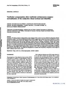
Fig. 1. Scalogram (Morlet wavelets) showing the sway frequency contents (SFC) of the postural responses with OK stimulation. In a representative CVD patient who recovered PC parameters after VRP, amplitudes, frequencies and time of recording (80 s) are shown. (A) Before treatment, showing SFC of up to 2.5 Hz and amplitudes at the low frequencies of up to 30 AUED. (B) After 2 months of the VRP, the patient showed a reduction in the SFC above 0.5 Hz (in line with the decrease in the SV) and also a reduction in the amplitude of the response at the low frequencies to 8 AUED (in line with the decrease in the CE).
146
H. Suarez et al.
Acta Otolaryngol 123
Table II. CE 6alues (cm 2) before the VRP, immediately after the VRP (post-treatment 1) and at longterm follow-up 1295 months later (post-treatment 2) Patient No.
Before treatment
Posttreatment 1
Posttreatment 2
1 2 3 4 5 6 7
12.31 4.20 2.80 11.07 9.98 4.36 8.54
6.70 2.39 3.71 7.82 3.76 3.15 5.32
16.62 4.66 4.63 14.80 5.22 5.22 10.24
Table III. SV 6alues (cm/s) before the VRP, immediately after the VRP (post-treatment 1) and at longterm follow-up 1295 months later (post-treatment 2) Patient No.
Before treatment
Posttreatment 1
Posttreatment 2
1 2 3 4 5 6 7
5.35 1.24 2.55 2.60 5.47 1.84 3.15
2.98 1.04 1.99 1.70 1.14 1.09 1.54
4.59 1.96 2.72 2.72 2.38 1.32 2.56
DISCUSSION The aim of VR in patients with CVD is to attempt to recover PC when some of the CNS structures involved in this plastic process have been damaged by the disease. Sensory information is usually altered (especially in the elderly) and frequently information from the sensory end organs is wrongly processed in the CNS. However, previous information (1, 2, 10) and the data presented here in demonstrate that a VRP can improve PC strategies in many CVD patients. The values of the CE and SV and the amplitudes and sway frequency contents of the scalograms are quantitative markers of PC evaluation (5). The correlation between the improvement in the clinical assessment (as assessed by means of the TEUSAC and DHI) and the lower values of the ‘‘markers’’ after the VRP allows us to perform quantitative measurements during the follow-up of these patients. With these behavioral ‘‘markers’’ of PC, we can measure the behavioral responses of PC after treatment and evaluate the instability of CVD patients and the risk of falls resulting from visual stimulation.The COP distribution is analyzed using the scalogram (Fig. 1) where we can observe in the 80 seconds of record, the frequencies involved and their amplitudes.
Fig. 3. Evolution of the SV values in the same seven patients as shown in Fig. 2: (1) before the VRP; (2) immediately after the VRP; and (3) at long-term follow-up 129 5 months after the VRP.
The loss of the adaptive mechanisms in those patients who discontinued the VRP, increasing their CE and SV values over time, illustrates a practical feature of the therapeutic approach in CVD patients. These patients need to maintain a controlled vestibular training program and to undergo periodic quantitative assessments of PC, in order for us to assess their level of instability and how they respond to rehabilitative treatment. Our results suggest that recovery of the postural behavior achieved after training cannot be maintained by itself. The data suggest failure of the mechanisms of neural learning and memory in these CVD patients, a fact that needs to be considered when a functional prognosis is needed and a therapeutic approach is designed. ACKNOWLEDGMENT This work was supported by a grant from BID-Conicyt (2/94).
REFERENCES 1. Shepard NT, Telian SA. Programmatic vestibular rehabilitation. Otolaryngol Head Neck Surg 1995; 112: 173–82. 2. Suarez H, Arocena M. Clinical assessment and balance training in cerebellar patients. In: Arenberg K, ed. Dizziness and balance disorders. Amsterdam: Kugler Publications, 1993: 737 –43. 3. Peterka RJ, Black FO, Schoenhoff MB. Age related changes in human vestibulo-ocular and optokinetic reflexes: pseudorandom rotation and caloric test. J Vestib Res 1990; 1: 49 – 59. 4. Tinetti ME, Williams CS, Gill TM. Health, functional, and psychological outcomes among older persons with chronic dizziness. J Am Geriatr Soc 2000; 48: 417 –21.
Acta Otolaryngol 123
5. Suarez H, Muse P, Suarez A, Arocena M. Assessment of the risk of fall, related to visual stimulation, in patients with central vestibular disorders. Acta Otolaryngol 2001; 121: 220 –4. 6. Jacobs GP, Newman C. The development of the Dizziness Handicap Inventory. Arch Otolaryngol Head Neck Surg 1990; 116: 424 –7. 7. Horak FB. Clinical measurements of postural control in adults. Phys Ther 1987; 67: 1881 –5. 8. Baloh R, Honrubia V. Electronystagmography. In: Baloh R, Honrubia V, eds. Clinical neurophysiology of the vestibular system. Philadelphia, PA: Davis, 1979: 125–61. 9. Rioul O, Vetterli M. Wavelets and signal processing. IEEE Signal Processing Mag 1991; 14: 38.
Postural control parameters in CVD patients
147
10. Shumway-Cook A, Horak FB. Rehabilitation strategies for patients with vestibular deficits. Neurol Clin 1990; 8: 441 – 7. Address for correspondence: Hamlet Suarez Laboratory of Audiology and Vestibular Pathophysiology School of Medicine C. de Guayaquil 1332 Montevideo 11400 Uruguay Tel.: + 598 2 487 10 20, ext. 242 Fax: +598 2 487 40 80 E-mail: [email protected]