J. Phys. Ther. Sci. 29: 16–19, 2017
The Journal of Physical Therapy Science Original Article
Changes in postural strategy during exercise against perturbation using the balance exercise assist robot: a pilot study Norihide Itoh, RPT, DMSc1–3)*, Shigeo Tanabe, RPT, PhD3), Satoshi Hirano, MD, DMSc4), Eiichi Saitoh, MD, DMSc4), Jumpei Kawabata, RPT5), Daisuke Imoto, RPT2), Yasuo Mikami, MD, PhD2), Toshikazu Kubo, MD, PhD1, 2, 6) 1) Department
of Advanced Rehabilitation Medicine, Graduate School of Medical Science, Kyoto Prefectural University of Medicine: 465 Kajii-cho, Kawaramachi-Hirokoji, Kamigyo-ku, Kyoto 602-8566, Japan 2) Department of Rehabilitation Medicine, Graduate School of Medical Science, Kyoto Prefectural University of Medicine, Japan 3) Faculty of Rehabilitation, School of Health Sciences, Fujita Health University, Japan 4) Department of Rehabilitation Medicine I, School of Medicine, Fujita Health University, Japan 5) Department of Rehabilitation, Fujita Health University Hospital, Japan 6) Department of Orthopaedics, Graduate School of Medical Science, Kyoto Prefectural University of Medicine, Japan
Abstract. [Purpose] To clarify the changes in postural strategy by evaluating leg joint motion and muscle activity before and after continuous exercise against perturbation using the Balance Exercise Assist Robot (BEAR). [Subjects and Methods] Nine healthy subjects (male 7, female 2; mean age 23 ± 1 years) performed a postural perturbation coping exercise only. In the task, the robot leaned and moved automatically. Participants were instructed to maintain their default upright position and they performed the exercise five times in a row (1 minute/trial). Changes in total movement distance, range of motion of each joint (hip, knee, ankle), and mean activity of each muscle for the first and fifth trials were compared. [Results] The total movement distance of BEAR and range of motion in the hip decreased significantly from the first trial to the last trial. No change in muscle activity was observed in the rectus femoris, biceps femoris, tibialis anterior or gastrocnemius. [Conclusion] The results for exercise against perturbation using BEAR in this study suggest that BEAR may be a promising method to improve the ankle strategy for maintaining a standing posture. Key words: Robot, Rehabilitation, Postural balance (This article was submitted Aug. 9, 2016, and was accepted Sep. 14, 2016)
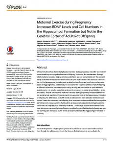
INTRODUCTION Recently, a variety of gait and balance training equipment that employs robotic technology has been introduced1–4). Robotic rehabilitation, compared with conventional practice tools, is highly useful in clinical practice as it can supply real-time data that permits immediate user feedback. We developed the Balance Exercise Assist Robot (BEAR) (Toyota Motor Corp., Toyota City, Japan) based on the personal transport assistance robot device ridden in the standing position (Fig. 1)1). Ozaki et al. performed three types of balance training therapy using the BEAR in eight patients with central nervous system disorders and found that dynamic balance and leg strength improved after the tasks compared with baseline1). However, the physical *Corresponding author. Norihide Itoh (E-mail:
[email protected]) ©2017 The Society of Physical Therapy Science. Published by IPEC Inc. This is an open-access article distributed under the terms of the Creative Commons Attribution Non-Commercial No Derivatives (by-nc-nd) License .
effects of BEAR have not been described. Therefore, this pilot study was designed to clarify the changes in postural strategy during exercise against perturbation using BEAR by evaluating leg joint motion and muscle activity.
SUBJECTS AND METHODS Nine healthy subjects (male 7, female 2; mean age 23 ± 1 years; height 171.8 ± 7.1 cm; weight 60.0 ± 7.4 kg) participated in this study. No subject had a history of neurological or orthopedic disease, or experience of exercise using the BEAR. All subjects provided written informed consent for the experimental procedure, which was approved by the human ethics committee of Fujita Health University (No. 15-148). This study was performed in accordance with the Declaration of Helsinki. Participants performed a postural perturbation coping exercise only. In the task, the robot leaned and moved automatically (sinusoidal wave with a frequency of 0.5 Hz and amplitude of 4°). Participants were instructed to maintain their default upright position and they performed the exercise five times in a row (1 minute/trial). In the first and fifth trials, the total movement distance of the BEAR was calculated. In addition, joint angles (hip flexion/extension, knee flexion/extension, and ankle dorsiflexion/plantar flexion) and muscle activity of the lower extremities were measured during both trials. The total movement distance of the robot was calculated from the coordinate information on the computer. All data for joint angles and electromyography (EMG) were measured on the right leg. Measurement time was fixed at 60 sec. Motion data of the lower extremity was collected with the KinemaTracer motion analysis system (KISSEI COMTEC Co., Ltd., Matsumoto, Japan) with four 60 Hz cameras. Each participant had five markers placed on their right leg to define the joint center and axes of motion: (1) on the acromia; (2) one-third distance from the great trochanter on a line joining the anterior superior iliac spine and great trochanter; (3) midpoint of the anteroposterior diameter of the lateral femoral epicondyle; (4) lateral malleolus; and (5) fifth metatarsal head. Each angle of hip, knee and ankle joint was calculated as the motion of the distal segment relative to the proximal segment. To obtain an accurate estimate of the individual ensemble average, 29 cycles were averaged and a normalized perturbation cycle was set at 200 points. For time-normalized data, the individual joint motions of the lower limb were averaged for each subject to obtain an ensemble average for every subject at each joint of motion. Surface EMG data were collected simultaneously from the rectus femoris, biceps femoris long head, anterior tibialis and gastrocnemius. The myoelectric signals were amplified 1,000-fold, filtered with a 0.03 Hz low-cut analog filter, and telemetered via a multichannel biotelemetry system (WEB-5000, Nihon Kohden Corp., Tokyo, Japan). Data acquisition was performed using a DAQ measurement system (USB-6229, National Instruments Japan Corp., Tokyo, Japan). The EMG data were recorded at a sampling frequency of 1.8 kHz and were band pass filtered between 10 and 500 Hz to remove low frequency motion artifact and high frequency noise via a third-order Butterworth filter using LabVIEW 8.5 custom-made software (National Instruments Japan Corp., Tokyo, Japan). Subsequently, the data were processed for rectification and integrated per 20 msec and quantified for electromyogram intensity as percent of maximum voluntary contraction (%MVC). Changes in total movement distance, range of motion of each joint (hip, knee, ankle), and mean activity of each muscle (rectus femoris, biceps femoris, tibialis anterior, gastrocnemius) for the first and fifth trials were compared using the Wilcoxon signed rank test. The significance level was set at p