Apr 26, 1982 - tides common to human epithelial amnion cells and lung fibro- blasts, whoserate of synthesis is sensitive to neoplastic transfor- mation [Bravo ...
Proc. NatL Acad. Sci. USA Vol. 79, pp. 4367-4370, July 1982
Cell Biology
Changes in the relative proportion of transformation-sensitive polypeptides in giant HeLa cells produced by irradiation with lethal doses of x-rays (two-dimensional gel electrophoresis/polypeptide markers/DNA synthesis/cell proliferation/cancer)
JAIME BELLATIN*, RODRIGO BRAVOt, AND JULIO E. CELIS Division of Biostructural Chemistry, Department of Chemistry, Aarhus University, DK-8000 Aarhus C, Denmark
Communicated by D. von Wettstein, April 26, 1982 ABSTRACT Irradiation of HeLa cells with 1,100 rads (1 rad = 0.01 J/kg = 0.01 Gy) of x-rays yielded a pure population of giant cells 5-7 days after irradiation. These cells do not divide but go through an intermittent DNA synthetic phase. The population of giant cells in S phase (8%) is considerably lower than that of control asynchronous HeLa cells (30%), but 80% of the giant cells go through S phase as determined by 48-hr labeling with [3H]thymidine. Previous studies with high-resolution two-dimensional gel electrophoresis identified 58 [asS]methionine-labeled polypeptides common to human epithelial amnion cells and lung fibroblasts, whose rate of synthesis is sensitive to neoplastic transformation [Bravo, R. & Celis, J. (1982) Clin. Chem. (Winston-Salem, NC) 28, 949-955]. These polypeptides also have been identified in HeLa cells and other transformed human cells such as Detroit 98, Chang liver, Fl-amnion, and WISH-amnion [Bravo, R. & Celis, J. (1982) Clin. Chem. (Winston-Salem, NC) 28, 949-955]. After irradiation of HeLa cells and giant cell formation, the relative proportions of most ofthe transformation-sensitive polypeptides (43 of 47) reverted to levels similar to those observed in nontumorigenic cells. This suggests that their relative proportions are dependent on the growth properties of the cells. In particular, the relative proportions of three polypeptides (designated 12g and 60dl in isoelectric focusing and 27b in nonequilibrium pH gradient electrophoresis) were not affected, indicating that their reduced amounts in transformed cells could reflect a fundamental change that develops during transformation.
Work in this laboratory has been devoted to the search for cellular polypeptides that could be involved in regulating cell proliferation and whose function may shed light on the process of neoplastic transformation (1-8). Fifty-eight polypeptides whose relative proportions are sensitive to transformation have been revealed so far among nearly 1,300 proteins analyzed in normal and transformed human lung fibroblasts and epithelial amnion cells (7). These polypeptides are present in both normal and transformed cells and are common to both cell types studied (7, 8). Similar analysis of the polypeptides synthesized by other transformed cell lines of human origin, such as HeLa, Detroit98, and Chang liver, showed that the relative proportions of the transformation-sensitive polypeptides are very similar to those observed in transformed human lung fibroblasts and epithelial amnion cells (7). From previous studies it seemed likely that the relative proportions of some of these polypeptides could reflect variations in the growth properties of the cells (1, 6, 7, 9). In this report we present a quantitative and qualitative analysis of the transformation-sensitive polypeptides in HeLa cells and in giant HeLa cells produced by irradiation with lethal doses of x-rays. The publication costs ofthis article were defrayed in part by page charge payment. This article must therefore be hereby marked "advertisement" in accordance with 18 U. S. C. §1734 solely to indicate this fact. 4367
Because giant cells do not proliferate but maintain a high degree of metabolic integrity (10-12), they represented a suitable system to search for transformation-sensitive polypeptides whose synthesis may be dependent on cell proliferation.
METHODS Cells. HeLa cells free of mycoplasma (GIBCO Bio-cult Limited; stock source, American Type Culture Collection) were grown routinely as monolayer cultures in Dulbecco's modified Eagle's medium (DME medium) containing 10% fetal calf serum and antibiotics (penicillin, 100 units/ml; streptomycin,
50 ,gg/ml).
X-Irradiation. HeLa cells were grown for irradiation on 9coverslips (Bellco Glass) placed in 0.30-ml flat-bottomed microtiter plates containing 0.2 ml of DME medium. The coverslip was removed from the wells and placed in a 3-cm Petri dish containing 2 ml of Hanks' balanced salt solution. The cells were irradiated with 1,100 rads (1 rad = 0.01 J/kg = 0.01 Gy) at room temperature with a Philips PW x-ray unit operated at 30 kV and 3-mA external filtration of 20 am of Ni (dose rate, 110 rads/min). Immediately after irradiation, the coverslips were transferred to fresh DME medium and were incubated at 37rC in 5% C02/95% air until a pure population of giant cells was obtained (usually between 5 and 7 days after irradiation). PH]Thymidine Incorporation in Autoradiography. Cells were labeled with 2 uCi (1 Ci = 3.7 x 10'° becquerels) of [3H]thymidine per ml (Amersham; specific activity, 40-60 Ci/ Thmol) for various times. They were then washed twice with Hanks' solution, fixed in 2.5% glutaraldehyde in Hanks' solution, washed with distilled H20, and air dried. The slides then were covered with Ilford K-2 nuclear emulsion and kept at 40C for 3 days before developing and staining with Giemsa stain. Labeling of Asynchronous Cells and Giant Cells with [35S]Methionine. HeLa cells or giant cells grown attached to 9mm coverslips and placed in 0.30-ml flat-bottomed microtiter plates (Nunc, Roskilde, Denmark) were labeled with 0.1 ml of homemade DME medium lacking methionine (NaHCO3, 1 g/ liter) and containing 10% dialyzed fetal calf serum (Flow Laboratories, McLean, VA), 100 ACi of [3S]methionine (Amersham, England; specific activity, 1,300 AuCi/mol), and cold methionine (final concentration, 1 mg/liter) (2, 5, 13, 14). The cells were usually labeled for 20 hr at 370C, during which time mm
Abbreviations: IEF, isoelectric focusing; NEPHGE, nonequilibrium pH gradient electrophoresis; DME medium, Dulbecco's modified Eagle's medium. * Present address: Medical Research Council Laboratory of Molecular Biology, Hills Road, Cambridge, England. t Present address: European Molecular Biology Laboratory, Postfach 10.2209 Meyerhofstrasse 1, 6900 Heidelberg, Federal Republic of Germany.
Cell Biology: Bellatin et aL
4368
Proc. Nad Acad. Sci. USA 79 (1982)
RESULTS
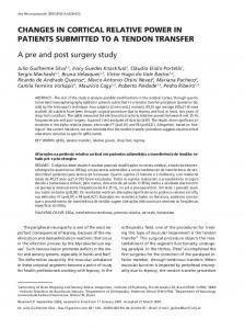
Characteristics of Giant HeLa Cells. Irradiation of HeLa cells with 1,100 rads of x-rays leads to the death of many cells before reaching the first mitosis (interphase death). However, a fraction of the cells continue to grow but fail to divide and so reach an average size many times that of control cells (9, 12) (Fig. 1). In this way a pure population ofgiant cells was obtained 5-7 days after irradiation (Fig. 1B). About 75% of the cell population consisted oflarge flat mononucleated cells, 15% of multinucleated cells (binuclear and higher), and 10% of micronucleated cells (Fig. 1B). These cells do not divide, as no metaphases or telophases could be observed in the cell populations (10), but go through S phase as determined by [3H]thymidine labeling. The fraction of giant cells in S phase was found to be 8% as compared to 30.3% in control cells (not shown). However, most giant cells went through S phase because 80% of these cells were labeled with [3H]thymidine during a 48-hr incubation period. Changes in the Relative Proportion of Transformation-Sensitive Polypeptides in Giant HeLa Cells. The two-dimensional polypeptide maps (IEF and NEPHGE) of control HeLa cells and giant HeLa cells labeled for 20 hr with [3S]methionine are shown in Figs. 2 and 3. The giant cells (pure population, Fig. 1B) were labeled 6 days after irradiation with 1,100 rads of xrays. Only 47 (27 acidic and 20 basic) of the 58 transformationsensitive polypeptides so far revealed in human cultured cells (7, 8) have been analyzed in this study and are indicated with arrows and arrowheads in Figs. 2 and 3. The number given to each polypeptide corresponds to that assigned in the HeLa protein catalogues (4, 8). The identity of a few polypeptides is known, and these correspond to vimentin (IEF 26), phosphovimentin (IEF 26e), cyclin (IEF 49), and a tropomyosin variant (IEF 52) (4, 7, 8). The spots were cut out from the gels for quantitative analysis, and the radioactivity was determined as described (14). In order to compare the radioactivities ofthe spots, the radioactivity of each spot was standardized against the total radioactivity applied to the gel. The ratio of label incorporated
4~~~~~~~~~4
9/
/,V4~~~~~00/.>
4.
o
B f illi.
';/z \4
. Irv4 -
_.
FIG. 1. Phase-contrast photomicrograph of control (A) and irradiated (B) HeLa cells. HeLa cells grown on 9-mm2 coverslips were irradiated with 1,100 rads of x-rays. Giant cells were photographed 6 days after irradiation. (x512.)
the microtiter plates were wrapped in Saran Wrap to avoid evaporation. Excess liquid was drained from the coverslips, and the samples were dissolved immediately in 20 pkl oflysis buffer (15). The procedures for two-dimensional gel electrophoresis [isoelectric focusing (IEF) and nonequilibrium pH gradient electrophoresis (NEPHGE)] (14-17) and quantitation of radioactive spots recovered from the gels (14) have been described in detail elsewhere. IEF
IEF
-
OXA L
_.
*
uz~~~~8l Z
-;.
-81
*.
Sm
. -~~~~ -
12tf
136
~~Bz3 0
8z36
1 14h __
5 tP 3.25I 33U~
*
***
.~~t a 19 4Sz66
_Ta
v/
/8z14
3
_
- 69 - 55
a
.26.
K 92 14h
sjb13Q O
~~~a -. ~~~~~~49 47k 48z52
*
_
FRm
_ 12g g _ .2/k
A/ ,3'2260
43
48z52
47k
I=
49
x
2_
48z66 t 48z90
56-.
56-'k
SOdi
30
60d1
t 63
63
FIG. 2. High-resolution two-dimensional gel electrophoresis (EEF) of [35S]methionine-labeled polypeptides from HeLa cells (A) and giant HeLa cells (B). HeLa cells grown on 9-mm2 coverslips were irradiated with 1,100 rads of x-rays. A pure population of giant cells was obtained 6 days after irradiation. Control and giant cells were labeled for 20 hr with [ISlmethionine. Only transformation-sensitive polypeptides are indicated (7, 8). Arrowheads, phosphorylated proteins (7); V, vimentin.
Cell Biology: Bellatin et aL
Proc. Natd Acad. Sci. USA 79 (1982)
-*
-NEPHGE
6r
NEPHGE
B
A
UI) 0
co
4369
la
I
92.5 69
-4
15
17-
55
-43
15
_
0 x
__
t4:3
_4 21 a
26i
26i
1*
if
27b
27b
._
-30
l 0
33
33
FIG. 3. High-resolution two-dimensional gel electrophoresis (NEPHGE) of [35S]methionine-labeled polypeptides from control HeLa cells (A) and giant HeLa cells (B). A pure population of giant cells was obtained 6 days after irradiation. Control and giant cells were labeled for 20 hr with [3'S]methionine. Only transformation-sensitive polypeptides are indicated.
in control HeLa cells compared to that in giant HeLa cells is given in Table 1. In other cases, however, the changes in relative proportions were established by inspecting films from many independent samples. The results in this case are expressed by using pluses. With the exception of IEF 12g and 60dl and NEPHGE 1z31 and 27b, the relative proportion of
all the other transformation-sensitive polypeptides analyzed changed substantially and reproducibly in the giant cells. These changes are based on the analysis of four independent samples and have been confirmed by carrying out similar studies on cells labeled for 4 hr. The polypeptides indicated with arrowheads in Fig. 2 [IEF 8m, 14h, and 26e (phosphovimentin)] are not listed in Table 1 and correspond to transformation-sensitive phosphoproteins (7). The relative proportions of IEF 8m and 14h decrease in giant HeLa cells while that of phosphovimentin increases (Fig. 2). Finally, it should be stressed that we have carried out similar studies on spontaneously transformed amnion cells (7) and giant amnion cells. Again a detailed qualitative analysis showed that with the exception of polypeptides IEF 12g and 60d1 and NEPHGE 1z31 and 27b, the intensities of all other transformation-sensitive polypeptides are similar to that observed in control normal amnion cells (7) (results not shown). Because the relative change in radioactivity in NEPHGE Wz31 is much smaller than those observed with the other three polypeptides, the importance of this change is difficult to evaluate.
DISCUSSION Because irradiation of cells with lethal doses of x-rays is known to produce a population of cells that do not divide but keep a certain degree of metabolic integrity ("giant cells") (10-12), it
of interest to compare the polypeptide pattern of giant HeLa cells with that of control HeLa cells. From our previous studies of normal and transformed cultured fibroblast and epithelial cells, it seemed likely that the relative proportions of many ofthe transformation-sensitive polypeptides could reflect changes in the growth properties of the cells (7). In this study we found that the relative proportions of 44 out of 47 transformation-sensitive polypeptides analyzed changed substantially and consistently in giant cells, suggesting that their relative proportions may be dependent on growth rate. In the case of IEF 49 (cyclin), this assumption has been confirmed by analyzing the relative proportion of this protein in rapidly growing and senescent human skin fibroblast cells (9). Interestingly, the analysis of giant cells showed that the relative proportions of three transformation-sensitive polypeptides (IEF 12g and 60d1 and NEPHGE 27b) could not be changed (6, 7, 17) and their reduced synthesis in transformed cells could reflect a fundamental change that develops during transformation. There is mounting evidence indicating that the synthesis of NEPHGE 27b [NEPHGE 27a in mouse (17)] decreases drastically in immortal cells, irrespective of whether these cells are tumorigenic or not (ref. 6; unpublished observations). It is interesting to note that at least at the present level of resolution and sensitivity achieved by two-dimensional gel electrophoresis (resolution and detection of nearly 1,300 polypeptides) (4, 8), we could not detect any new major polypeptide appearing in the giant cells. Thus, it would seem likely that the drastic changes in growth properties and of cell morphology accompanying giant cell formation may be brought about by changes in the relative proportions (abundances) of proteins was
4370
Proc. Nad Acad. Sci. USA 79 (1982)
Cell Biology: Bellatin et aL
Table 1. Basic and acidic human proteins-sensitive to neoplastic transformation: Relative proportions in giant HeLa cells Coordinates Giant-HeLa Giant HeLa Coordinates in HeLa cells, relative in HeLa cells, relative Giant/control catalogue* proportiont Polypeptide proportiont cell ratio* Polypeptide catalogue* NEPHGE EEF if ig 1z24 1z31 5 6c 7g '8 8a lOa lOb 13 14 '15 17 19 '21 26i 27b 33
92/0.22
92/0.18 71/0.51 71/0.30 64/0.70 61/0.82 59.5/0.49 57/0.24
57/0.43 54.5/0.76 54.5/0.86 49/0.67 48.5/0.24 47/1.00
-41/1.22 39.5/0;40 -'35/1.45 28.2/0.71 27/1.11 18/0.68
Decreases Decreases Increases Unaffected Decreases Decreases Increases Increases Increases Decreases Decreases Decreases Unaffected Increases Decreases Increases Decreases Increases Unaffected Decreases
(+/+ +) (+/+ + +) (+ +/+)
(0.57)
(+/+ + +) (+/+ + +) (1.40) (+ +/+) (+/+ +) (+/+ +) (0.57) (0:81) (1.50) (0.60) (1.94) (0.48) (+ +/+) (0.60)
2 8z14 8z36 10 12 12g 13e 13g 13q 14 251 26 (vimentin) 33r 39 47k 48z52
48z66 48z90 49 (cyclin) 52 (tropomyosin related) 53 56 60dl 63
110/0.44
84/0.32 76/1.27 72/1.28 68/0:85 68/0.89 67/1.03 66/0.76 66:5/1.03 66/0.86 55/0.25 54/1.19 50/0.70
45.5/0.91 43/1.08 35/0.68 38.5/0.92 38/1.06 36/1.48 35/1.43 32/1.28 31/1.41 -26/0.84 20/0.64
Increases Decreases Decreases Decreases Decreases Unaffected Decreases Decreases Decreases Decreases Decreases Increases
Increases Decreases Increases Increases Decreases Decreases Decreases Increases Increases Increases Unaffected Decreases
Giant/control cell ratiot (+ +/+)
(+/+ +) (0.68) (0.67)
(0.59) (0.50) (+/+ +) (0.50) (0.67) (0.41) (2.28) (+ +/+) (0.50) (+ +/+) (+ +/+) (+/+ +) (+/+ +) (0.21) (2.56) (1.66) (+ +/+)
(0.47)
* From refs. 4 and 8. tAllof the gels were normalized to the same total radioactivity. * In all cases, with the exception of polypeptides NEPHGE 1z31-and 27b and EEF 12g and 60dl, the ratio of giant HeLa cells to HeLa-cells iasimilar to that observed in normal versus transformed human fibroblasts and epithelial cells (see ref. 7). Numerical ratios are given where the spots were quantitated. +, + +, and + + + indicate increasing levels of radioactivity as determined by visual inspection of the fluorograms.
-already synthesized by HeLa cells. This situation is very similar to that observed in pairs of normal and transformed cells of human, mouse, and hamster origin (1, 5, 7). In that case we have proposed that transformation may be due to the abnormal expression of-normal genes (5, 7, 9).
6. Bravo, R., Schifer, R., Willecke, K., Macdonald-Bravo, H., Fey, S. J. & Celis, J. E. (1982) Proc. NatL Acad. Sci. USA 79, 2281-2285. 7. Bravo, R. & Celis, J. E. (1982) Clin. Chemn. (Winston-Salem, NC)
We thank our colleagues S. J. Fey, P. Mose Larsen, -and A. Celis for helpful discussion. J. B. was a recipient of a Danish International Development Agency Fellowship. R.B. was supported by a fellowship from the'Danish Medical and Natural Science Research Councils. This work was supported in part by grants from Euratom and the Danish Medical and Natural Science Research Councils, the Danish Cancer Society, the Carlsberg Foundation, and Novo.
9.
1. Bravo, R. & Celis, J. E. (1980) Exp. Cell Res. 127, 249-260. 2. Bravo, R. & Celis, J. E. (1980) J. Cell BwIlO 84, 795-802. 3. Bravo, R., Fey, S. J. & Celis, J. E. (1981) Carcinogenesis 2, .769-782. 4. Bravo, R., Bellatin, J. &.Celis, J. 'E. (1981) Cell BoL Int. Rep. 5, 93-96. 5. Celis, J. E. & Bravo, R. (1981) Trends Biochein. Sci. 6, 197-201.
28, 949-955. 8. Bravo, R. & Celis, J. E.
10. 11. 12. 13. 14.
15. 16. 17.
(1982) Clin. Ch/wn. (Winston-Salem, NC) 28, 766-781. Bravo, R., Fey, S. J., Bellatin, J., Mose Larsen, P., Arevalo, J. & Celis, J. E. (1981) Exp. .Cell Res. 136, 311-319. Puck, T. T. & Marcus, P. L. (1956) J. Exp. Med. 103, 653-666. Tolmach, L. J. & Marcus, P. I. (1960) Exp. Cell Res. 20, 350-X-360. K. I., Okada, S. (1970) in Radiation Biochemistry, eds. Altman, Gerber, G. B. & Okada, S. (Academic, New York), Vol. 1, pp. 239-246. Bravo, R., Fey, S. J., Small, J. V., Mose Larsen, P. & Celis, J. E. (1981) Cell 25, 195-207. Bravo, R., Small, J. V., 'Fey, S. J., Mose Larsen, P. & Celis, J. E. (1982)J. Mol. Biol 154, 121-143. O'Farrell, P. (1975) J. Biolu /hem. 50, 4007-4021. O'Farrell, P. Z., Goodman, J. M. & O'Farrell, P. H. (1977) Cell 12, 1133-1142. Fey, S. J., Bravo, R., Mose Larsen, P., Bellatin, J. & Celis, J. E.(1981) Cell BioL It. Rep. 5, 491-500.