saturated with buffer and will permit the required flow of current or liquid for electro and convection blotting. 2.1. Nitrocellulose. Membranes. 2.1.1. Synthesis of ...
Chapter 3 Introduction to Protein Blotting Biji T. Kurien and R. Hal Scofield Summary Protein blotting is a powerful and important procedure for the immunodetection of proteins following electrophoresis, particularly proteins that are of low abundance. Since the inception of the protocol for protein transfer from an electrophoresed gel to a membrane in 1979, protein blotting has evolved greatly. The scientific community is now confronted with a variety of ways and means to carry out this transfer. Key words: Western blotting, Sodium dodecyl sulfate polyacrylamide gel electrophoresis, Nitrocellulose membrane, Polyvinylidene difluoride membrane
1. Introduction The transfer of macromolecules (proteins or nucleic acids) to microporous membranes is referred to as “blotting,” and this term encompasses both “spotting” (manual sample deposition) and transfer from planar gels. Proteins that are resolved on sodium dodecyl sulfate polyacrylamide gel electrophoresis (SDS PAGE) gels are typically transferred to adsorbent membrane supports under the influence of an electric current in a procedure that is known as western blotting (WB) or protein blotting (1, 2). Nucleic acids are routinely transferred from agarose gels, to a membrane support, through capillary action (Southern blotting). Protein blotting evolved from DNA (Southern) blotting (3) and RNA (northern) blotting (4). The term “western blotting” was coined to describe (5) this procedure to retain the “geographic” naming tradition initiated by Southern’s paper (3). The blotted
Biji T. Kurien and R. Hal Scofield (eds.), Methods in Molecular Biology, Protein Blotting and Detection, vol. 536 © Humana Press, a part of Springer Science + Business Media, LLC 2009 DOI: 10.1007/978-1-59745-542-8_3
9
10
Kurien and Scofield
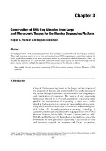
proteins form an exact replica of the gel and have proved to be the starting step for a variety of experiments. The subsequent employment of antibody probes directed against the membranebound proteins (immunoblotting) has revolutionized the field of immunology (Fig. 1). Dot blotting refers to the analysis of proteins applied directly to the membrane rather than after transfer from a gel. The utility of the high resolving power of SDS PAGE (6) was limited in purpose, owing to the fact that the separated proteins in the gel matrix were difficult to access with molecular probes, until the advent of protein blotting. Protein transfer with subsequent immunodetection has found wide application in the fields of life sciences and biochemistry. This procedure (1, 2) is a powerful tool to detect and characterize a multitude of proteins, especially those proteins that are of low abundance. It offers the following specific advantages: (a) wet membranes are pliable and are easy to handle compared with gels, (b) easy accessibility of the proteins immobilized on the membrane to different ligands, (c) only small amount of
Western Blotting and Detection
SDS-PAGE
E
A
Western blot
Develop color
B
D
Blot from gel Add primary antibody
C Add enzyme conjugate
Fig. 1. Schematic of western blotting and detection procedure: (A) Unstained SDS PAGE gel prior to western blot. The bands shown are hypothetical. (B) Exact replica of SDS PAGE gel obtained as a blot following western transfer. (C) Primary antibody binding to a specific band on the blot. (D) Secondary antibody conjugated to an enzyme (alkaline phosphatase or horse radish peroxidase) binding to primary antibody. (E) color development of specific band (reproduced from (10) with permission from Elsevier).
Introduction to Protein Blotting
11
reagents is required for transfer analysis, (d) multiple replicas of a gel are possible, (e) prolonged storage of transferred patterns, prior to use, becomes possible, and (f) the same protein transfer can be used for multiple successive analyses (7–9). Protein blotting has been evolving constantly, since its inception, and now the scientific community is faced with a multitude of ways and means of transferring proteins (10). Nonetheless, western blot sensitivity is dependent on efficiency of blotting or transfer, retention of antigen during processing, and the final detection/amplification system used. Results are compromised if there are deficiencies in any of these steps (11). 1.1. Blotting Efficiency
2. Immobilizing Supports for Protein Transfer
The efficient transfer of proteins from a gel to a solid membrane support depends greatly on the nature of the gel, the molecular mass of the proteins being transferred, and the membrane used. Running the softest gel, in terms of acrylamide and cross-linker that yields the required resolution, is the best option. Transfer becomes more complete and faster with the use of thinner gels. However, the use of ultrathin gels may cause handling problems, and a 0.4-mm thickness represents the lower practical limit (12). Proteins with a high molecular mass blot poorly following SDS PAGE, resulting in low levels of detection on immunoblots. However, the efficiency of transfer of such proteins has been facilitated with heat, special buffers, and partial proteolytic digestion of the proteins prior to transfer (11, 13–17).
A wide range of solid phases are available for immobilization, ranging from the truly solid phase such as glass or plastic to latex and cellulose that are porous. The most common phases used for blotting comprise microporous surfaces and membranes such as cellulose, nitrocellulose (NC), polyvinylidine diflouride, cellulose acetate, polyethane sulfone, and nylon. The unique properties of microporous surfaces that make them suitable for traditional assays such as western blotting are (a) large volume-to-surface area ratio, (b) high binding capacity, (c) short- and long-term storage of immobilized molecules, (d) ease of processing by allowing a solution phase to interact with the immobilized molecule, (e) lack of interference with the detection strategy, and (f) reproducibility. These properties are useful for the high-thoroughput assays used in the postgenomic era as well (2, 4, 14, 18, 19). Typically, these microporous surfaces are used in the form of membranes or sheets with a thickness of 100 μm and possessing an average pore size that ranges from 0.05 to 10 μm in diameter.
12
Kurien and Scofield
The interaction of biomolecules with each of these membranes is not completely understood, except for the fact that it is generally known to be noncovalent (20, 21). Regardless of the type of membrane used, it must be borne in mind that exceeding the protein binding capacity of the membrane used tends to reduce the signal obtained in immunoblotting. Excess protein, weakly associated with the membrane, is readily accessible to react with the primary antibody or any other ligand in solution (e.g., lectin). However, the resulting antibody–protein complexes will easily wash off during further processing of the membrane. Such a scenario would not have prevailed if the protein had initially made good contact with the membrane (18). 2.1. Nitrocellulose Membranes
Nitrocellulose (NC) is perhaps the most versatile of all the surfaces mentioned earlier for the immobilization of proteins, glycoproteins, or nucleic acids (3, 4, 19). In addition to traditional blotting, NC is used in high-throughput array, immunodiagnostic as well as mass spectrometry-coupled proteomic applications, filtration/concentration, ion exchange, and amino acid sequencing in addition to traditional blotting procedures. It was Southern who first demonstrated (in 1975) the usefulness of NC to capture nucleic acids. Towbin in 1979 (1) and Burnette in 1981 (5) showed that NC could also be used for proteins. This unique polymer derived from cellulose has been used as the most common immobilization surface in biological research for over 65 years. Since high-throughput methodologies for proteomics and genomics rely heavily on traditional concepts of molecular immobilization followed by hybridization binding or analysis, NC continues to be useful in postgenomic era technology (19).
2.1.1. Synthesis of Nitrocellulose from Cellulose
Treatment of cellulose with nitric acid results in the hydroxyl moieties on each sugar unit of cellulose being substituted by nitrate groups, resulting in NC. Organic solvents readily dissolve dry NC resulting in the formation of a lacquer. When the solvents are evaporated the polymer is deposited as a thin film. By including a nonsolvent such as water in the lacquer pores, nonsolvent can be introduced into the film to create a microporous membrane. Pore formation is a consequence of differential evaporation of the nonsolvent and the solvent. Therefore, pore size and porosity can be readily controlled by the amount of the nonsolvent in the lacquer (2). The pore size of 0.45 μm refers to the average effective diameter of the irregular long and tortuous channels that traverse the membrane. The pores of 0.45 μm in NC membranes account for about 80% of the filter’s volume reaching an average density of 450 × 106/cm2 (18). In the blotting process, the membrane needs to be porous to allow it to be saturated with buffer and will permit the required flow of current or liquid for electro and convection blotting.
Introduction to Protein Blotting
13
2.1.2. Immobilization Mechanism
Even though the exact mechanism by which biomolecules interact with NC is unknown, several lines of evidence suggest that the interaction is noncovalent and hydrophobic. One evidence favoring hydrophobic interaction is the fact that since most proteins at pH values above 7 are negatively charged, it is surprising that NC which is also negatively charged can bind proteins efficiently. An additional fact is that nonionic detergents (such as Triton X-100) are effective in removing bound antigens from NC (8). High concentrations of salt and low concentrations of methanol increase immobilization efficiency (22). NC is unique, when compared with other microporous membranes, in its ability to distinguish between single- and double-stranded nucleic acids, small and large proteins, short and long nucleic acids, and complexed versus uncomplexed molecules (22). It can be stained with amido black (4), Coomassie Brilliant Blue (CBB) (1), aniline blue black, Ponceau S, fast green, or toluidine blue. Amido black staining can detect a 25-ng dot of bovine serum albumin readily with acceptable background staining. The background staining tends to be higher with CBB while Ponceau S gives a very clean pattern but with slightly less sensitivity than amido black.
2.1.3. Disadvantages of NC
One clear disadvantage of NC is the fact that it cannot be stripped and reprobed multiple times owing to its fragile nature. It also has a tendency to become brittle when dry. In addition, small proteins tend to move through NC membranes and only a small fraction of the total amount actually binds. Using membranes with smaller pores can obviate this (12). Gelatin-coated NC has been used for quantitative retention (10, 23). In supported NC (e.g., Hybond-C Extra), the mechanical strength of the membrane has been improved by incorporating a polyester support web, thereby making handling easier.
2.2. Polyvinylidene Difluoride
Polyvinylidene difluoride (PVDF) is a linear polymer with repeating –(CF2-CH2)– units. The use of “di” in polyvinylidene difluoride is redundant (including its use here) and its use needs to be discouraged (2). Polyvinylidene fluoride or polyvinylidene difluoride refers to the same membrane first made available for protein blotting by Millipore in June of 1986. The product was renamed as Immobilon-P™ Transfer Membrane after being initially referred to as Immobilon™ PVDF transfer membrane to differentiate it from other PVDF and non-PVDF-based blotting membranes referred to collectively as Immobilon family and marketed by Millipore. Immobilon-PSQ membrane with a 0.2-μm pore size suitable for proteins with a molecular weight less than 20,000 (to prevent blow through) and Immobilon-FL membrane optimized for all fluorescence applications also form part of the Immobilon family of PVDF membranes, added recently. Sequelon (24), a PVDF-based
14
Kurien and Scofield
sequencing membrane, sold by Milligen/BioSearch, a Millipore subsidiary is advantageous because of high protein binding capacity, physical strength, and chemical stability. 2.2.1. Immobilization Mechanism
Proteins transferred to the Immobilon-P membrane during western transfer are retained well on the membrane surface throughout the immunodetection process via a combination of dipole and hydrophobic interactions. The antigen binding capacity of the membrane is 170 μg/cm2 for bovine serum albumin and this is proportionate with the binding capacity of NC. In addition, the Immobilon-P membrane has very good mechanical strength and like Teflon™ (a related fluorocarbon polymer) it is compatible with a range of chemicals and organic solvents [acetonitrile, trifluoroacetic acid, hexane, ethylacetate, and trimethylamine (2, 25)]. Blotting mechanics are not different from those seen with NC, except that it is necessary to prewet the membrane in either methanol or ethanol before using with aqueous buffers. This is because PVDF is highly hydrophobic and there is no added surfactant in PVDF.
2.2.2. Advantages of PVDF
One of the advantages of electroblotting proteins onto PVDF membranes is that replicate lanes from a single gel can be used for various purposes such as N-terminal sequencing, proteolysis/ peptide separation/internal sequencing along with western analysis. Proteins blotted to PVDF membranes can be stained with amido black, India ink, or silver nitrate (26). These membranes are also amenable to staining with CBB, thus allowing excision of proteins for N-terminal protein sequencing, a procedure first demonstrated by Matsudaira in 1987 (25).
2.3. Activated Paper
Activated paper (diazo groups) binds proteins covalently but is disadvantageous in that the coupling method is incompatible with many gel electrophoresis systems. Linkage is through primary amines, and therefore systems that use gel buffers without free amino groups must be used with this paper. In addition, the paper is expensive and the reactive groups have a limited half-life once the paper is activated.
2.4. Nylon Membranes
Nylon-based membranes are thin and smooth surfaced as NC but with much better durability. Two kinds of membranes are available commercially: Gene Screen and Zetabind (ZB). ZB is a nylon matrix (polyhexamethylene adipamine or Nylon 66) modified by the addition of numerous tertiary amino groups during the manufacturing process (extensive cationization). It has excellent mechanical strength and also offers the potential of very significant (yet reversible) electrostatic interactions between the membrane and polyanions. Nylon shows a greater protein binding capacity compared with NC (480 μg vs. 80-μg BSA bound/cm2). In addition, nylon
Introduction to Protein Blotting
15
offers the advantages of more consistent transfer results and a significantly increased sensitivity compared with other membranes (7, 18). This effect is possible owing to the extra potential difference created by the positive charge of ZB. 2.4.1. Disadvantages of Nylon
The high binding capacity of these membranes, however, produces higher nonspecific binding. Another problem with using nylon membranes is that they bind strongly to the commonly used anionic dyes such as CBB, amido black 10B (18), aniline blue black, Ponceau S, fast green, or toluidine blue. SDS, dodecyl trimethylammonium bromide, or Triton X-100 at low concentrations (0.1% in water) remove the dyes from the membrane while simultaneously destaining the transferred proteins, with SDS being the best. Destaining of this membrane is thus not possible, unlike NC, and therefore the background remains as high as the signal (8). On account of these problems, NC membranes have remained the best compromise for most situations. However, an immunological stain and India ink have been used to detect proteins on ZB (27, 28) and NC membranes. Nylon membranes, especially the positively charged ZB membranes, have been found very useful in binding the negatively charged DNA. As a consequence it has been used more for DNA blotting than for protein blotting.
3. Buffers Used in Transfer Protocols Commonly used buffers for western blotting are (a) Towbin system buffer [25 mM Tris, 192 mM glycine, 20% methanol (v/v), none to 0.01% SDS (1)] and (b) CAPS buffer system [CAPS: 10 mM 3-(cyclohexyl-amino)-1-propanesulfonic acid, 10% methanol (v/v), pH 11] for transfer to PVDF popularized by Matsudaira (24) for use prior to in situ blot sequencing. Transfer buffers without SDS are better, in general, when using Immobilon-P, since proteins have been reported to pass through the plane of the membrane in the presence of SDS (29, 30). However, for proteins that have a tendency to precipitate, SDS should be in the buffer (150,000) best results are obtained without added methanol. Nonmethanolic transfer is also advised when enzyme activity needs to be preserved as well as when transferring conformation-sensitive antibodies. PAGE gels tend to swell in low ionic strength buffers in the absence of methanol. The “bands” may become distorted if this swelling is allowed to occur during protein transfer. Preswelling of the gel by incubating it in transfer buffer for 30 min to 1 h prior to transfer has been shown to prevent this problem (5, 8).
4. Settings (Current/Voltage) for Protein Transfer
Some of the issues to be considered before electrotransfer include deciding on whether to use constant voltage or constant current and the use of tank of semidry electroblotting units. The use of constant voltage provides the best driving force (that is, potential difference) during transfer (2). The buffer composition changes as salts are eluted from the gels, resulting in an increase in current and a drop in resistance (8, 18). However, joule heating can cause an accompanying rise in current. Ohm’s law states that voltage (V) = current (I) × resistance (R). A transfer using constant voltage leads to an increase in current and a decrease in resistance while a transfer using constant current leads to decrease in voltage as well as resistance (I = V/R). When current reaches over 500 mA in a constant voltage setting, heating can be a problem in tank buffer systems and the use of cooling elements has been recommended in such a scenario. However, constant voltage transfer can be efficiently carried out using heated buffer, from which methanol was omitted, to transfer high molecular weight proteins (17, 32). Semidry blotters have been used to rapidly transfer proteins electrophoretically without excessive heat, using small volumes of buffer, short electrode distances, and planar electrodes that also serve as heat sinks (33). Low molecular weight proteins are preferentially eluted from the gel into the plane of the blotting membrane when a planar gel having electrophoretically resolved protein is exposed to a current
Introduction to Protein Blotting
17
perpendicular to its surface. As a result, large molecular weight proteins will be undertransferred under conditions optimized for transfer of low molecular weight polypeptides. On the other hand, a prolonged transfer will help the movement of large molecular weight species with accompanying loss of smaller species consequent to “blow through.” A second sheet of membrane as a “backup” is useful to capture proteins that span a large molecular weight range. The use of gradient electric fields to reduce overall current use and allow the quantitative transfer of a wide range of proteins has been suggested (18). Another approach involves a two-step electrotransfer beginning with elution of low molecular weight proteins at low current (1 mA/cm2) for an hour followed by transfer at high current density (3.5–7.5 mA/cm2), which aids the elution of high molecular weight proteins (34). Recent work has shown the utility of heated buffer to transfer high molecular weight proteins rapidly (17, 32).
5. Techniques to Transfer Proteins from Gel to Membrane
5.1. Simple Diffusion
Transfer of proteins from SDS-PAGE or native gels to nitrocellulose or PVDF membranes has been achieved by (a) simple diffusion, (b) vacuum-assisted solvent flow, and (c) “western” blotting or electrophoretic elution (4, 12, 35–39). Diffusion blotting was originally developed for transferring proteins separated by isoelectric focusing on thin gels to membranes and this was later expanded to other gel systems (32, 40–46). In this procedure a membrane is placed on the gel surface with a stack of dry filter papers on top of the membrane. A glass plate and an object with a certain weight are usually placed on this assembly to enable the diffusion process. However, since there is no quantitative transfer of protein this protocol has not gained widespread acceptance. A waning interest in diffusion transfer was resuscitated when it was demonstrated that up to 12 blots can be obtained from a single gel by sandwiching it between two membranes sequentially (see Chapter “Non-electrophoretic bi-directional transfer of a single SDS-PAGE gel with multiple antigens to obtain twelve immunoblots”) (Fig. 2) (31). Nonelectrophoretic membrane lifts from SDS-PAGE gels for immunoblotting, obtained by using this method, are very useful for identification of proteins by mass spectrometry (47, 48). The gel can be stained with Coomassie following diffusion blotting. The antigens on the blot are detected by immunostaining, and the immunoblotted target band can be compared with the Coomassie-stained gel by superimposing the blot and the stained gel,
18
Kurien and Scofield
Gel
Glass plate
Filter paper
Membrane
Plastic container
Moist paper towel
Clamp
Fig. 2. Bidirectional, nonelectrophoretic transfer of proteins from SDS-PAGE gels to NC membranes to obtain up to 12 blots. The PAGE gel is sandwiched between two membranes, filter paper, and glass plates and incubated at 37°C for varying periods of time to obtain up to 12 blots (reproduced from (10) with permission from Elsevier).
allowing the identification of the band to be excised for tryptic digestion and subsequent matrix-assisted laser desorption time of flight mass spectrometric analysis. The main advantage of diffusion blotting compared with electroblotting is that several transfers or imprints can be obtained from the same gel and different antisera can be tested on identical imprints. Subsequently, quantitative information regarding protein transfer during diffusion blotting was obtained using 14C-labeled proteins. A 3-min diffusion blotting was shown to allow a transfer of 10% compared with electroblotting. Diffusion blotting of the same gels carried out multiple times for prolonged periods at 37°C causes the gel to shrink. This can be overcome by using gels cast on plastic supports (44, 45). Zymography or activity gel electrophoresis has also been studied with regard to the utility of diffusion. This involves the electrophoresis of enzymes (either nucleases or proteases) through discontinuous polyacrylamide gels containing enzyme substrate (either type III gelatin or β-casein). Following electrophoresis, SDS is removed from the gel by washing in 2.5% Triton X-100. This allows the enzyme to renature, and the substrate to be degraded. Staining of the gel with CBB (in the case of proteins) allows the bands of enzyme activity to be detected as clear bands of lysis against a blue background (49). An additional immunoblotting analysis using another gel is often required in this procedure to examine a particular band that is involved. Diffusion blotting has been used to circumvent the use of a second gel for this purpose (45) . The activity gel was blotted onto PVDF for immunostaining and the remaining gel after blotting was used for routine “activity staining.” Since the blot and the activity staining are derived from the same gel, the signal localization in the gel and the replica can be easily aligned for comparison.
Introduction to Protein Blotting
19
Diffusion blotting transfers 25–50% of the proteins to the membrane compared with electroblotting (45). However, the advantage of obtaining multiple blots from the same gel could outweigh the loss in transfer and actually be compensated for by using sensitive detection techniques. The gel remains on its plastic support, which prevents stretching and compression; this ensures identical imprints and facilitates more reliable molecular mass determination. If only a few imprints are made, sufficient protein remains within the gel for general protein staining. These advantages make diffusion blotting the method of choice when quantitative protein transfer is not required. 5.2. Vacuum Blotting
This method was developed (50) as an alternative to diffusion blotting and electroblotting. The suction power of a pump connected to a slab gel dryer system was used to drive the separated polypeptides from the gel to the nitrocellulose membrane. Both low and high molecular weight proteins could be transferred using this method. Since small molecular weight proteins (±14,000) are not well adsorbed by the 0.45-μm membrane nitrocellulose, membranes with a small pore size (0.2 or 0.1 μm) should be used when using low molecular weight proteins. The gel can dry out if the procedure is carried out over 45 min and in such a scenario enough buffer should be used. In some instances low-concentration polyacrylamide gels stick to the membrane following transfer. Rehydrating the gel helps detaching the nitrocellulose membrane from the gel remnants.
5.3. Electroblotting
Electroblotting is the most commonly used procedure to transfer proteins from a gel to a membrane. The main advantages are the speed and the completeness of transfer compared with diffusion or vacuum blotting. Electroelution can be achieved either by (a) complete immersion of a gel-membrane sandwich (Fig. 3) in a buffer
Support pads
Gel Filter paper
Transfer membrane Support pads
Positive electrode
Fig. 3. The western blot transfer assembly (reproduced from (10) with permission from Elsevier).
20
Kurien and Scofield
(wet transfer) or by (b) placing the gel-membrane sandwich between absorbent papers soaked in transfer buffer (semidry transfer). The transfer conditions as such are dependent on gel type, the immobilization membrane, the transfer apparatus used as well as the protein themselves. SDS gels, urea gels (4), lithium dodecyl sulfate-containing gels, nondenaturing gels, two-dimensional gels, and agarose gels have been used for protein blotting (electrophoretic) (18). The electric charge of the protein should be determined and the membrane should be placed on the appropriate side of the gel. When using urea gels the membrane should be placed on the cathode side of the gel (4). Proteins from SDS PAGE gels are eluted as anions and therefore the filter should be placed on the anode side of the gel. 5.3.1. Wet Transfer
In this procedure, the sandwich is placed in a buffer tank with platinum wire electrodes. A large number of different apparatuses are available to efficiently transfer proteins (or other macromolecules) transversely from gel to membrane. Most of these, however, are based on the design of Towbin et al. (1), that is, they have vertical stainless steel/platinum electrodes in a large tank.
5.3.2. “Semidry” Transfer
In semidry transfer, the gel-membrane sandwich is placed between carbon plate electrodes. Semidry or horizontal blotting uses two plate electrodes (stainless steel or graphite/carbon) for uniform electrical field over a short distance, and sandwiches between these up to six gel/membrane/filter paper assemblies, all well soaked in the transfer buffer. The assembly is clamped or otherwise secured on its side, and electrophoretic transfer is effected in this position, using as transfer buffer only the liquid contained in the gel and filter papers or other pads in the assembly. The advantages to this procedure over the conventional upright protocol are that (a) gels can be blotted simultaneously, (b) electrodes can be cheap carbon blocks, and (c) less power is required for transfer (and therefore a simpler power pack). As will be seen in the following chapters, protein blotting has been evolving constantly and now the scientific community is faced with a plethora of ways and means of transferring and detecting proteins.
References 1. Towbin, H., Staehelin, T., and Gordon, J. (1979) Electrophoretic transfer of proteins from polyacrylamide gels to NC sheets: procedure and applications. Proc Natl Acad Sci USA 76, 4350–4354. 2. LeGendre, N. (1990). Immobilon-P transfer membrane: applications and utility in protein biochemical analysis. Biotechniques 9 (6 Suppl), 788–805. Review.
3. Southern, E.M. (1975). Detection of specific sequences among DNA fragments separated by gel electrophoresis. J Mol Biol 98, 503–517. 4. Alwine, J. C., Kemp, D. J., Stark, G.R. (1977) Method for detection of specific RNAs in agar gels by transfer to diazobenzyloxymethyl-paper and hybridization with DNA probes. Proc Natl Acad Sci USA 74, 5350–5354.
Introduction to Protein Blotting 5. Burnette, W.N. (1981) “Western blotting”: electrophoretic transfer of proteins from sodium dodecyl sulfate–polyacrylamide gels to unmodified NC and radiographic detection with antibody and radioiodinated protein A. Anal Biochem 112, 195–203. 6. Laemmli, U.K. (1970) Cleavage of structural proteins during assembly of the head of bacteriophage T4. Nature 227, 680–685. 7. Kost, J., Liu, L-S., Ferreira, J., and Langer, R. (1994) Enhanced protein blotting from PhastGel media to membranes by irradiation of low-intensity. Anal Biochem 216, 27–32. 8. Gershoni, J.M. and Palade, G.E. (1982) Electrophoretic transfer of proteins from sodium dodecyl sulfate-polyacrylamide gels to a positively charged membrane filter. Anal Biochem 124, 396–405. 9. Gershoni, J.M. (1988) Protein blotting: a manual. Methods Biochem Anal33, 1–58. Review. 10. Kurien, B.T. and Scofield, R.H. (2006) Western blotting. Methods 38, 283–293. 11. Karey, K.P. and Sirbasku, D.A. (1989) Glutaraldehyde fixation increases retention of low molecular weight proteins (growth factors) transferred to nylon membranes for western blot analysis. Anal Biochem 178, 255–259. 12. Harlow, E. and Lane, D. (1988) Immunoblotting. In: Antibodies. A Laboratory Manual, Harlow, E. and Lane, D. (eds.). Cold Spring Harbor Laboratory, New York, p. 485. 13. Renart, J., Reiser, J., and Stark, G.R. (1979) Transfer of proteins from gels to diazobenzyloxymethyl paper and detection with anti-sera: a method for studying antibody specificity and antigen structure. Proc Natl Acad Sci USA, 76, 3116–3120. 14. Elkon, K.B., Jankowski, P.W., and Chu, J.L. (1984) Blotting intact immunoglobulins and other high-molecular-weight proteins after composite agarose-polyacrylamide gel electrophoresis. Anal Biochem 140, 208–213. 15. Gibson, W. (1981). Protease-facilitated transfer of high-molecular-weight proteins during electrotransfer to NC. Anal Biochem 118, 1–3. 16. Bolt, M.W. and Mahoney, P.A. (1997) High efficiency blotting of proteins of diversesizes following sodium dodecyl sulfate-polyacrylamide gel electrophoresis. Anal Biochem 247, 185–192. 17. Kurien, B.T. and Scofield, R.H. (2002) Heat mediated, ultra-rapid electrophoretic transfer of high and low molecular weight proteins to NC membranes. J Immunol Methods 266, 127–133.
21
18. Gershoni, J.M. and Palade, G.E. (1983) Protein blotting: principles and applications. Anal Biochem 131, 1–15. 19. Thornton, D.J., Carlstedt, I., and Sheehan, J.K. (1996) Identification of glycoproteins on nitrocellulose membranes and gels. Mol Biotechnol 5, 171–176. 20. Tonkinson, J.L. and Stillman, B. (2002) NC: a tried and true polymer finds utility as a post-genomic substrate. Front Biosci 7, c1–c12. Review. 21. Lauritzen, E., Masson, M., Rubin, I., Bjerrum, O.J., and Holm, A. (1993) Peptide dot immunoassay and immunoblotting: electroblotting from aluminum thin-layer chromatography plates and isoelectric focusing gels to activated NC. Electrophoresis 14, 852–859. 22. Masson, M., Lauritzen, E., and Holm, A. (1993) Chemical activation of NC membranes for peptide antigen–antibody binding studies: direct substitution of the nitrate group with diaminoalkane. Electrophoresis 14, 860–865. 23. Too, C.K., Murphy, P.R., and Croll, R.P. (1994) Western blotting of formaldehydefixed neuropeptides as small as 400 daltons on gelatin-coated NC paper. Anal Biochem 219, 341–348. 24. Coull, J.M., Dixon, J.D., Laursen, R.A., Koester, H., and Pappin, D.J.C. (1989) Development of membrane supports for the solid-phase sequence analysis of proteins and peptides. In: Methods in Protein Sequence Analysis, Witmann-Liebold, B. (ed.). Springer, Berlin, pp. 69–78. 25. Matsudaira, P. (1987) Sequence from picomole quantities of proteins electroblotted onto polyvinylidene difluoride membranes. J Biol Chem 262,10035–10038. 26. Pluskal, M.F., Przekop, M.B., Kavonian, M.R., Vecoli, C., and Hick, D.A. (1986) Biotechniques 4, 272–282. 27. Kittler, J.M., Meisler, N.T., Viceps-Madore, D., Cidlowski, J.A., Thanassi, J.W. (1984) A general immunochemical method for detecting proteins on blots. Anal Biochem 137, 210–216. 28. Hughes, J.H., Mack, K., and Hamparian, V.V. (1988) India ink staining of proteins on nylon and hydrophobic membranes. Anal Biochem 173, 18–25. 29. Tovey, E.R. and Baldo, B.A. (1989) Protein binding to NC, nylon and PVDF membranes in immunoassays and electroblotting. J Biochem Biophys Methods 19, 169–183. 30. Xu, Q.Y. and Shively, J.E. (1988) Microsequence analysis of peptides and proteins. VIII. Improved electroblotting of proteins
22
31.
32.
33.
34.
35.
36. 37. 38.
39.
40.
41.
Kurien and Scofield onto membranes and derivatized glass-fiber sheets. Anal Biochem 170, 19–30. Nielsen, P.J. (1982) The phosphorylation of ribosomal protein S6 in rat tissues following cycloheximide injection, in diabetes, and after denervation of diaphragm. A simple immunological determination of the extent of S6 phosphorylation on protein blots. J Biol Chem 257, 12316–12321. Kurien, B.T. and Scofield, R.H. (1997) Multiple immunoblots after non-electrophoretic bidirectional transfer of a single SDS-PAGE gel with multiple antigens. J Immunol Methods 205, 91–94. Kyhse-Andersen, J. (1984) Electroblotting of multiple gels: a simple apparatus without buffer tank for rapid transfer of proteins from polyacrylamide to nitrocellulose. J Biochem Biophys Methods 10, 203–209. Otter, T., King, S.M., and Witman, G.B. (1987) A two-step procedure for efficient electro transfer of both high-molecular weight (greater than 400,000) and low-molecular weight (less than 20,000) proteins. Anal Biochem 162, 370–377. Harper, D.R., Kit, M.L., and Kangro, H.O. (1990) Protein blotting: ten years on. J Virol Methods 30, 25–39. Review. Egger, D. and Bienz, K. (1994) Protein (western) blotting. Mol Biotechnol 1, 289–305. Wisdom, G.B. (1994) Protein blotting. Methods Mol Biol 32, 207–213. Kurien, B.T. and Scofield, R.H. (2003) Protein blotting: a review. J Immunol Methods 274, 1–15. Review. Kurien, B.T. and Scofield, R.H. (2005) Blotting techniques, In: Encyclopedia of Analytical Science, Second Edition (Worsfold, P.J., Townshend, A., and Poole, C.F., eds.). Elsevier, Oxford, p. 425. Reinhart, M.P. and Malamud, D. (1982) Protein transfer from isoelectric focusing Gels: the native blot. Anal Biochem 123, 229–235. Jagersten, C., Edstrom, A., Olsson, B., and Jacobson, G. (1988) Blotting from PhastGel media after horizontal sodium dodecyl
42.
43.
44.
45.
46.
47.
48.
49.
50.
sulfate-polyacrylamide gel electrophoresis. Electrophoresis 9, 662–665. Kazemi, M. and Finkelstein R.A. (1990) Checkerboard immunoblotting (CBIB): an efficient, rapid, and sensitive method of assaying multiple antigen/antibody cross-reactivities. J Immunol Methods 128, 143–146. Heukeshoven, J. and Dernick, R. (1995). Effective blotting of ultrathin polyacrylamide gels anchored to a solid matrix. Electrophoresis 16, 748–756. Olsen, I. and Wiker, H.G. (1998) Diffusion blotting for rapid production of multiple identical imprints from sodium dodecyl sulfate polyacrylamide gel electrophoresis on a solid support. J Immunol Methods 220, 77–84. Chen, H. and Chang, G.D. (2001) Simultaneous immunoblotting analysis with activity gel electrophoresis in a single polyacrylamide gel. Electrophoresis 22, 1894–1899. Bowen, B., Steinberg, J., Laemmli, U.K., and Weintraub, H. (1980) The detection of DNA-binding proteins by protein blotting. Nucleic Acids Res 8, 1–20. Kurien, B.T. and Scofield, R.H. (2000) Association of neutropenia in systemic lupus erythematosus with anti-Ro and binding of an immunologically cross-reactive neutrophil membrane antigen. Clin Exp Immunol 120, 209–217. Kurien, B.T., Matsumoto, H., and Scofield, R.H. (2001) Purification of tryptic peptides for mass spectrometry using polyvinylidene fluoride membrane. Indian J Biochem Biophys 38, 274–276. Bischoff, K.M., Shi, L., and Kennelly, P.J. (1998) The detection of enzyme activity following sodium dodecyl sulfate-polyacrylamide gel electrophoresis. Anal Biochem 260, 1–17. Review. Peferoen, M., Huybrechts, R., and De Loof, A. (1982) Vacuum-blotting: a new simple and efficient transfer of proteins from sodium dodecyl sulfate-polyacrylamide gels to NC. FEBS Lett 145, 369–372.