This is original (non-published) author’s version of the book chapter forthcoming in: A.I. Yashin et al., Biodemography of Aging, The Springer Series on Demographic Methods and Population Analysis 40, Springer 2016. http://link.springer.com/chapter/10.1007%2F978-94-017-7587-8_5
Chapter 5. Factors that May Increase Vulnerability to Cancer and Longevity in Modern Human Populations Svetlana V. Ukraintseva*, Konstantin G. Arbeev, Igor Akushevich, Alexander M. Kulminski, Eric Stallard, and Anatoliy I. Yashin. *
[email protected] Duke University, NC 27708 Abstract Cancer incidence rates for all disease sites combined and life expectancy have increased over time in many countries around the world. These increases were concurrent with economic progress and the spread of the Western lifestyle. Overall cancer risk and longevity are currently higher in more than in less developed regions of the world. What caused this global increase in cancer risk, beyond known carcinogenic exposures? Could life in affluent societies make people more susceptible to cancer? And could the increases in cancer risk and longevity be favored by the same factors linked to economic prosperity and the related lifestyle? In this chapter, we address these important questions. We discuss the global epidemiological evidence and results of human and animal studies to show that the higher overall cancer risk in the more developed world might be a result of a higher proportion of individuals more susceptible to cancer, rather than merely the result of a higher carcinogenic burden in respective populations. This proportion could increase over time under the influence of several factors linked to economic development and the Western lifestyle, including improved medical and living conditions that allow for survival of people with less efficient immune systems and some novel exposures that are not carcinogenic themselves but may increase one’s vulnerability to established carcinogens. Some factors associated with the Western lifestyle (e.g., food enriched with growth factors and delayed childbirth) may favor both longevity and vulnerability to cancer. This suggests that trade-offs between cancer and aging may potentially contribute to concurrent increases in longevity and cancer risks in modern human populations.
1
Content: 5.1 Introduction: Economic Prosperity, Longevity, and Cancer Risk 5.2 The Proportion of People Who Are More Susceptible to Cancer May be Higher in the More Developed World 5.2.1 Improved Survival of Frail Individuals 5.2.2 Avoiding or Reducing Traditional Exposures 5.2.3 Burden of Novel and Nontraditional Exposures 5.3 Some of the Factors Associated with High Economic Development and Western Lifestyle May Antagonistically Influence Aging and Vulnerability to Cancer 5.3.1 Cancer and Aging: A Trade-off? 5.3.2 Increased Exposure to Growth Factors 5.3.3 Later Menopause 5.3.4 Giving Birth at a Later Age 5.4 Conclusion References
5.1 Introduction: Economic Prosperity, Longevity, and Cancer Risk1 According to the IARC (International Agency for Research on Cancer, WHO) data, SEER, and other epidemiological sources, the overall cancer incidence rate is generally higher in the more developed regions of the world (Figures 5.1-5.3). It has also increased during the second half of the 20th century around the globe in association with economic progress and the spread of the Western lifestyle (Figures 5.2, 5.4) (CI5, 1966–2013; Ferlay et al. 2013; Howlader et al. 2014; Ries et al. 2002; Jemal et al. 2008, 2011; Ukraintseva and Yashin, 2005, Ukraintseva et al. 2008). The higher cancer risk in more developed countries is largely attributed to the higher incidence rates of many common cancer sites (especially, lung, male prostate, female breast, colon, melanoma, kidney, pancreas, leukemia, non-Hodgkin lymphoma (NHL), male bladder, and female thyroid and uterus) in these countries (Figure 5.1) (CI5, 1966–2013; GLOBOCAN, 2012). After a long-term increase, the incidence rates for all cancer sites combined showed a deceleration or a decline starting in the 1990s in some developed countries, especially in the U.S., and mostly in males (CI5, 1966-2013; Ries et al. 2000; Ferlay et al. 2013; Howlader et al. 2014; Edwards et al. 2014) (Figure 5.2).
1
By “cancer risk” in this chapter, we refer to the risk for all cancers combined, if not stated otherwise. In this regard, one needs first to explain why we believe that it is appropriate to discuss common risk factors for overall cancer, considering that “cancer” is the generic term for more than 100 diseases, each characterized by specific etiology, pathogenesis, and tissue localization. The development of cancer has multiple causes, including genetic predisposition, infectious agents, and exposure to chemical or physical carcinogens. If so, then how could we discuss the risk factors for overall cancer? As far as cancer is concerned, this is justified because most cancers share common key features or hallmarks. They include uncontrolled abnormal growth of cells, their potential immortality due to evasion of apoptosis, de-differentiation, and capacity for invasion and metastasis (Ukraintseva and Yashin, 2003; Hanahan and Weinberg 2000, 2011). These common features suggest that there may exist common risk factors for the different cancers. For example, chronic inflammation might be one such factor, because it facilitates almost all cancer features described above (Coussens and Werb 2002; Coussens et al. 2013). In this chapter, we mainly discuss common risk factors for cancer, especially those linked to economic prosperity and the Western lifestyle and those that may influence both individual vulnerability to cancer and aging/longevity in humans.
2
In the U.S., the decline was largely due to decreasing rates of some of the common cancer sites (male lung and prostate, female breast and cervix, and colon and stomach in both sexes) (Howlader et al. 2014; Edwards et al. 2014; Jemal et al. 2008, 2013). The reference is usually made to declining exposure to tobacco smoking for lung cancer, use of screening with removal of precancerous polyps for colorectal cancer, controlling the H. pylori for stomach cancer and papilloma virus infection for cervix cancer, reduced HRT use for breast cancer, and decreased detection due to recent leveling off of the screening for breast and prostate cancers. However, for the majority of other common cancer sites the incidence rates continued to increase in the U.S., for which explanations have not been fully elucidated (Jemal et al. 2008, 2013; Ukraintseva et al. 2005, 2008; Edwards et al. 2014). The overall cancer risk has continued to increase in most countries, especially in quickly developing ones, and in countries with a relatively recent history of rapid economic growth and adoption of the Western lifestyle (e.g., Japan, Singapore, and some East European Countries). This increase involved multiple cancer sites, such as thyroid, melanoma, kidney, pancreas, leukemia, liver, myeloma, male NHL, female uterus, and childhood cancer, among others (CI5, 1966–2013; Ukraintseva and Yashin 2005; Ukraintseva et al. 2008; Jemal et al. 2011; Ferlay et al. 2013; Edwards et al. 2014).
Figure 5.1a. Age-standardized cancer rates (per 100,000): All cancers but skin. More vs. less developed regions GLOBOCAN 2012 (IARC), http://globocan.iarc.fr, Section of Cancer Surveillance (accessed 9/15/2014).
3
Figure 5.1b. Age-standardized cancer rates (per 100,000): Top 20 cancers. More vs. less developed regions. Males. GLOBOCAN 2012 (IARC), http://globocan.iarc.fr (accessed 9/28/2014). Shown in comparison with the past chart from GLOBOCAN 2000 representing 1990s (smaller figure on the left).
Figure 5.1c. Age-standardized cancer rates (per 100,000): Top 20 cancers. More vs. less developed regions. Females. GLOBOCAN 2012 (IARC), http://globocan.iarc.fr (accessed 9/28/2014). Shown in comparison with the past chart from GLOBOCAN 2000 representing 1990s (smaller figure on the left).
4
(b) men
(a) women
Figure 5.2a. Trends in all-cancer incidence rates in selected countries: Age-standardized rate (world) per 100,000: (a) men, (b) women. GLOBOCAN 2012 (IARC), http://globocan.iarc.fr , Section of Cancer Surveillance (accessed 15/9/2014).
Figure 5.2b. All cancer age-adjusted incidence rates, USA 19752011 (SEER, 2014).
5
Cancer incidence rates, different countries, females
2000
Incidence per 100,000
1500
India Thailand Belarus Denmark Sweden Australia
1000
500
0 0
20
40 Age
60
80
Figure 5.3. Age-patterns of cancer incidence rate (all sites, but skin), males and females, 1988-1992, average annual (CI5, 19662013). More developed regions in comparison with less developed ones.
Currently, the overall cancer incidence rate (age-adjusted) in the less developed world is roughly half that seen in the more developed world (Figure 5.1) (Jemal et al. 2011; Ferlay et al. 2013). The age curves of the cancer incidence rates displayed in Figure 5.3 suggest that factors linked to economic prosperity may be more important contributors to the differences in cancer risk between more and less developed regions than ethnic, geographic, and climate related factors. For countries with similar levels of economic development but different climate and ethnic characteristics (e.g., West Germany vs. Australia), the cancer rate patterns look much more similar than for the countries that share the same geographic location, climate, and ethnic distribution, but differ in the level of economic development (e.g., East vs. West Germany before reunification). This suggests that different countries may share common factors linked to economic prosperity that could be primarily responsible for the modern increases in overall cancer risk. What are these factors?
6
Cancer incidence rate in Japan 1960-62 vs1988-92, by sex
3000
Cancer incidence rate in the USA (Connecticut), 1960-62 vs 1988-92, by sex 4500.0 US 60-62 M Incidence per 100 000
Incidence per 100 000
JapM60-62 JapF60-62 2000
JapM88-92 JapF88-92
1000
US 60-62F US 88-92 M
3000.0
US 88-92 F
1500.0
0.0
0 0
10
20
30
40 50 Age
60
70
0
80
5 10 15 20 25 30 35 40 45 50 55 60 65 70 75 80 85
Age
Cancer icidence rates in male cohorts, Japan 3000
1899M
Incidence per 100 000
1909M 1919M 1929M
2000
1939M
1000
0 0
5
10
15
20
25
30
35
40
45
50
55
60
65
70
75
80
85
Age
Figure 5.4. Age-patterns of cancer incidence rates (all sites combined, average annual) in the same country in different time periods or different cohorts (CI5I, 1966 -2013).
Traditional explanations of the higher overall cancer incidence rates in the more developed world involve population aging, improved cancer diagnostics, and elevated exposure to carcinogens. Population aging (increases in the proportion of older people) may indeed partly explain the rise in the global cancer burden (Jemal et al. 2011); however, it cannot explain increases in agespecific cancer incidence rates over time (Figure 5.4). Improved diagnostics and elevated exposures to carcinogens may explain increases in rates for selected cancer sites, but they cannot fully explain the increase in the overall cancer risk, nor incidence rate trends for most individual cancers (Jemal et al. 2008, 2013). Could life in affluent societies make people more susceptible to cancer, so that the increased overall cancer risk there would be a result of, on average, higher individual vulnerability to cancer rather than merely the result of improved diagnostics and a higher carcinogenic burden? Human longevity (measured both by increases in life expectancy and increases in proportions of the longest lived people) also dramatically increased during the second half of the 20 th century, along with economic progress and the spread of the Western lifestyle, with a dominance of adult and oldest-old mortality reduction (Vaupel et al. 1998; Canudas-Romo 2010). Typical explanations of the modern rise in human longevity and in the proportion of centenarians, especially in developed countries, include saving lives due to better medical and living 7
conditions (Finch et al. 2014), as well as a possible increase in the fraction of people who biologically age slower (Yashin et al. 2001a,b). Longevity and the overall cancer risk are thus both higher in affluent societies. Could it be that the same factors linked to economic prosperity and Westernization actually promote both? And could some of these factors also intervene in physiological aging processes in humans? Answering these questions is vital for understanding the mechanisms of both aging and cancer development. Here we propose that the association between the overall cancer risk and the economic progress and spread of the Western lifestyle could in part be explained by the higher proportion of individuals more susceptible to cancer in the populations of developed countries, and discuss several mechanisms of such an increase in the proportion of the vulnerable. We also hypothesize that some of the factors that may enhance susceptibility to cancer in affluent societies may also favor longevity, possibly through beneficial effects on physical and reproductive aging. Below we discuss current evidence in support of this view.
5.2 The Proportion of People Who Are More Susceptible to Cancer May be Higher in the More Developed World Improved diagnostics and increased exposure to carcinogenic factors do not appear to fully explain the observed association between cancer risk and economic prosperity. An alternative explanation could be that people in more developed countries may be on average more susceptible to cancer, so that at the same level of a carcinogenic exposure, the more susceptible individuals would end up with a higher risk of cancer than the less susceptible ones. There are epidemiological, demographic, and biological indicators of the possibility of such a scenario, and we will discuss relevant examples in this section. We will combine these examples into several categories according to potential mechanisms connecting economic progress/Westernization with the increase in the proportion more susceptible to cancer in the respective populations. These mechanisms include but are not limited to: (i) Improved survival of frail individuals. Better medical and living conditions in developed countries contribute to “relaxation” of environmental selection and allow for survival of individuals with less efficient immune systems, who would otherwise have died in the past. The less efficient immune systems may in turn be less capable of controlling cancer, making these individuals more vulnerable to it. (ii) Avoiding or reducing traditional exposures. Excessive disinfection and hygiene typical of the developed world can diminish exposure to some factors that were abundant in the past, such as dirt, unsanitary conditions, and diverse microbial communities. Such exposures can be essential for proper training of the immune system, especially in youth, and for forming adequate immune responses later in life. Insufficiently or improperly trained immune systems may be less capable of resisting cancer.
8
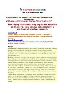
(iii) Burden of novel exposures. Some new medicines, cleaning agents, foods, etc., that are not carcinogenic themselves may still affect the natural ways of processing carcinogens in the body, and through this increase a person’s susceptibility to established carcinogens. Also, organismal resources are not unlimited, so that the increased burden of novel, even individually harmless, exposures on the xenobiotic processing system may reduce its capacity to address real threats and thus increase the body’s vulnerability to cancer. (iv) Some of the factors linked to economic prosperity and the Western lifestyle (e.g., delayed childbirth and food enriched with growth factors) may antagonistically influence aging and cancer risk. That is, such factors may attenuate some phenotypes of physical and reproductive aging, and, at the same time, increase the body’s vulnerability to cancer. The latter suggests a trade-off between cancer and aging that may contribute to concurrent increases in cancer risk and longevity in modern populations. 5.2.1. Improved Survival of Frail Individuals More developed countries have higher living standards and quality of medical care. These achievements, however, may lead to a “relaxation” of environmental selection, thereby facilitating the survival of individuals with various genetic and immune deficiencies, who would likely have died in the past. These survivors may contribute to a higher proportion of people who are more vulnerable to diseases (including cancer) in the populations of developed countries. Below are several epidemiological indicators of the possibility of such a scenario. Improved Survival during Childhood There was a dramatic decline in infant and childhood mortality in developed countries during the last century. For example, the infant mortality rate in the United States was about 6 percent of live births in 1935, 3 percent in 1950, 1.3 percent in 1980, and 0.6 percent in 2010. That is, it declined ten-fold over the course of 75 years (Singh and van Dyck, 2010; Health U.S. 2013). Most newborns in developed countries now reach reproductive age. This decline in mortality was largely due to radical improvements in the survival of infants with birth defects and infectious diseases, particularly with severe respiratory and intestinal infections (Singh and van Dyck, 2010). Contrarily, childhood mortality (up to 5 years of age) in some regions of the less developed countries such as India was until recently nearly 20 percent. This indicates that the pressure of environmental selection could be much higher in the least developed countries compared to the most developed ones. However, the better survival in the more developed world has its shadowy side. Because almost all children (including those with immunity deficiencies) survive, the proportion of the children who are inherently more vulnerable could be higher in the more developed countries. This is consistent with a typically higher proportion of children with chronic inflammatory immune disorders such as asthma and allergy in the populations of developed countries compared to less developed ones (Pearce and Douwes 2006). People with such disorders may be more susceptible to some cancers (Ukraintseva et al. 2010; Josephs et al. 2013). Cancer Incidence in Countries with Shorter and Longer Histories of Economic Growth If improved living conditions do facilitate survival of people who are more susceptible to cancer, then developed countries with shorter histories of economic prosperity should have lower overall cancer incidence rates than countries with a longer history of economic growth, particularly at old ages. This is because the older individuals in the rapidly developing countries have 9
experienced an improved quality of life only recently, whereas they faced more difficult living conditions earlier in their life. In such circumstances, robust individuals were more likely to survive the environmental selection and reach old age. This should result in a lower proportion of individuals who are susceptible to cancer among the elderly in recently developed countries compared to countries with longer histories of economic growth. Figure 5.5 supports this prediction. It shows that the age-specific cancer incidence rates in Japan, Singapore, and Kuwait (the "younger" developed countries) are lower than in the United States, the United Kingdom, and Switzerland (the "older" developed countries), especially at older ages, despite the similar quality of cancer diagnostics in these countries nowadays. Cancer incidence rate, all sites combined (1988-1992), males
Incidence per 100000
6000
Japan USA Singapore WGermany Kuwait Switzerlnd
4000
2000
0 0
5
10 15 20 25 30 35 40 45 50 55 60 65 70 75 80 85 Age
Figure 5.5. Comparison of age trajectories for the overall cancer incidence rate among developed countries with different histories of economic development (CI5I, 19662013). One can see that countries with more recently developed economies (Japan, Singapore, and Kuwait) have lower cancer incidence rates than countries with the longer history of economic growth, such as the USA, Switzerland, and West Germany, especially at old ages.
5.2.2. Avoiding or Reducing Traditional Exposures The better living conditions in developed countries have a downside in excessive hygiene and body cleansing. Excessive disinfection and hygiene may prevent or diminish some exposures that were abundant in the past, such as dirt, unsanitary conditions, or diverse microbial communities, among others. Until recently, these exposures were an inherent part of human living and our immune system learned to develop by interacting with them (Sing and Sing 2010). Over-reduction of such traditional exposures may result in an insufficiently/improperly trained immune system early in life, which could make it less able to resist diseases, including cancer later in life, thus contributing to the increased proportion of vulnerable individuals in adult populations of developed countries. There is accumulating evidence of the important role of these effects in cancer risk. An earlier study by a National Cancer Institute team suggested that improved public hygiene conditions, as measured by a decreased prevalence of hepatitis A virus infection, were also 10
associated with higher incidence rates of acute lymphoblastic leukemia (ALL) in children (Smith et al. 1998). More recently, it was shown that long-term exposure to microbial endotoxins can stimulate an anti-cancer immune response and reduce the risk of lung cancer by 40% (with a 20-year lag time), while the lack of such exposure increases the risk (Mastrangelo et al. 2005; Astrakianakis et al. 2007). Excessive body cleansing during childhood also decreases exposure to helminths (traditional in the past) that is important for the proper development of immunoregulatory mechanisms (Oikonomopoulou et al. 2014). The excessive cleansing and avoiding contact with dirt and dust may lead not only to reduced exposure to particular microorganisms, but also to reduced exposure to a diversity of molecules found in dirt and dust, some of which may play a role in immune priming and immune system development in the long term (Sing and Sing, 2010). A number of studies have connected excessive disinfection and lack of antigenic stimulation (especially in childhood) of the immune system in Westernized communities with increased risks of both chronic inflammatory diseases and cancer (Krämer et al. 1999; Lange et al. 2003; Hajdarbegovic et al. 2012; Oikonomopoulou et al. 2013; Francescone et al. 2014; Sheflin et al. 2014). For example, it was shown that some changes in traditional exposures may lead to microbial dysbiosis in the human body, which in turn may promote chronic inflammation (e.g., in the gut) and favor cancer development (Sheflin et al. 2014). Since chronic inflammation plays an important role in cancer development (Coussens and Werb, 2002; Coussens et al. 2013), these studies warrant deeper research towards understanding the impact of reduced traditional exposures on cancer development through inflammatory mechanisms. Differences in cancer rates among migrants to the same country provide additional epidemiological indicators of the importance of early environmental exposures in shaping susceptibility to cancer later in life. The IARC data on migrants to Israel (CI5I, 1966–2013) allow for comparison of the age trajectories of cancer incidence rates between adult Jews who live in Israel but were born in other countries (Figure 5.6).
Figure 5.6. Comparison of age trajectories of the overall cancer incidence rate among Jews born in more and less developed regions of the world (CI5I, 19662013).
11
The age curves of cancer incidence in Figure 5.6 show that Jews born in less developed regions (Africa and Asia) have overall lower cancer risk than those born in the more developed regions (Europe and America). The discrepancy is unlikely to be due to differences in cancer diagnostics because at the moment of diagnosis all these people were citizens of the same country with the same standard of medical care. These results suggest that surviving childhood and growing up in a less developed country with diverse environmental exposures might help form resistance to cancer that lasts even after moving to a high risk country. 5.2.3. Burden of Novel and Nontraditional Exposures Many behavioral and dietary habits, new medicines, foods, and chemicals, are typical of economically prosperous countries but not common in the less developed ones. Some of the novel and non-traditional exposures associated with the economic prosperity and Western lifestyle, not being formally carcinogenic, might affect the natural ways of processing carcinogens in the body, or favor chronic inflammation, and through this increase a person’s susceptibility to cancer. Other factors that are not carcinogenic when considered individually, together might create an excessive burden on the body’s xenobiotic processing system. This burden may reduce the capacity of this system to address real threats, and thus increase the body’s susceptibility to cancer. Here we present some recent evidence in support of this view. Many of the relevant examples are provided by the IARC Monographs on the Evaluation of Carcinogenic Risks to Humans (IARC Monographs, 1972–2014), a valuable WHO resource. Western Pattern of Food Consumption The Western pattern of food consumption is characterized by a high content of animal protein (from meat, eggs, milk, cheese, etc.), fat, and purified sugar, as well as a low content of crude plants and grains in the everyday diet. A number of human and animal studies suggest a causal connection between the spread of Western dietary habits and changes in vulnerability to some cancers (Watson and Collins, 2011; Mosby et al. 2012). For example, individuals with relatively high consumption of animal protein may face a significantly increased risk of colon cancer (Willett 1989, Ananthakrishnan et al. 20015; Carr et al. 2015). Kagawa et al. (1978) described the traditional pattern of food consumption in Japan in the past as characterized by a high proportion of crude grain (barley) and a low proportion of any kind of animal protein. Since the 1950s, this diet has been gradually replaced by one that includes a high proportion of protein and a low proportion of barley. Other components of the Japanese diet (e.g., vegetables) did not change much during the same period. The colon cancer incidence rate has been increasing in Japan since then, while the rate of stomach cancer has been decreasing (CI5I, 1966–2013). The mechanism could involve a trade-off between reduced damage to the stomach and increased microbial dysbiosis. That is, on the one hand, an excessive amount of crude fibers in food can harm the stomach’s mucous membrane and promote inflammation, thus potentially increasing the stomach’s vulnerability to cancer. On the other hand, a decreased fiber intake may diminish this particular harm, but at the same time it may suppress intestinal motility and thus elevate the risk of colon cancer. This is because the motility prevents the intestine from festering, and the festering creates an environment conducive to the development of microbial dysbiosis in the intestine. The dysbiosis, in turn, may favor carcinogenic production by the colon bacteria, such as E.coli (Falk et al., 1998; Parsonnet, 1999). An increased intake of meat promotes food festering in the intestine accompanied by the bacterial imbalance and respective increase in internal carcinogenic exposure (Parsonnet, 1999). A number of recent studies strongly support the idea that changes in the traditional pattern of 12
food consumption may lead to microbial dysbiosis (both on the skin and in the gut), which in turn may favor cancer development through inflammatory and other mechanisms (Francescone et al. 2014; Sheflin et al. 2014). New Medicines and Other Chemicals Some pharmaceuticals and dietary supplements that are prevalent in developed but not in developing countries may influence vulnerability to cancer, although they have not been individually shown to be carcinogenic. The following illustration shows how it could happen. A well-studied chemical, benzpyrene, is a non-direct carcinogen. It needs to be metabolically processed in the body before it can become harmful. First, oxidative enzymes (e.g., cytochrome P450) decompose benzpyrene into intermediate products of the metabolism. Several of these products (e.g., phenol) are already carcinogenic. Other substances (e.g., glutathione) bind these carcinogenic products to deactivate them and take them out of the metabolism. If there is the right balance between cytochrome P450 and glutathione in a cell, then the carcinogen is quickly deactivated without harming the organism. However, if the amount of glutathione is not in the right balance with the amount of cytochrome P450, the carcinogen deactivation process is delayed or incomplete. As a result, carcinogenic metabolites may accumulate in the body and increase the chances of developing cancer. The right balance of chemical players in the processing of carcinogens could, therefore, be an important factor in vulnerability to cancer (Diggs et al. 2013). The high burden of new substances on the body’s systems processing xenobiotics may disturb the delicate balance of events and processes leading to neutralization of carcinogens and through this to increased vulnerability to cancer. Research on the simultaneous exposures to carcinogenic and non-carcinogenic compounds provides support for this mechanism. Paracetamol (Acetaminophen, Tylenol, Contac) is a non-prescription antipyretic which has been used extensively in developed countries since 1946. The drug is not classifiable by the IARC for carcinogenicity to humans. However, animal experiments have shown that paracetamol increases the incidence of renal adenomas induced by an established carcinogen, N-nitrosoethyl-Nhydroxyethylamine (IARC Monographs, Vol. 50). That is, paracetamol, being not harmful on its own, may enhance the body’s susceptibility to an established carcinogen, so that a lower level of carcinogenic exposure may be required to induce tumor growth in the more susceptible individual. Antibiotics. People who are frequently treated with antibiotics may have decreased diversity of microbial community, especially in the gut and on the skin. The decreased microbial diversity itself was shown to be associated with increased risks of several cancers, most notably with colon cancer (Modi et al. 2014; Ahn et al. 2013). The mechanism could be that the decreased microbial diversity after treatment with antibiotics creates the conditions for bacterial imbalance in the colon. As mentioned above, this imbalance (dysbiosis) may result in the suppression of the bifidobacteria and the promotion of E.coli bacteria (Falk et al. 1998). The latter have a propensity to transform normal metabolic products (e.g., bile acids) into internal carcinogens, thereby increasing the risk of colon cancer (Parsonnet, 1999). Antibiotics may also influence the metabolism of external carcinogens. Metronidazole, an antibiotic which can destroy H. pylori and decrease the risk of stomach cancer, also increases the incidence of colon cancer in rats, induced by the administration of an established carcinogen (Sloan et al. 1983; IARC Monographs, Suppl. 7). Chloramphenicol, an antibiotic broadly used since the 1950s, increased 13
the incidence of lymphomas induced by an established carcinogen in mice, while the drug alone did not show clear carcinogenic effect (IARC Monographs, Vol. 50). On the other hand, antibiotics may sometimes reduce cancer risks. This may be particularly relevant to stomach cancer, whose risk is typically lower in the more developed world. A number of studies discussed in the IARC Monographs on the Evaluation of Carcinogenic Risks to Humans (1972–2014) established an association between seropositivity for the H. pylori bacteria and stomach cancer. An estimate of the relative risk was about four times the natural risk of this cancer (IARC Monographs, Vol. 61). A possible mechanism involves the cancer promoting effects of chronic inflammation which accompanies the infection. The prevalence of H. pylori infection is substantially lower in developed countries than in developing ones. In both, the prevalence is higher in the lower socioeconomic classes. A progressive reduction in the rate of this infection in successive birth cohorts in the developed countries (IARC Monographs, Vol. 61) is held to be the result of improved hygiene and the spread of antibiotics which can destroy H. pylori bacteria. Antibiotic treatment may therefore decrease the risk of stomach cancer. So, the increase in colon cancer and the decrease in stomach cancer risks, which occurred along with economic progress, could in part be explained not only by the change in food patterns, but also by the population-wide exposure to antibiotics. Hormone replacement therapy (HRT). Menopausal and early postmenopausal HRT with various combinations of estrogen and progestin is more common in developed than in developing countries. At the peak of its use in 1999, approximately 20 million women in the developed world used HRT, including about half of all women aged 50–65 years in the U.S. HRT is thought to be in part responsible for differences in incidence rates of female hormone-dependent cancers between more and less developed countries (IARC Monographs, Vol. 72, Vol. 91). Postmenopausal HRT use in the U.S. has dropped since 2002, particularly for continuous HRT, following the report of adverse effects by the Women’s Health Initiative’s estrogen plus progestin trial. Prescriptions for HRT declined from 61 million prescriptions in 2001 to 21 million in 2004. This was followed by a decline in the risk of estrogen-receptor-positive breast cancer. For example, the age-adjusted incidence rate of breast cancer in women who were 50 years of age or older fell 6.7 percent in the United States in 2003 (IARC Monographs, Vol. 91; Ravdin et al. 2007; Ukraintseva et al. 2008). Oral contraceptives. Another female hormonal treatment, oral contraception for pregnancy prevention, is even more prevalent than postmenopausal therapy. Oral contraceptives usually include both estrogen and progesterone. This treatment has been popular in developed countries since the 1960s. Today, worldwide, more than 100 million women, an estimated 10% of all women of reproductive age, use combined hormonal contraceptives. Current use of these drugs is greatest in developed countries (16%) and is lower in developing ones (6%). They have been shown to increase the risk of breast and liver cancer, while being protective against endometrial and ovarian cancers in women (IARC monographs, Vol. 91). Starting in the 1970s, the decline in female endometrial and ovarian cancer incidence rates, as well as the increase in incidence rates of breast and liver cancers in the United States, could in part be related to the nationwide exposure of American women to hormonal contraceptives. The mechanism of the effect of estrogen, alone and in combination with progestin, on the female organism is very complex and depends on age, stage of ontogeny, and target tissue. Cancer 14
promoting properties could in part be related to the estradiol-associated stimulation of growth hormone release (Veldhuis et al. 2004), which could potentially lead to excessive cell proliferation, while cancer protective properties could be linked to increased regenerative potential and competitive ability of normal host cells surrounding a transformed cell or latent tumor (Ukraintseva and Yashin 2003, 2004) or to other mechanisms. What is clear is that HRT and the estrogen-progestogen contraceptives are not simply carcinogens but may differentially influence susceptibility to cancer in different tissues and periods of life. Household Chemicals. Chemicals used at home or in small businesses, such as components of plasticware, cleaning agents, flame retardants, and others, are normally tested for carcinogenic properties before market introduction, and therefore are unlikely to be directly carcinogenic. However, the pre-market testing usually does not take into account that the products may occasionally be consumed with drink or food, or through the skin. The problem is that the clearance of such chemicals from surfaces (e.g., glasses, plates, or clothes from washing detergents) can be poor, and their residuals may enter the body and potentially accumulate in amounts sufficient to harm it. Research on this important topic is emerging though still rather limited. It is increasingly recognized that many household chemicals that are common in developed countries may have “endocrine-disrupting” effects, meaning that they may interfere with hormonal processes in the body and consequently increase an organism’s susceptibility to various health disorders, including cancer, especially in people who lack detoxifying enzymes (e.g., De Coster and van Larebeke, 2012; The 2013 Berlaymont Declaration on Endocrine Disrupters). Examples of relevant chemicals that are currently discussed as having endocrinedisrupting properties with possible health consequences, include (but are not limited to): Bisphenol A (BPA), some flame retardants, phthalates, pesticides, solvents, household cleaning products, air fresheners, hair dyes, cosmetics, and sunscreens (e.g., Travier et al. 2002; Zota et al. 2010; De Coster and van Larebeke, 2012; The 2013 Berlaymont Declaration on Endocrine Disrupters). Quantity versus Quality There may be several biological mechanisms by which exposure to large numbers of new, individually harmless chemicals may increase susceptibility to cancer. Some may involve cumulative effects, “cocktail effects”, and synergistic interactions of chemicals (Cedergreen 2014). For example, BPA and estrogen may both interact with estrogen receptors (Gao et al. 2015), so their cumulative effect on these receptors may be more “endocrine-disrupting” and influential to cell proliferation and migration than when they act alone. There may also be less specific mechanisms, in which variety and quantity of the chemicals rather than their qualities play a major role. Indeed, in developed countries, people are typically exposed to many new chemicals at once. When these chemicals end up in the body all together, they may create an excess burden on the body’s xenobiotic processing system. We speculate that the unusually large numbers of unfamiliar compounds entering the xenobiotic processing system may reduce its efficiency to respond to real threats, and thus potentially increase the body’s vulnerability to cancer, even in absence of particular oncogenic effects of individual compounds. This potential mechanism deserves further investigation.
15
5.3 Some of the Factors Associated with Economic Development and the Western Lifestyle May Antagonistically Influence Aging and Vulnerability to Cancer 5.3.1. Cancer and Aging: A Trade-off? One paradoxical feature of economic progress is the concurrent increase in longevity and overall cancer risk in affluent societies. As discussed above, some factors linked to economic development and the Western lifestyle may contribute to increased vulnerability to cancer. Here we show that some of such factors may influence both cancer and aging/ontogeny related traits (e.g., growth, reproductive period, and physical senescence), sometimes antagonistically. Those factors that increase vulnerability to cancer but also attenuate some phenotypes of physical and reproductive aging might contribute to increases in both cancer risk and longevity in modern human populations. Below we provide evidence from human and animal studies supporting such a possibility. 5.3.2. Increased Exposure to Growth Factors People in developed countries have virtually unlimited access to dense nutritious food, such as meat, fat, and sweets, which may promote growth (increase in height and weight) and affect the metabolism of internal growth factors (Kaklamani et al. 1999; Giovannucci et al. 2004; Larsson et al. 2005; Bujnowski et al. 2011; TeMorenga et al. 2012). Increased height, weight, and levels of internal growth factors are in turn considered to play an important role in cancer development and have been associated with risks of several common cancers (Giovannucci et al. 2004; Renehan et al. 2004; Pollak et al. 2008; Batty et al. 2009; Moore et al. 2009; Key et al. 2010; Yoshimoto et al. 2011; Kabat et al. 2011; Mellemkjær et al. 2012; Davis et al. 2013). Potential mechanisms may involve enhanced cell growth and proliferation, and the anti-apoptotic effects of growth factors favoring survival of transformed cells and latent tumors (van der Veeken et al. 2009; Bruchim et al. 2009; Arnaldez and Helman, 2012). As for aging, the higher levels of internal growth factors, such as IGF-1, estrogens, and some others, have been linked to attenuation of phenotypes of physical senescence, including elderly frailty, sarcopenia, muscle atrophy, heart failure, hip fractures, as well as to better muscle regeneration and healing (e.g., Ruiz-Torres and Soares de Melo Kirzner 2002; Vasan et al. 2003; Roubenoff et al. 2003; Vinciguerra et al. 2010; Conti et al. 2011; Musaro 2012; Thornton 2013; Yeap et al. 2013; Locatelli et al. 2014; Levine et al. 2014). In rodent models, a reduction in growth factors/IGF-I signaling is often correlated with increased longevity (e.g., Bartke et al. 2003). This increase in longevity is largely attributed to reduced cancer risk in the laboratory animals (Ikeno et al. 2009). At the same time, the overexpression of IGF-1 was shown to attenuate the aging-associated cardiac, cerebrovascular, and cognitive decline in older animals (Torella et al. 2004; Trejo et al. 2007; Sonntag et al. 2013). Mouse studies have shown that high protein intake and upregulated GHR-IGF-1 signaling favor the incidence and progression of several cancers (e.g., breast and melanoma tumors); however, a low protein diet had detrimental effects in the very old (Levine et al. 2014).
16
Overall, data support the possibility of trade-offs between the effects of growth factors on certain cancer and aging-related phenotypes. The pro-cancer properties of growth factors could be related to upregulated growth and proliferation, and anti-apoptotic effects. Anti-aging properties could be related to decelerated muscle loss, better tissue regeneration, and cell survival. Growth factors may especially contribute to cancer risk and mortality before the oldest old ages (90+), when the incidence rate reaches its peak in the population for most cancers (Ukraintseva and Yashin, 2003a; Ukraintseva et al. 2008; Akushevich et al. 2012). They may also contribute to extreme longevity because higher levels of growth factors can be particularly beneficial for survival at very old ages, when physical senescence and related disorders (e.g., heart failure) become leading contributors to mortality risk. Also, several major diseases that could potentially benefit from higher levels of growth factors (e.g., stroke and AD) reach maximal incidence risk at oldest old ages (90+) when cancer risk is already declining (Ukraintseva and Yashin, 2003a; Johnsen et al. 2005; Ukraintseva et all. 2008; Akushevich et al. 2012; Duron et al. 2012; Dong et al. 2014). This suggests that the timing of exposure may be important for the pro-cancer or prolongevity effects of growth factors. 5.3.3. Later Menopause The median age of menopause is generally higher in more developed countries. This age typically varies from 44–49 years in less developed regions (e.g., Mexica, India, Africa) to 50– 54 years in more developed ones (e.g., the UK, the United States) (MacMahon 1966; McKinaly et al. 1972; Garrido-Latorre et al. 1996; Kriplani and Banerjee 2005; Dratva et al. 2009). It also tends to be higher in upper socio-economic groups within the same country (e.g., Hardy and Kuh, 2005). Later menopause has been associated with elevated risks of female hormone-related cancers in postmenopausal women, especially with breast, ovarian, and endometrial tumors (Franceschi et al. 1991; Ossewaarde et al. 2005; Mondul et al. 2005; Collaborative group, 2012). At the same time, later menopause was linked to increased overall survival and longevity in several large studies. For example, the natural menopause that occurred at ages 50–54 vs. 40–44 years was associated with longer survival in large cohorts of Dutch and American women (Ossewaarde et al. 2005; Mondul et al. 2005). This longer survival was accompanied by significantly increased mortality from breast, endometrial, and ovarian cancers, as well as reduced mortality from pneumonia, influenza, and falls (which are common causes of death in the very old). Later menopause was linked to a lower total mortality risk, and to reductions in cardiovascular deaths and heart failure, in several other studies (Snowdon et al. 1989; Jansen et al. 2002; de Kleijn et al. 2002; Rahman et al. 2015). The postponed menopause can also be accompanied by signs of slower physiological aging. For example, it was significantly associated with slower cognitive aging, especially with better memory in naturally postmenopausal elderly women (McLay et all. 2003; Tierney et al. 2013). 5.3.4. Giving Birth at Later Age Age at childbirth has increased across world populations along with economic progress and adoption of the Western lifestyle. This age is typically higher in younger compared to older
17
generations in developed countries (Morabia and Costanza, 1999; Savage et al. 2013; Baghurst et al. 2014). Older age at childbirth (first, last, and average) has been associated with elevated risks of several human cancers in mothers, especially with breast cancer and melanoma (Ewertz et al. 1990; Wohlfahrt and Melbye, 2001; Li et al. 2014). For example, women who gave their first birth after age 35 had a risk increase for breast cancer by 40 percent compared to mothers who experienced their first birth before age 20. And the relative risk for melanoma was 1.47 in women of the oldest versus youngest age at first birth in a meta-analysis (Ewertz et al. 1990; Li et al. 2014). On the other hand, an older age at birth, especially that of the last child, shows a positive association with mother’s survival toward very old age (Helle et al. 2005; McArdle et al. 2006; Sun et al. 2015). For example, women who had their last child after age 33 had twice the odds for survival to the top 5th percentile of survival for their birth cohorts compared with women who had their last child by age 29 years (Sun et al. 2015). Our unpublished study of 2,401 Danish Twins aged 75+ (LSADT 1995-2000) also revealed that women who were in their 40s at the time of birth of their last child had about 22% lower death rates after age 75 compared to women who had their last child before age 40. In line with these data, experimental animal studies found that selection for late reproductive ability results in increased longevity of Drosophila flies after just a few generations of such selection (e.g., Rose and Charlesworth, 1981). Older parental age (both maternal and paternal) was also linked to increased cancer risks in the offspring, especially of leukemia, brain tumors, and breast and prostate cancers (Hemminki et al. 1999; Zhang et al. 1999; Hodgson et al. 2004). Increasing parental age in developed countries may therefore also contribute to a higher vulnerability to cancer in these countries. Notably, in laboratory rodents, a higher father’s age was associated with increased susceptibility to an established carcinogen in mice offspring (Anisimov and Gvardina, 1995). It is not clear so far if having an older parent carries anti-aging/pro-longevity benefits for the offspring. One potential mechanism of how the late birth may do both (i.e., increase vulnerability to cancer and attenuate physical aging in offspring) might involve trade-off-like effects of internal growth factors, such as estrogens and IGF-1, on the cancer and aging phenotypes (Yang et al. 2005; Levine et al. 2014). It was shown that children of mothers who were 30–35 years of age at childbirth were taller and displayed a 19% increase in IGF-I concentrations compared to offspring of mothers who gave birth prior to age 30 (Savage et al. 2013). An association has also been found between older paternal age at birth and longer leukocyte telomere length in the offspring which may indicate postponed replicative senescence (Prescott et al. 2012), but supportive studies are rather limited.
5.4 Conclusion In this chapter, we discussed factors responsible for the higher cancer risk in the more developed world, and for concurrent increases in cancer risk and longevity in association with economic progress and a Western lifestyle. We suggested that in populations of developed countries, the proportion of individuals more susceptible to cancer may be higher than in less developed
18
regions of the world, and that this may contribute to the typically higher overall cancer risk in such countries. We provided evidence from human and animal studies suggesting that several factors associated with advanced economic development and a Western lifestyle could favor an increase in the proportion of individuals who are more susceptible to cancer in the respective populations. Such factors include (but are not limited to) dramatic improvements in medical care and living conditions, which may lead to a “relaxation” of environmental selection and improved survival of frail individuals in affluent societies. They may also lead to an insufficiently/inadequately trained immune system early in life, which may make an individual more vulnerable to cancer later in life. Other factors (e.g., some new foods, medicines, and other chemicals) may also increase a person’s susceptibility to cancer. Not being directly or individually carcinogenic, these factors may affect the metabolism of established carcinogens, or act synergistically, and through this influence cancer risks. Quantities and variety of the chemicals rather than their individual qualities may also play an important role. People in developed countries are exposed to many new chemicals. We hypothesized that the large number of unfamiliar compounds may reduce the efficiency of the xenobiotic processing system, and through this increase the body’s vulnerability to cancer, even in the absence of particular oncogenic effects of individual compounds. This potential mechanism deserves further investigation. Some of the factors associated with economic prosperity and a Western lifestyle may influence both aging and vulnerability to cancer, sometimes oppositely. Current evidence supports a possibility of trade-offs between cancer and aging-related phenotypes (Ukraintseva et al. 2015), which could be influenced by delayed reproduction and exposures to growth factors (Levine et al. 2014; Li et al. 2014; Sun et al. 2015). The latter may be particularly beneficial at very old age. This is because the higher levels of growth factors may attenuate some phenotypes of physical senescence, such as decline in regenerative and healing ability, sarcopenia, frailty, elderly fractures and heart failure due to muscles athrophy. They may also increase the body’s vulnerability to cancer, e.g., through growth promoting and anti-apoptotic effects (Ukraintseva and Yashin, 2003a,b, 2004; Pollak et al. 2004). The increase in vulnerability to cancer due to growth factors can be compatible with extreme longevity because cancer is a major contributor to mortality mainly before age 85, while senescence-related causes (such as physical frailty) become major contributors to mortality at oldest old ages (85+). In this situation, the impact of growth factors on vulnerability to death could be more deleterious in middle-to-old life (~before 85) and more beneficial at older ages (85+). The complex relationships between aging, cancer, and longevity are challenging (Ukraintseva and Yashin, 2003a; Ukraintseva et al. 2015). This complexity warns against simplified approaches to extending longevity without taking into account the possible trade-offs between phenotypes of physical aging and various health disorders, as well as the differential impacts of such trade-offs on mortality risks at different ages. Acknowledgement Research reported in this chapter was partly supported by the National Institute on Aging of the National Institutes of Health under Award Numbers R01AG046860 and P01AG043352. The content is solely the responsibility of the authors and does not necessarily represent the official views of the National Institutes of Health.
19
References Ahn J, Sinha R, Pei Z, Dominianni C, Wu J, Shi J, Goedert JJ, Hayes RB, Yang L. Human gut microbiome and risk for colorectal cancer. J Natl Cancer Inst. 2013 Dec 18;105(24):1907-11. doi: 10.1093/jnci/djt300. Epub 2013 Dec 6. PubMed PMID: 24316595; PubMed Central PMCID: PMC3866154. Akushevich I, Kravchenko J, Ukraintseva S, Arbeev K, Yashin AI. Age patterns of incidence of geriatric disease in the U.S. elderly population: Medicare-based analysis. J Am Geriatr Soc. 2012 Feb;60(2):323-7. doi: 10.1111/j.1532-5415.2011.03786.x. Epub 2012 Jan 27. PubMed PMID: 22283485; PubMed Central PMCID: PMC3288526. Ananthakrishnan AN, Du M, Berndt SI, Brenner H, Caan BJ, Casey G, Chang-Claude J, Duggan D, Fuchs CS, Gallinger S, Giovannucci EL, Harrison TA, Hayes RB, Hoffmeister M, Hopper JL, Hou L, Hsu L, Jenkins MA, Kraft P, Ma J, Nan H, Newcomb PA, Ogino S, Potter JD, Seminara D, Slattery ML, Thornquist M, White E, Wu K, Peters U, Chan AT. Red Meat Intake, NAT2, and Risk of Colorectal Cancer: A Pooled Analysis of 11 Studies. Cancer Epidemiol Biomarkers Prev. 2015 Jan;24(1):198-205. doi: 10.1158/10559965.EPI-14-0897. Epub 2014 Oct 23. PubMed PMID: 25342387; PubMed Central PMCID: PMC4294960. Anisimov VN, Gvardina OE. N-nitrosomethylurea-induced carcinogenesis in the progeny of male rats of different ages. Mutat Res. 1995 Feb;316(3):139-45. PubMed PMID: 7862177. Arnaldez FI, Helman LJ. Targeting the insulin growth factor receptor 1. Hematol Oncol Clin North Am. 2012 Jun;26(3):527-42, vii-viii. doi: 10.1016/j.hoc.2012.01.004. Epub 2012 Feb 28. Review. PubMed PMID: 22520978; PubMed Central PMCID: PMC3334849. Astrakianakis G, Seixas NS, Ray R, Camp JE, Gao DL, Feng Z, Li W, Wernli KJ, Fitzgibbons ED, Thomas DB, Checkoway H. Lung cancer risk among female textile workers exposed to endotoxin. J Natl Cancer Inst. 2007 Mar 7;99(5):357-64. PubMed PMID: 17341727. Baghurst P, Robson S, Antoniou G, Scheil W, Bryce R. The association between increasing maternal age at first birth and decreased rates of spontaneous vaginal birth in South Australia from 1991 to 2009. Aust N Z J Obstet Gynaecol. 2014 Jun;54(3):237-43. doi: 10.1111/ajo.12182. Epub 2014 Feb 8. PubMed PMID: 24506445. Bartke A, Chandrashekar V, Dominici F, Turyn D, Kinney B, Steger R, Kopchick JJ. Insulin-like growth factor 1 (IGF-1) and aging: controversies and new insights. Biogerontology. 2003;4(1):1-8. Review. PubMed PMID: 12652183. Batty GD, Shipley MJ, Gunnell D, Huxley R, Kivimaki M, Woodward M, Lee CM, Smith GD. Height, wealth, and health: an overview with new data from three longitudinal studies. Econ Hum Biol. 2009 Jul;7(2):137-52. doi: 10.1016/j.ehb.2009.06.004. Epub 2009 Jun 28. Review. PubMed PMID: 19628438. 20
Bleyer A, Welch HG. Effect of three decades of screening mammography on breast-cancer incidence. N Engl J Med. 2012 Nov 22;367(21):1998-2005. doi: 10.1056/NEJMoa1206809. PubMed PMID: 23171096. Bruchim I, Attias Z, Werner H. Targeting the IGF1 axis in cancer proliferation. Expert Opin Ther Targets. 2009 Oct;13(10):1179-92. doi: 10.1517/14728220903201702. Review. PubMed PMID: 19663648. Bujnowski D, Xun P, Daviglus ML, Van Horn L, He K, Stamler J. Longitudinal association between animal and vegetable protein intake and obesity among men in the United States: the Chicago Western Electric Study. J Am Diet Assoc. 2011 Aug;111(8):11501155.e1. doi: 10.1016/j.jada.2011.05.002. PubMed PMID: 21802560; PubMed Central PMCID: PMC3158996. CI5, 1966-2013. Cancer Incidence in Five Continents. Volumes I-X (1966-2013). IARC Scientific Publications. Lyon: IARC (International Agency for Research on Cancer). http://ci5.iarc.fr/CI5plus/Default.aspx; http://ci5.iarc.fr/CI5I-X/Pages/references.aspx , last assessed on 09/28/2014. (The reference time period for Volumes I-X: 1965–2007). Canudas-Romo V. Three measures of longevity: time trends and record values. Demography. 2010 May;47(2):299-312. PubMed PMID: 20608098; PubMed Central PMCID: PMC3000019. Carr PR, Walter V, Brenner H, Hoffmeister M. Meat subtypes and their association with colorectal cancer: systematic review and meta-analysis. Int J Cancer. 2015 Jan 12. doi: 10.1002/ijc.29423. [Epub ahead of print] PubMed PMID: 25583132. Cedergreen N. Quantifying synergy: a systematic review of mixture toxicity studies within environmental toxicology. PLoS One. 2014 May 2;9(5):e96580. doi: 10.1371/journal.pone.0096580. eCollection 2014. PubMed PMID: 24794244; PubMed Central PMCID: PMC4008607. Collaborative Group on Hormonal Factors in Breast Cancer. Menarche, menopause, and breast cancer risk: individual participant meta-analysis, including 118 964 women with breast cancer from 117 epidemiological studies. Lancet Oncol. 2012 Nov;13(11):1141-51. doi: 10.1016/S1470-2045(12)70425-4. Epub 2012 Oct 17. PubMed PMID: 23084519; PubMed Central PMCID: PMC3488186. Conti, E., Musumeci, M.B., De Giusti, M., Dito, E., Mastromarino, V., Autore, C., and Volpe, M. (2011). IGF-1 and atherothrombosis: relevance to pathophysiology and therapy. Clinical Science 120, 377-402. Coussens LM, Werb Z. Inflammation and cancer. Nature. 2002 Dec 19-26;420(6917):860-7. Review. PubMed PMID: 12490959; PubMed Central PMCID: PMC2803035. Coussens LM, Zitvogel L, Palucka AK. Neutralizing tumor-promoting chronic inflammation: a magic bullet? Science. 2013 Jan 18;339(6117):286-91. doi: 10.1126/science.1232227. 21
Review. Erratum in: Science. 2013 Mar 29;339(6127):1522. PubMed PMID: 23329041; PubMed Central PMCID: PMC3591506. Cremers RG, Karim-Kos HE, Houterman S, Verhoeven RH, Schröder FH, van der Kwast TH, Kil PJ, Coebergh JW, Kiemeney LA. Prostate cancer: trends in incidence, survival and mortality in the Netherlands, 1989-2006. Eur J Cancer. 2010 Jul;46(11):2077-87. doi: 10.1016/j.ejca.2010.03.040. Epub 2010 May 12. PubMed PMID: 20471247. Davis E, Jacoby P, de Klerk NH, Cole C, Milne E. Western Australian children with acute lymphoblastic leukemia are taller at diagnosis than unaffected children of the same age and sex. Pediatr Blood Cancer. 2011 May;56(5):767-70. doi: 10.1002/pbc.22832. Epub 2011 Jan 18. PubMed PMID: 21246704. De Coster S, van Larebeke N. Endocrine-disrupting chemicals: associated disorders and mechanisms of action. J Environ Public Health. 2012;2012:713696. Epub 2012 Sep 6. Review. PubMed PMID: 22991565; PubMed Central PMCID: PMC3443608. de Kleijn MJ, van der Schouw YT, Verbeek AL, Peeters PH, Banga JD, van der Graaf Y. Endogenous estrogen exposure and cardiovascular mortality risk in postmenopausal women. Am J Epidemiol. 2002 Feb 15;155(4):339-45. PubMed PMID: 11836198. De Rooij MJ, Rampen FH, Schouten LJ, Neumann HA. Total skin examination during screening for malignant melanoma does not increase the detection rate. Br J Dermatol. 1996 Jul;135(1):42-5. PubMed PMID: 8776357. Diggs DL, Myers JN, Banks LD, Niaz MS, Hood DB, Roberts LJ 2nd, Ramesh A. Influence of dietary fat type on benzo(a)pyrene [B(a)P] biotransformation in a B(a)P-induced mouse model of colon cancer. J Nutr Biochem. 2013 Dec;24(12):2051-63. doi: 10.1016/j.jnutbio.2013.07.006. PubMed PMID: 24231098; PubMed Central PMCID: PMC3904801. Dong X, Chang G, Ji XF, Tao DB, Wang YX. The relationship between serum insulin-like growth factor I levels and ischemic stroke risk. PLoS One. 2014 Apr 11;9(4):e94845. doi: 10.1371/journal.pone. 0094845. eCollection 2014. PubMed PMID: 24728374; PubMed Central PMCID: PMC3984250. Dratva J, Gómez Real F, Schindler C, Ackermann-Liebrich U, Gerbase MW, Probst-Hensch NM, Svanes C, Omenaas ER, Neukirch F, Wjst M, Morabia A, Jarvis D, Leynaert B, Zemp E. Is age at menopause increasing across Europe? Results on age at menopause and determinants from two population-based studies. Menopause. 2009 Mar-Apr;16(2):38594. doi: 10.1097/gme.0b013e31818aefef. PubMed PMID: 19034049. Edwards, B. K., Noone, A.-M., Mariotto, A. B., Simard, E. P., Boscoe, F. P., Henley, S. J., Jemal, A., Cho, H., Anderson, R. N., Kohler, B. A., Eheman, C. R. and Ward, E. M. (2014), Annual Report to the Nation on the status of cancer, 1975-2010, featuring prevalence of comorbidity and impact on survival among persons with lung, colorectal, breast, or prostate cancer. Cancer, 120: 1290–1314. doi: 10.1002/cncr.28509 22
Ewertz M, Duffy SW, Adami HO, Kvåle G, Lund E, Meirik O, Mellemgaard A, Soini I, Tulinius H. Age at first birth, parity and risk of breast cancer: a meta-analysis of 8 studies from the Nordic countries. Int J Cancer. 1990 Oct 15;46(4):597-603. PubMed PMID: 2145231.
Falk PG, Hooper LV, Midtvedt T, Gordon JI. Creating and maintaining the gastrointestinal ecosystem: what we know and need to know from gnotobiology. Microbiol Mol Biol Rev. 1998 Dec;62(4):1157-70. Review. PubMed PMID: 9841668; PubMed Central PMCID: PMC98942. Ferlay J, Soerjomataram I, Ervik M, Dikshit R, Eser S, Mathers C, Rebelo M, Parkin DM, Forman D, Bray, F. GLOBOCAN 2012 v1.0, Cancer Incidence and Mortality Worldwide: IARC CancerBase No. 11 [Internet]. Lyon, France: IARC (International Agency for Research on Cancer); 2013. Available from: http://globocan.iarc.fr, accessed on 12/25/2014. Finch CE, Beltrán-Sánchez H, Crimmins EM. Uneven futures of human lifespans: reckonings from gompertz mortality rates, climate change, and air pollution. Gerontology. 2014;60(2):183-8. doi: 10.1159/000357672. Epub 2013 Dec 24. PubMed PMID: 24401556; PubMed Central PMCID: PMC4023560. Franceschi S, La Vecchia C, Booth M, Tzonou A, Negri E, Parazzini F, Trichopoulos D, Beral V. Pooled analysis of 3 European case-control studies of ovarian cancer: II. Age at menarche and at menopause. Int J Cancer. 1991 Aug 19;49(1):57-60. PubMed PMID: 1874570. Francescone R, Hou V, Grivennikov SI. Microbiome, inflammation, and cancer. Cancer J. 2014 May-Jun;20(3):181-9. doi: 10.1097/PPO.0000000000000048. PubMed PMID: 24855005; PubMed Central PMCID: PMC4112188. Gao H, Yang BJ, Li N, Feng LM, Shi XY, Zhao WH, Liu SJ. Bisphenol a and hormoneassociated cancers: current progress and perspectives. Medicine (Baltimore). 2015 Jan;94(1):e211. PubMed PMID: 25569640. Garrido-Latorre F, Lazcano-Ponce EC, López-Carrillo L, Hernández-Avila M. Age of natural menopause among women in Mexico City. Int J Gynaecol Obstet. 1996 May;53(2):15966. PubMed PMID: 8735297. Giard RW, Coebergh JW, Casparie-van Velsen IJ. [A marked increase in the rate of diagnosed prostate cancer in the Netherlands during 1990-1996]. Ned Tijdschr Geneeskd. 1998 Aug 29;142(35):1958-62. Dutch. PubMed PMID: 9856189. Giovannucci E, Rimm EB, Liu Y, Willett WC. Height, predictors of C-peptide and cancer risk in men. Int J Epidemiol. 2004 Feb;33(1):217-25. PubMed PMID: 15075172.
23
Hajdarbegovic E, Verkouteren J, Balak D. Non-melanoma skin cancer: the hygiene hypothesis. Med Hypotheses. 2012 Dec;79(6):872-4. doi: 10.1016/j.mehy.2012.09.012. Epub 2012 Oct 13. PubMed PMID: 23073119. Hanahan D, Weinberg RA. The hallmarks of cancer. Cell. 2000 Jan 7;100(1):57-70. Review. PubMed PMID: 10647931. Hanahan D, Weinberg RA. Hallmarks of cancer: the next generation. Cell. 2011. Mar 4;144(5):646-74. Review. PubMed PMID: 21376230. Hardy R, Kuh D. Social and environmental conditions across the life course and age at menopause in a British birth cohort study. BJOG. 2005 Mar;112(3):346-54. PubMed PMID: 15713152. Health U.S. 2013. National Center for Health Statistics. Health, United States, 2013: With Special Feature on Prescription Drugs. Hyattsville, MD. 2014. Helle S, Lummaa V, Jokela J. Are reproductive and somatic senescence coupled in humans? Late, but not early, reproduction correlated with longevity in historical Sami women. Proc Biol Sci. 2005 Jan 7;272(1558):29-37. PubMed PMID: 15875567; PubMed Central PMCID: PMC1634941. Hemminki K, Kyyrönen P, Vaittinen P. Parental age as a risk factor of childhood leukemia and brain cancer in offspring. Epidemiology. 1999 May;10(3):271-5. PubMed PMID: 10230837. Hirsch FR, Merrick DT, Franklin WA. Role of biomarkers for early detection of lung cancer and chemoprevention. Eur Respir J. 2002 Jun;19(6):1151-8. Review. PubMed PMID: 12108871. Hodgson ME, Newman B, Millikan RC. Birthweight, parental age, birth order and breast cancer risk in African-American and white women: a population-based case-control study. Breast Cancer Res. 2004;6(6):R656-67. Epub 2004 Sep 22. PubMed PMID: 15535848; PubMed Central PMCID: PMC1064078. Howlader N, Noone AM, Krapcho M, Garshell J, Miller D, Altekruse SF, Kosary CL, Yu M, Ruhl J, Tatalovich Z,Mariotto A, Lewis DR, Chen HS, Feuer EJ, Cronin KA (eds). SEER Cancer Statistics Review, 1975-2011, National Cancer Institute. Bethesda, MD, http://seer.cancer.gov/csr/1975_2011/, based on November 2013 SEER data submission, posted to the SEER web site, April 2014, accessed on 09/15/2014. IARC Monographs on the Evaluation of Carcinogenic Risks to Humans. Volumes 1 - 108, and Supplements 1-7, published during 1972-2014. IARC Working Group on the Evaluation of Carcinogenic Risks to Humans. Lyon: IARC Press. The link to the monographs on the IARC/WHO web site: http://monographs.iarc.fr/ENG/Monographs/PDFs/index.php, as on Jan 5, 2015.
24
Ikeno Y, Hubbard GB, Lee S, Cortez LA, Lew CM, Webb CR, Berryman DE, List EO, Kopchick JJ, Bartke A. Reduced incidence and delayed occurrence of fatal neoplastic diseases in growth hormone receptor/binding protein knockout mice. J Gerontol A Biol Sci Med Sci. 2009 May;64(5):522-9. doi: 10.1093/gerona/glp017. Epub 2009 Feb 19. PubMed PMID: 19228785; PubMed Central PMCID: PMC2667132. Jemal A, Bray F, Center MM, Ferlay J, Ward E, Forman D. Global cancer statistics. CA Cancer J Clin. 2011 Mar-Apr;61(2):69-90. doi: 10.3322/caac.20107. Epub 2011 Feb 4. Erratum in: CA Cancer J Clin. 2011 Mar-Apr;61(2):134. PubMed PMID: 21296855. Jemal A, Thun MJ, Ries LA, Howe HL, Weir HK, Center MM, Ward E, Wu XC, Eheman C, Anderson R, Ajani UA, Kohler B, Edwards BK. Annual report to the nation on the status of cancer, 1975-2005, featuring trends in lung cancer, tobacco use, and tobacco control. J Natl Cancer Inst. 2008 Dec 3;100(23):1672-94. doi: 10.1093/jnci/djn389. Epub 2008 Nov 25. PubMed PMID: 19033571; PubMed Central PMCID: PMC2639291. Jemal A, Simard EP, Dorell C, Noone AM, Markowitz LE, Kohler B, Eheman C, Saraiya M, Bandi P, Saslow D, Cronin KA, Watson M, Schiffman M, Henley SJ, Schymura MJ, Anderson RN, Yankey D, Edwards BK. Annual Report to the Nation on the Status of Cancer, 1975-2009, featuring the burden and trends in human papillomavirus(HPV)associated cancers and HPV vaccination coverage levels. J Natl Cancer Inst. 2013 Feb 6;105(3):175-201. doi: 10.1093/jnci/djs491. Epub 2013 Jan 7. PubMed PMID: 23297039; PubMed Central PMCID: PMC3565628. Johnsen SP, Hundborg HH, Sørensen HT, Orskov H, Tjønneland A, Overvad K, Jørgensen JO. Insulin-like growth factor (IGF) I, -II, and IGF binding protein-3 and risk of ischemic stroke. J Clin Endocrinol Metab. 2005 Nov;90(11):5937-41. Epub 2005 Aug 30. PubMed PMID: 16131586. Josephs DH, Spicer JF, Corrigan CJ, Gould HJ, Karagiannis SN. Epidemiological associations of allergy, IgE and cancer. Clin Exp Allergy. 2013 Oct;43(10):1110-23. doi: 10.1111/cea.12178. Review. PubMed PMID: 24074329. Kabat GC, Anderson ML, Heo M, Hosgood HD 3rd, Kamensky V, Bea JW, Hou L, Lane DS, Wactawski-Wende J, Manson JE, Rohan TE. Adult stature and risk of cancer at different anatomic sites in a cohort of postmenopausal women. Cancer Epidemiol Biomarkers Prev. 2013 Aug;22(8):1353-63. doi: 10.1158/1055-9965.EPI-13-0305. Epub 2013 Jul 25. PubMed PMID: 23887996. Kagawa Y. Impact of Westernization on the nutrition of Japanese: changes in physique, cancer, longevity and centenarians. Prev Med. 1978 Jun;7(2):205-17. PubMed PMID: 674107. Key TJ, Appleby PN, Reeves GK, Roddam AW, Endogenous Hormones and Breast Cancer Collaborative Group. Insulin-like growth factor 1 (IGF1), IGF binding protein 3 (IGFBP3), and breast cancer risk: pooled individual data analysis of 17 prospective studies. Lancet Oncol. 2010 Jun;11(6):530-42. doi: 10.1016/S1470-2045(10)70095-4. Epub 2010 May 14. PubMed PMID: 20472501; PubMed Central PMCID: PMC3113287. 25
Kaklamani VG, Linos A, Kaklamani E, Markaki I, Koumantaki Y, Mantzoros CS. Dietary fat and carbohydrates are independently associated with circulating insulin-like growth factor 1 and insulin-like growth factor-binding protein 3 concentrations in healthy adults. J Clin Oncol. 1999 Oct;17(10):3291-8. PubMed PMID: 10506632. Krämer U, Heinrich J, Wjst M, Wichmann HE. Age of entry to day nursery and allergy in later childhood. Lancet. 1999 Feb 6;353(9151):450-4. PubMed PMID: 9989715. Kriplani A, Banerjee K. An overview of age of onset of menopause in northern India. Maturitas. 2005 Nov-Dec;52(3-4):199-204. PubMed PMID: 16257611. Lange JH, Rylander R, Fedeli U, Mastrangelo G. Extension of the "hygiene hypothesis" to the association of occupational endotoxin exposure with lower lung cancer risk. J Allergy Clin Immunol. 2003 Jul;112(1):219-20. PubMed PMID: 12847509. Larsson SC, Wolk K, Brismar K, Wolk A. Association of diet with serum insulin-like growth factor I in middle-aged and elderly men. Am J Clin Nutr. 2005 May;81(5):1163-7. PubMed PMID: 15883443. Levine ME, Suarez JA, Brandhorst S, Balasubramanian P, Cheng CW, Madia F, Fontana L, Mirisola MG, Guevara-Aguirre J, Wan J, Passarino G, Kennedy BK, Wei M, Cohen P, Crimmins EM, Longo VD. Low protein intake is associated with a major reduction in IGF-1, cancer, and overall mortality in the 65 and younger but not older population. Cell Metab. 2014 Mar 4;19(3):407-17. doi: 0.1016/j.cmet.2014.02.006. PubMed PMID: 24606898; PubMed Central PMCID: PMC3988204. Li Z, Gu M, Cen Y. Age at first birth and melanoma risk: a meta-analysis. Int J Clin Exp Med. 2014 Dec 15;7(12):5201-9. eCollection 2014. PubMed PMID: 25664022; PubMed Central PMCID: PMC4307469. Locatelli V, Bianchi VE. Effect of GH/IGF-1 on Bone Metabolism and Osteoporsosis. Int J Endocrinol. 2014;2014:235060. doi: 10.1155/2014/235060. Epub 2014 Jul 23. Review. PubMed PMID: 25147565; PubMed Central PMCID: PMC4132406. McKinlay S, Jefferys M, Thompson B. An investigation of the age at menopause. J Biosoc Sci. 1972 Apr;4(2):161-73. PubMed PMID: 5030375. Mastrangelo G, Grange JM, Fadda E, Fedeli U, Buja A, Lange JH. Lung cancer risk: effect of dairy farming and the consequence of removing that occupational exposure. Am J Epidemiol. 2005 Jun 1;161(11):1037-46. PubMed PMID: 15901624. MacMahon B, Worcester J. Age at menopause. United States--1960-1962. Vital Health Stat 11. 1966 Oct;(19):1-20. PubMed PMID: 5298639. McArdle PF, Pollin TI, O'Connell JR, Sorkin JD, Agarwala R, Schäffer AA, Streeten EA, King TM, Shuldiner AR, Mitchell BD. Does having children extend life span? A genealogical study of parity and longevity in the Amish. J Gerontol A Biol Sci Med Sci. 2006 Feb;61(2):190-5. PubMed PMID: 16510865. 26
McKean-Cowdin R, Feigelson HS, Ross RK, Pike MC, Henderson BE. Declining cancer rates in the 1990s. J Clin Oncol. 2000 Jun;18(11):2258-68. PubMed PMID: 10829046. McLay RN, Maki PM, Lyketsos CG. Nulliparity and late menopause are associated with decreased cognitive decline. J Neuropsychiatry Clin Neurosci. 2003 Spring;15(2):161-7. PubMed PMID: 12724456. Mellemkjær L, Christensen J, Frederiksen K, Baker JL, Olsen A, Sørensen TI, Tjønneland A. Leg length, sitting height and postmenopausal breast cancer risk. Br J Cancer. 2012 Jun 26;107(1):165-8. doi: 10.1038/bjc.2012.244. Epub 2012 Jun 7. PubMed PMID: 22677900; PubMed Central PMCID: PMC3389429. Modi SR, Collins JJ, Relman DA. Antibiotics and the gut microbiota. J Clin Invest. 2014 Oct 1; 124(10):4212-8. doi: 10.1172/JCI72333. Epub 2014 Oct 1. PubMed PMID: 25271726. Mondul AM, Rodriguez C, Jacobs EJ, Calle EE. Age at natural menopause and cause-specific mortality. Am J Epidemiol. 2005 Dec 1;162(11):1089-97. Epub 2005 Oct 12. PubMed PMID: 16221806. Moore SC, Rajaraman P, Dubrow R, Darefsky AS, Koebnick C, Hollenbeck A, Schatzkin A, Leitzmann MF. Height, body mass index, and physical activity in relation to glioma risk. Cancer Res. 2009 Nov 1;69(21):8349-55. doi: 10.1158/0008-5472.CAN-09-1669. Epub 2009 Oct 6. PubMed PMID: 19808953; PubMed Central PMCID: PMC2783605. Morabia A, Costanza MC. International variability in ages at menarche, first livebirth, and menopause. World Health Organization Collaborative Study of Neoplasia and Steroid Contraceptives. Am J Epidemiol. 1998 Dec 15;148(12):1195-205. Erratum in: Am J Epidemiol 1999 Sep 1;150(5):546. PubMed PMID: 9867266. Mosby TT, Cosgrove M, Sarkardei S, Platt KL, Kaina B. Nutrition in adult and childhood cancer: role of carcinogens and anti-carcinogens. Anticancer Res. 2012 Oct;32(10):417192. Review. PubMed PMID: 23060538. Musaro, A. (2012). To the heart of the problem. mIGF-1: local effort for global impact. Aging 4, 377-378. Oikonomopoulou K, Brinc D, Kyriacou K, Diamandis EP. Infection and cancer: revaluation of the hygiene hypothesis. Clin Cancer Res. 2013 Jun 1;19(11):2834-41. doi: 10.1158/10780432.CCR-12-3661. Epub 2013 Mar 27. Review. PubMed PMID: 23536438. Ossewaarde ME, Bots ML, Verbeek AL, Peeters PH, van der Graaf Y, Grobbee DE, van der Schouw YT. Age at menopause, cause-specific mortality and total life expectancy. Epidemiology. 2005 Jul;16(4):556-62. PubMed PMID: 15951675. Parsonnet J (editor). Microbes and Malignancy: Infection as a cause of human cancers. 465 pp. New York, Oxford University Press, 1999. ISBN: 0-19-510401-3.
27
Pawson IG. Growth and development in high altitude populations: a review of Ethiopian, Peruvian, and Nepalese studies. Proc R Soc Lond B Biol Sci. 1976 Aug 27;194(1114):8398. Review. PubMed PMID: 11483. Pearce N, Douwes J. The global epidemiology of asthma in children. Int J Tuberc Lung Dis. 2006 Feb;10(2):125-32. Review. PubMed PMID: 16499249. Pollak M. Insulin and insulin-like growth factor signaling in neoplasia. Nat Rev Cancer. 2008 Dec;8(12):915-28. doi: 10.1038/nrc2536. Review. Erratum in: Nat Rev Cancer. 2009 Mar;9(3):224. PubMed PMID: 19029956. Poppleton PK, Brown PE. The secular trend in puberty: has stability been achieved? Br J Educ Psychol. 1966 Feb;36(1):95-100. PubMed PMID: 5908264. Prescott J, Du M, Wong JY, Han J, De Vivo I. Paternal age at birth is associated with offspring leukocyte telomere length in the nurses' health study. Hum Reprod. 2012 Dec;27(12):3622-31. doi: 10.1093/humrep/des314. Epub 2012 Aug 30. PubMed PMID: 22940768; PubMed Central PMCID: PMC3501241. Rahman I, Åkesson A, Wolk A. Relationship between age at natural menopause and risk of heart failure. Menopause. 2015 Jan;22(1):12-6. doi: 10.1097/GME.0000000000000261. PubMed PMID: 24824644. Renehan, A.G., Zwahlen, M., Minder, C., O'Dwyer, S.T., Shalet, S.M., and Egger, M. (2004). Insulin-like growth factor (IGF)-I, IGF binding protein-3, and cancer risk: systematic review and meta-regression analysis. Lancet 363, 1346-1353. Ries LA, Wingo PA, Miller DS, Howe HL, Weir HK, Rosenberg HM, Vernon SW, Cronin K, Edwards BK. The annual report to the nation on the status of cancer, 1973-1997, with a special section on colorectal cancer. Cancer. 2000 May 15;88(10):2398-424. PubMed PMID: 10820364. Rose MR, Charlesworth B. Genetics of life history in Drosophila melanogaster. II. Exploratory selection experiments. Genetics. 1981 Jan;97(1):187-96. PubMed PMID: 6790341; PubMed Central PMCID: PMC1214383. Roubenoff R, Parise H, Payette HA, Abad LW, D'Agostino R, Jacques PF, Wilson PW, Dinarello CA, Harris TB. Cytokines, insulin-like growth factor 1, sarcopenia, and mortality in very old community-dwelling men and women: the Framingham Heart Study. Am J Med. 2003 Oct 15;115(6):429-35. PubMed PMID: 14563498. Ruiz-Torres A, Soares de Melo Kirzner M. Ageing and longevity are related to growth hormone/insulin-like growth factor-1 secretion. Gerontology. 2002 Nov-Dec;48(6):4017. PubMed PMID: 12393957. Sandler DP, Wilcox AJ, Horney LF. Age at menarche and subsequent reproductive events. Am J Epidemiol. 1984 May;119(5):765-74. PubMed PMID: 6720673.
28
Savage T, Derraik JG, Miles HL, Mouat F, Hofman PL, Cutfield WS. Increasing maternal age is associated with taller stature and reduced abdominal fat in their children. PLoS One. 2013;8(3):e58869. doi: 10.1371/journal.pone.0058869. Epub 2013 Mar 20. PubMed PMID: 23527040; PubMed Central PMCID: PMC3604016. Sheflin AM, Whitney AK, Weir TL. Cancer-promoting effects of microbial dysbiosis. Curr Oncol Rep. 2014 Oct;16(10):406. doi: 10.1007/s11912-014-0406-0. PubMed PMID: 25123079; PubMed Central PMCID: PMC4180221. Sing D, Sing CF. Impact of direct soil exposures from airborne dust and geophagy on human health. Int J Environ Res Public Health. 2010 Mar;7(3):1205-23. doi: 10.3390/ijerph7031205. Epub 2010 Mar 19. Review. PubMed PMID: 20617027; PubMed Central PMCID: PMC2872320. Singh GK, van Dyck PC. Infant Mortality in the United States, 1935-2007: Over Seven Decades of Progress and Disparities. A 75th Anniversary Publication. Health Resources and Services Administration, Maternal and Child Health Bureau. Rockville, Maryland: U.S. Department of Health and Human Services; 2010. http://www.mchb.hrsa.gov/ Sloan DA, Fleiszer DM, Richards GK, Murray D, Brown RA. Increased incidence of experimental colon cancer associated with long-term metronidazole therapy. Am J Surg. 1983 Jan;145(1):66-70. PubMed PMID: 6295197. Smith MA, Simon R, Strickler HD, McQuillan G, Ries LA, Linet MS. Evidence that childhood acute lymphoblastic leukemia is associated with an infectious agent linked to hygiene conditions. Cancer Causes Control. 1998 May;9(3):285-98. PubMed PMID: 9684709. Snowdon DA, Kane RL, Beeson WL, Burke GL, Sprafka JM, Potter J, Iso H, Jacobs DR Jr, Phillips RL. Is early natural menopause a biologic marker of health and aging? Am J Public Health. 1989 Jun;79(6):709-14. PubMed PMID: 2729468; PubMed Central PMCID: PMC1349628. Sonntag WE, Deak F, Ashpole N, Toth P, Csiszar A, Freeman W, Ungvari Z. Insulin-like growth factor-1 in CNS and cerebrovascular aging. Front Aging Neurosci. 2013 Jul 2;5:27. doi: 0.3389/fnagi.2013.00027. eCollection 2013. PubMed PMID: 23847531; PubMed Central PMCID: PMC3698444. Sun F, Sebastiani P, Schupf N, Bae H, Andersen SL, McIntosh A, Abel H, Elo IT, Perls TT. Extended maternal age at birth of last child and women's longevity in the Long Life Family Study. Menopause. 2015 Jan;22(1):26-31. doi: 10.1097/GME.0000000000000276. PubMed PMID: 24977462; PubMed Central PMCID:PMC4270889. Tanner JM. Menarcheal age. Science. 1981 Nov 6;214(4521):604, 606. PubMed PMID: 7292001.
29
Te Morenga L, Mallard S, Mann J. Dietary sugars and body weight: systematic review and metaanalyses of randomised controlled trials and cohort studies. BMJ. 2012 Jan 15;346:e7492. doi: 10.1136/bmj.e7492. Review. PubMed PMID: 23321486. The
2013 Berlaymont Declaration on Endocrine http://www.ipcp.ethz.ch/IPCP_Berlaymont.html, accessed on Jan 5, 2015.
Disrupters
Thornton MJ. Estrogens and aging skin. Dermatoendocrinol. 2013 Apr 1;5(2):264-70. doi: 10.4161/derm.23872. Review. PubMed PMID: 24194966; PubMed Central PMCID: PMC3772914.
Tierney MC, Ryan J, Ancelin ML, Moineddin R, Rankin S, Yao C, MacLusky NJ. Lifelong estrogen exposure and memory in older postmenopausal women. J Alzheimers Dis. 2013;34(3):601-8. doi: 10.3233/JAD-122062. PubMed PMID: 23246919. Torella D, Rota M, Nurzynska D, Musso E, Monsen A, Shiraishi I, Zias E, Walsh K, Rosenzweig A, Sussman MA, Urbanek K, Nadal-Ginard B, Kajstura J, Anversa P, Leri A. Cardiac stem cell and myocyte aging, heart failure, and insulin-like growth factor-1 overexpression. Circ Res. 2004 Mar 5;94(4):514-24. Epub 2004 Jan 15. PubMed PMID: 14726476. Travier N, Gridley G, De Roos AJ, Plato N, Moradi T, Boffetta P. Cancer incidence of dry cleaning, laundry and ironing workers in Sweden. Scand J Work Environ Health. 2002 Oct;28(5):341-8. PubMed PMID: 12432988. Trejo JL, Piriz J, Llorens-Martin MV, Fernandez AM, Bolós M, LeRoith D, Nuñez A, TorresAleman I. Central actions of liver-derived insulin-like growth factor I underlying its procognitive effects. Mol Psychiatry. 2007 Dec;12(12):1118-28. Epub 2007 Sep 11. PubMed PMID: 17848918. Ukraintseva SV, Arbeev KG, Yashin AI. Epidemiology of hormone-associated cancers as a reflection of age. Adv Exp Med Biol. 2008;630:57-71. Review. PubMed PMID: 18637485. Ukraintseva S.V, Yashin A.I. Individual aging and cancer risk: How are they related? Demo Research. 2003a 9-8: 163-196. DOI: 10.4054/DemRes.2003.9.8. Ukraintseva SV, Yashin AI. Opposite phenotypes of cancer and aging arise from alternative regulation of common signaling pathways. Ann N Y Acad Sci. 2003b Dec;1010:489-92. Review. PubMed PMID: 15033776. Ukraintseva SV, Yashin AI. Cancer as "rejuvenescence". Ann N Y Acad Sci. 2004 Jun;1019:200-5. PubMed PMID: 15247014. Ukraintseva S.V., Yashin A.I. Economic Progress as Cancer Risk Factor: Part I. Puzzling facts of cancer epidemiology. Max-Plank Institute 2005, WP-2005-021. Available from: 30
http://www.demogr.mpg.de/en/projects_publications/publications_1904/mpidr_working_ papers/economic_progress_as_cancer_risk_factor_i_puzzling_facts_of_cancer_epidemiol ogy_2015.htm accessed on 09/15/2014. Ukraintseva S, Yashin A, Arbeev K, Kulminski A, Akushevich I, Wu D, Joshi G, Land KC, Stallard E. Puzzling role of genetic risk factors in human longevity: "risk alleles" as prolongevity variants. Biogerontology. 2015 Aug 26. [Epub ahead of print] PubMed PMID: 26306600. van der Veeken J, Oliveira S, Schiffelers RM, Storm G, van Bergen En Henegouwen PM, Roovers RC. Crosstalk between epidermal growth factor receptor-and insulin-like growth factor-1 receptor signaling: implications for cancer therapy. Curr Cancer Drug Targets. 2009 Sep;9(6):748-60. Review. PubMed PMID: 19754359. Vasan, R.S., Sullivan, L.M., D'Agostino, R.B., Roubenoff, R., Harris, T., Sawyer, D.B., Levy, D., and Wilson, P.W.F. (2003). Serum insulin-like growth factor I and risk for heart failure in elderly individuals without a previous myocardial infarction: The Framingham Heart Study. Annals of Internal Medicine 139, 642-648. Vaupel JW, Carey JR, Christensen K, Johnson TE, Yashin AI, Holm NV, Iachine IA, Kannisto V, Khazaeli AA, Liedo P, Longo VD, Zeng Y, Manton KG, Curtsinger JW. Biodemographic trajectories of longevity. Science. 1998 May 8;280(5365):855-60. Review. PubMed PMID: 9599158. Veldhuis JD, Anderson SM, Patrie JT, Bowers CY. Estradiol supplementation in postmenopausal women doubles rebound-like release of growth hormone (GH) triggered by sequential infusion and withdrawal of somatostatin: evidence that estrogen facilitates endogenous GH-releasing hormone drive. J Clin Endocrinol Metab. 2004 Jan;89(1):1217. PubMed PMID: 14715838. Vinciguerra, M., Musaro, A., and Rosenthal, N. (2010). Regulation of Muscle Atrophy in Aging and Disease. In Protein Metabolism and Homeostasis in Aging, N. Tavernarakis, ed., pp. 211-233. Watson AJ, Collins PD. Colon cancer: a civilization disorder. Dig Dis. 2011;29(2):222-8. doi: 10.1159/000323926. Epub 2011 Jul 5. Review. PubMed PMID: 21734388. Willett W. The search for the causes of breast and colon cancer. Nature. 1989 Mar 30;338(6214):389-94. Review. PubMed PMID: 2648156. Wohlfahrt J, Melbye M. Age at any birth is associated with breast cancer risk. Epidemiology. 2001 Jan;12(1):68-73. PubMed PMID: 11138822. Yang J, Anzo M, Cohen P. Control of aging and longevity by IGF-I signaling. Exp Gerontol. 2005 Nov;40(11):867-72. Epub 2005 Sep 8. Review. PubMed PMID: 16154307. Yashin AI, Ukraintseva SV, De Benedictis G, Anisimov VN, Butov AA, Arbeev K, Jdanov DA, Boiko SI, Begun AS, Bonafe M, Franceschi C. Have the oldest old adults ever been frail 31
in the past? A hypothesis that explains modern trends in survival. J Gerontol A Biol Sci Med Sci. 2001a ct;56(10):B432-42. Review. PubMed PMID: 11584028. Yashin AI, Begun AS, Boiko SI, Ukraintseva SV, Oeppen J. The new trends in survival improvement require a revision of traditional gerontological concepts. Exp Gerontol. 2001b Dec;37(1):157-67. PubMed PMID: 11738156. Yeap BB, Paul Chubb SA, Lopez D, Ho KK, Hankey GJ, Flicker L. Associations of insulin-like growth factor-I and its binding proteins and testosterone with frailty in older men. Clin Endocrinol (Oxf). 2013 May;78(5):752-9. doi: 10.1111/cen.12052. PubMed PMID: 23009592. Yoshimoto N, Nishiyama T, Toyama T, Takahashi S, Shiraki N, Sugiura H, Endo Y, Iwasa M, Fujii Y, Yamashita H. Genetic and environmental predictors, endogenous hormones and growth factors, and risk of estrogen receptor-positive breast cancer in Japanese women. Cancer Sci. 2011 Nov;102(11):2065-72. doi: 10.1111/j.1349-7006.2011.02047.x. Epub 2011 Aug 24. PubMed PMID: 21790896. Zhang Y, Kreger BE, Dorgan JF, Cupples LA, Myers RH, Splansky GL, Schatzkin A, Ellison RC. Parental age at child's birth and son's risk of prostate cancer. The Framingham Study. Am J Epidemiol. 1999 Dec 1;150(11):1208-12. PubMed PMID: 10588081. Zigeuner RE, Lipsky K, Riedler I, Auprich M, Schips L, Salfellner M, Pummer K, Hubmer G. Did the rate of incidental prostate cancer change in the era of PSA testing? A retrospective study of 1127 patients. Urology. 2003 Sep;62(3):451-5. PubMed PMID: 12946745. Zota AR, Aschengrau A, Rudel RA, Brody JG. Self-reported chemicals exposure, beliefs about disease causation, and risk of breast cancer in the Cape Cod Breast Cancer and Environment Study: a case-control study. Environ Health. 2010 Jul 20;9:40. doi: 10.1186/1476-069X-9-40. PubMed PMID: 20646273; PubMed Central PMCID: PMC2918587.
32