180. APPENDIX. 191. CURRICULUM VITAE. 192 ...... ice were reco n stitu te d with. B. M infe cte d with. CD. 8. -GFP. L u c len tiviru. s a n d im a g e d a. s d e.
Characterizing Changes in T Cell Composition and Function Within Prostate Tumors Following Androgen Ablation and Non-invasive Imaging of Anti-Tumor Immune Responses
By
ELIZABETH JOY AKINS A Dissertation Submitted to the Graduate Faculty of WAKE FOREST UNIVERSITY GRADUATE SCHOOL OF ARTS AND SCIENCES In Partial Fulfillment of the Requirements For the Degree of DOCTOR OF PHILOSOPHY Molecular Medicine and Translational Science August 2009 Winston-Salem, North Carolina
Approved By: Purnima Dubey, Ph.D., Advisor Examining Committee: Mark Willingham M.D., Chairman Kevin High, M.D. Mark Miller, Ph.D. George Kulik, Ph.D.
TABLE OF CONTENTS Page ACKNOWLEDGEMENTS
iii
LIST OF ABBREVIATIONS
iv
LIST OF TABLES AND FIGURES
ix
ABSTRACT
xii
CHAPTER I
INTRODUCTION
CHAPTER II
SEQUESTRATION OF EFFECTOR CYTOLYTIC T CELLS WITHIN PROSTATE TUMOR STROMA AFTER ANDROGEN ABLATION IS ALLEVIATED BY CONCOMITANT REGULATORY T CELL DEPLETION 83
CHAPTER III
NON-INVASIVE IMAGING OF ANTI-TUMOR IMMUNE RESPONSES
127
SUMMARY
180
CHAPTER IV
1
APPENDIX
191
CURRICULUM VITAE
192
ii
ACKNOWLEDGEMENTS
But by God's grace I am what I am, and his grace shown to me was not wasted. ~I Corinthians 15:10 (ISV) My time at Wake Forest has had many ups and downs. Both professionally and personally, there have been those who helped me move forward and bridge gaps. To Purnima: I didn’t know what hard work was until I worked with you. Thank you for teaching me the importance of diligence, thoroughness, and humility. Wherever I end up, and whatever I do, I owe these and coutless other lessons to your guidance and mentorship. To my committee members, both past and present: Thank you for all of the guidance and assistance over the years. Without you all, this day would not have come. To my lab mates: Thanks for the laughs - You made the good times better and the bad times bearable. Your friendship has helped to carry me through and I look forward to many more years of laughs and camaraderie. To my family: You know more intimately than most my struggle to keep at this grad school thing. Thank you for your valiant efforts to convince me that it would all be worth it in the end. Thank you for your prayers and encouragement. I love you all more than you know. To my grad school guardian angel: Bass, I thank God everyday for sending you to me as I embarked on the most difficult part of my Ph.D. journey. Thank you for holding my hand, walking beside me, and pushing me when necessary. I am forever grateful that you stayed with me until I could see the light at the end of the tunnel for myself. You couldn’t be here to see the final product, but I know you were with me in spirit. Thank you for your unwavering faith that persistence would prevail, even when I had no faith of my own. I love you. Rest in peace, baby.
iii
LIST OF ABBREVIATIONS 2-ME, β-mercaptoethanol AEC, 3-amino-9-ethylcarbazole AP, Anterior Prostate APC, Antigen Presenting Cell AR, Androgen Receptor ARG, arginase ATP, Adenosine Tri-Phosphate BCG, Bacillus Calmette-Guérin BLI, Bioluminescent Imaging BM, Bone Marrow BMT, Bone Marrow Transfer BPH, Benign Prostatic Hyperplasia CCD, Charged Coupled Device CCL, chemokine (C-C motif) ligand CD, Cluster of Differentiation cPPT, central polypurine tract CTL, Cytotoxic T Lymphocyte CTLA-4, Cytotoxic T Lymphocyte Antigen-4 CZ, Central Zone DAB, 3, 3'-Diaminobenzidine DC, Dendritic Cell DHT, dihydrotestosterone iv
DLP, Dorsolateral Prostate DNA, deoxyribonucleic acid EBV, Epstein Barr Virus FDG, 18F-fluoro-deoxy-glucose FHBG, 18F-labeled 9-[4-fluoro-3-(hydroxymethyl)butyl]guanine FIAU, fluoro-2'-deoxy-1-ß-D-arabinofuranosyl-5-iodouracil FLT-3L, FMS-like tyrosine kinase 3 ligand FMDV, Foot and Mouth Disease Virus FoxP3, Forkhead box protein 3 GFP, Green Fluorescent Protein GFPLuc, Green Fluorescent Protein – Luciferase fusion protein GMCSF, Granulocyte Macrophage Colony Stimulating Factor H&E, Hematoxylin and Eosin HBSS, Hank’s Balanced Salt Solution HGPIN, high grade PIN HIF, Hypoxia Inducible Factor HIV, Human Immunodeficiency Virus HSV1-TK, herpes simplex virus-1 thymidine kinase IFN-γ, interferon gamma IHC, immunohistochemistry IL, interleukin IMDM, Iscove’s Modified Dulbecco’s Medium KLK4, kallikrein-related peptidase 4 v
LAMP, lysosomal-associated membrane protein L-ARG, L-arginine LGPIN, low grade PIN LIGHT, Lymphotoxin-like, exhibits Inducible expression, and competes with HSV Glycoprotein D (gD) for HVEM, a receptor expressed by T lymphocytes LTR, Long Terminal Repeat MCA, 3-methylcholanthrene MDSC, myeloid derived suppressor cell MHC, Major Histocompatability Complex MNU, N-Nitroso-N-methylurea MRI, Magnetic Resonance Imaging NFAT, Nuclear Factor of Activated T cells NIH, National Institutes of Health NK, Natural Killer Nkx3.1, NK3 homeobox 1 NOD, Non-Obese Diabetic NOS, Nitric Oxide Synthase PAP, Prostatic Acid Phosphatase PAS, Periodic Acid Schiff PBCre4, probasin-cre-4 PCR, Polymerase Chain Reaction PET, Positron Emission Tomography PIN, Prostatic Intraepithelial Neoplasia vi
PSA, Prostate Specific Antigen PSCA, Prostate Stem Cell Antigen PSMA, Prostate Specific Membrane Antigen PTEN, phosphatase and tensin homolog on chromosome 10 PZ, Peripheral Zone RAG, Recombinase Activating Gene RLU, Relative Light Units RNA, Ribonucleic acid ROI, Region of Interest RPMI, Roswell Park Memorial Institute SCF, Stem Cell Factor SCID, Severe Combined Immune Deficiency SPECT, Single photon emission computed tomography STEAP, Six-Transmembrane Epithelial Antigen of the Prostate SV40, Simian Virus 40 TCR, T cell Receptor TGF-β, Transforming Growth Factor- beta TIL, Tumor Infiltrating Lymphocytes TNF-α, Tumor Necrosis Factor alpha TRAMP, Transgenic Adenocarcinoma of Mouse Prostate Treg, regulatory T cell TURP, Transurethral Resection of the Prostate TZ, Transitional Zone vii
UbcP, human ubiquitin promoter UGS, urogenital system VP, Ventral Prostate VSV-G, Vesicular Stomatitis Virus-protein G WPRE, Woodchuck Hepatitis Virus Posttransciptional Regulatory Element
viii
LIST OF TABLES AND FIGURES CHAPTER I FIGURE 1
Human and Mouse Prostate Anatomy
FIGURE 2
The PTEN KO mouse breeding scheme
4 37
CHAPTER II FIGURE 1
Conditional PTEN knockout mice retain adenocarcinoma following castration
90
FIGURE 2
Castration induces a transient increase in T cell localization to the prostate
92
Table S1
Comparison of T cell density in lymphoid aggregate and adjacent prostate tissue
93
TABLE 1
Androgen ablation increases the CD8+:FoxP3+ cell ratio in the 94 prostates of tumor bearing mice
FIGURE 3
Androgen ablation does not alter Granzyme B+, CD8+ T cells proportions
96
FIGURE 4
Both effector CD8+ T cells and FoxP3+ T cells are localized to prostate tumor stroma after androgen ablation
98
Figure S1
Anti-CD25 treatment depletes CD4+CD25+ cells in the spleen 100 by 2 days post injection
FIGURE 5
Treg depletion changes T cell distribution within the prostate tumor microenvironment
102
FIGURE 6
Treg depletion enhances granzyme B+ cells in the spleen
104
FIGURE 7
Anti-CD25 treatment increases Gr1+ cells within the prostate
106
CHAPTER III TABLE 1
Primer sequences used to obtain various tissue specific promoters ix
132
FIGURE 1
Despite demonstrable luciferase activity in CD8-GFPLuc infected splenocytes, GFP is not detectable
138
FIGURE 2
C57BL/6 mice exhibit luciferase signal in lymphoid organs after transfer of bone marrow infected with CD8-GFPLuc lentivirus
140
TABLE 2
Luciferase expression patter in splenocytes of mice reconstituted with CD8-GFPLuc
141
FIGURE 3
Lymphocytes return to normal proportion following bone marrow transfer
142
FIGURE 4
Free and flag-tagged luciferase are difficult to detect by flow cytometry
145
FIGURE5
FMDV 2A sequence does result in individual proteins, but does not result in detectable GFP
146
FIGURE 6
The CD8-GFPLuc construct expression was not restricted to CD8 cells in vivo and inclusion of the CD4 splicing module did not restore specificity
147
TABLE 3
Constructs made in attempt to mark CD8+ T cells of functional 151 status of T cells
FIGURE 7
Splenocytes infected with NFAT–CD8-GFPLuc lentivirus 152 expresses significant levels of GFP following TCR stimulation
FIGURE 8
Upregulation of luciferase signal upon in vivo T cell stimulation
153
FIGURE 9
Expression of NFAT-CD8-GFPLuc construct is not restricted to CD8+ T cells.
155
FIGURE 10 The perforin promoter construct displays relative specificity for CD8+ T cells
156
FIGURE 11 Mice reconstituted with CD8-GFPLuc infected bone marrow mount a robust response to the 8101-RE tumor which can be followed non-invasively
159
x
FIGURE 12 Localization of bone marrow derived cells to progressively growing 8101-PRO1A tumors is detected in mice reconstituted with CD8-GFPLuc infected bone marrow
162
FIGURE 13 Activated lymphocytes localize to both a regressing and a progressing tumor
164
FIGURE 14 Prostate specific PTEN knockout mice have increased non-invasive signal at the prostate.
166
xi
ABSTRACT Elizabeth Joy Akins CHARACTERIZING CHANGES IN T CELL COMPOSITION AND FUNCTION WITHIN PROSTATE TUMORS FOLLOWING ANDROGEN ABLATION AND NON-INVASIVE IMAGING OF ANTI-TUMOR IMMUNE RESPONSES Dissertation under the direction of Purnima Dubey, Ph.D., Assistant Professor of Pathology
Prostate cancer is the second leading cause of cancer deaths in American men and accounts for approximately 25% of all new cancer diagnoses. Currently, there is no effective therapy for the androgen independent disease that inevitably arises following androgen ablation therapy. The goal of cancer immunotherapy is to use the cells of the immune system to eradicate tumors.
This is an
especially attractive option for the treatment of androgen independent prostate cancer because as an immunogenic, non-vital organ, collateral prostate tissue damage is not life threatening. We set out to characterize the prostate infiltrating lymphocytes following androgen ablation as well as at later time points using standard immunohistochemical techniques.
This method allowed us to
determine not only localization of cells to the organ, but also whether the cells gain access to the tumor epithelium.
While IHC is a very useful tool, we
recognize the value of having an instantaneous and responsive means of monitoring the dynamic nature of immune responses. Molecular imaging is a method of detecting cells or cellular processes non-invasively within whole, living animals. This methodology has the potential to contribute significantly to the xii
study of immunotherapy in preclinical models as well as in patients. Here we not only characterize the T cell localization and function in PTEN knockout prostate tumors, but also demonstrate the usefulness and feasibility of using molecular imaging as a tool to complement in vitro and ex vivo analysis of anti-tumor immune responses.
xiii
Chapter I
Introduction
A portion of the following chapter is part of a manuscript which was published in The Journal of Nuclear Medicine, and is reprinted with permission. Stylistic variations are due to the requirements of the journal. E. Akins and P. Dubey prepared the manuscript.
1
The goal of cancer immunotherapy is to use the cells of the immune system to eradicate tumors.
This is an especially attractive option for the
treatment of androgen independent prostate cancer for which there is currently no effective therapy.
However, the consequences of previous and concurrent
hormonal therapy on the effectiveness of such treatment options remain to be elucidated.
Therefore, we set out to characterize the lymphocyte infiltrate
following androgen ablation.
This was accomplished by performing standard
immunohistochemical techniques on tissues excised at various stages of androgen independent disease. Using this method, we were able to determine the number, proportion, and functional capacity of T cells within the prostate. Because of the dynamic nature of immune responses, an instantaneous and responsive means of monitoring immune status is desirable.
Molecular
imaging has the potential to contribute significantly to the study of immunotherapy in preclinical models as well as in patients. Therefore, we sought to use molecular imaging to study antitumor responses in mouse models of cancer, including prostate cancer. The following introductory section will provide the background necessary to understand the various degrees of prostate pathology, how the immune system functions, the basic principles of tumor immunology, and how the technique of molecular imaging can enhance cancer immunotherapy studies. These topics will provide the rationale for my work and reveal its importance in the field of prostate tumor immunotherapy.
2
Prostate Anatomy The prostate gland is an accessory male sex organ located in the pelvis between the external sphincter and the bladder and seminal vesicles (Myers, 2002). Development of the prostate begins in fetal life and continues through the onset of puberty.
The urogenital sinus gives rise to the prostate gland with
organogenesis beginning at about 17.5 days of gestation in mice (Staack et al., 2003). The initiation of prostate development is dependent on circulating androgens, which are produced by the testes of the fetus.
Significantly,
dihydrotestosterone (DHT), a metabolite of testosterone, is necessary for the development and function of the mature prostate (Marker et al., 2003). For this reason ablation of testosterone secretion by any means inhibits the ability of prostate cells to grow and thrive. This is the basis for endocrine treatment of prostate tumors. Both the human and mouse prostate contain three major cell types: secretory epithelial, basal epithelial and stromal smooth muscle cells.
Less
common neuroendocrine cells are also present (Roy-Burman et al., 2004). The human prostate is organized into a compact structure roughly the size a walnut (Fig. 1A). It is surrounded by a fibrous capsule and is comprised of three zones: peripheral (PZ), central (CZ), and transitional (TZ). In contrast to the human gland, the mouse prostate is organized into four paired lobes (Fig. 1B). The mouse anterior prostate (AP) is analogous to the human CZ. The dorsal and lateral prostates commonly referred to as dorsolateral (DLP), corresponds to the 3
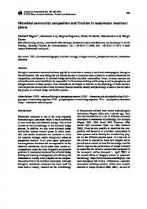
Figure 1: Human and Mouse Prostate Anatomy. (A) Human prostate (adapted from Roy-Burman et al., 2004) and (B) mouse prostate anatomy (adapted from Cunha et al., 1987)) are shown. (C) H&E staining of biopsy tissue specimens of (i) normal, (ii) BPH, (iii) PIN, and (iv) adenocarcinoma of the prostate (adapted from Singh et al., 2006).
4
human PZ. The ventral prostate (VP) has no analogous human zone and the TZ has no counterpart in the mouse. Because the majority of prostate cancers arise in the human PZ, mouse models target tumorigenesis to the DLP (Roy-Burman et al., 2004). Even though spontaneous prostate cancer is rare in the mouse, this species has been used extensively as a model in the study of normal prostate and prostate pathologies. The similarities mentioned above make the mouse a good model for human prostate disease.
New methods of manipulating the
mouse genome have facilitated the development of a number of models that recapitulate the various stages of prostate pathology, which are described below. Prostate Pathology There are several different types of prostate pathology with varying morbidity and mortality. Prostatitis, or inflammation of the prostate, is extremely common. It affects even young men and can be acute or chronic (Haverkamp et al., 2008).
Benign prostatic hyperplasia (BPH) is another common prostate
disorder characterized by nodules in the prostate gland which can lead to many lower urinary tract symptoms (Edwards, 2008). Prostatic intraepithelial neoplasia (PIN) is considered a precancerous lesion and is often a predictor of invasive adenocarcinoma (Ayala and Ro, 2007). Finally, invasive adenocarcinoma with or without metastasis can be androgen dependent or androgen independent (Damber and Aus, 2008). Any or all of these pathological stages may be present within a single tumor demonstrating the extremely heterogeneous nature of 5
prostate cancer. Prostatitis. Inflammation of the prostate or prostatitis can be acute or chronic. Acute prostatitis is caused by infection, while the etiology of chronic prostatitis is most often unknown.
Significantly, there is increasing evidence that chronic
inflammation may play a role in carcinogenesis of the prostate as well as other organs (Haverkamp et al., 2008). While studies showing a causal relationship between prostate inflammation and prostate cancer are lacking, correlations of proinflammatory cytokines, chemokines and matrix metalloproteinases with prostate cancer have been observed (Haverkamp et al., 2008). Furthermore, clinical studies have shown that men diagnosed with prostatitis are more likely to develop adenocarcinoma within 5 years than those with no history of prostate inflammation (Haverkamp et al., 2008). For this reason the presence, as well as the causes, of prostatitis are of importance in the study of prostate cancer. Benign Prostatic Hyperplasia. Benign prostatic hyperplasia (BPH) is a common ailment of elderly men. It has been reported that as many has 90% of men in their seventies suffer from BPH. Typically, BPH is diagnosed based on lower urinary tract difficulties.
Common treatments for BPH include watchful waiting,
α-blockers, and 5-α reductase inhibitors. If the condition is severe, and medical treatment fails to ameliorate symptoms, transurethral resection of the prostate (TURP) may be an option (Edwards, 2008). Prostatic intraepithelial neoplasia.
Prostatic intraepithelial neoplasia (PIN) is
defined as cellular enhancement of normal glandular structure accompanied by 6
cytological atypia in individual cells. PIN can be classified as either low grade (LGPIN) or high grade (HGPIN). A diagnosis of HGPIN is a strong predictor of the presence of carcinoma, with cancer being found in about one-third of cases. In fact, a retrospective study showed that in patients having 4 or more biopsy cores containing HGPIN, there was a 75% chance of finding invasive adenocarcinoma on re-biopsy (Ayala and Ro, 2007).
In addition to studies
showing co-localization of HGPIN and adenocarcinoma, studies have also identified gene modifications common to HGPIN and low grade prostate cancer, supporting the notion that there is a transition from PIN to invasive cancer (Ashida et al., 2004). For these reasons, PIN is considered to be a precancerous lesion of the prostate. Invasive adenocarcinoma.
Prostate cancer is the second leading cause of
cancer deaths in American men (Jemal et al., 2008). Prostate adenocarcinoma accounts for approximately 25% of all new cancer diagnoses among American men. Prostate cancer is detected by digital rectal examination of the prostate (Smith et al., 2003) and diagnosis is based on histological and pathological examination of prostate tissue obtained by several needle biopsies (Damber and Aus, 2008).
Figure 1C shows sample H&E stained tissue sections from
prostates with different pathology grades. Again, it is important to note that all of these histological types may be present within a single tumor. Once prostate cancer has been diagnosed, the determination of the tumor grade is made according to the Gleason system. The Gleason grading system is based on the patterns of histology present in tissue sections stained with 7
standard H&E. The two most prevalent patterns are given a score between 1 and 5. These two scores are subsequently added to give the Gleason score. Lower scores are given to better differentiated, less aggressive tumors and higher scores to less differentiated, more aggressive tumors. The Gleason score is perhaps the most powerful predictor of patient outcome with high Gleason score indicating a poor prognosis and lower scores indicating better prognosis (Humphrey, 2004). In contrast to the human prostate cancer grading system, rodent cancer is often assigned a grade based on the level of tissue differentiation.
Using this system, tumors are assigned a grade of well-
differentiated, moderately differentiated, or poorly differentiated (Gingrich et al., 1997). Since 1986, serum prostate specific antigen (PSA) has been used as a screening method for prostate cancer (approved for use in 1994). PSA is a serine protease in the kallikrein family that liquefies semen within the prostate. However, in various states of pathology, including prostate cancer, PSA is instead released from the prostate into the blood where its concentration can increase by as much as 105 fold.
Men found to have PSA levels ≥4ng/ml are
then submitted for further testing. This fast, easy, inexpensive screening for prostate cancer has resulted in a significant increase in the apparent incidence of prostate cancer in the United States. Additionally, grade migration has occurred with more cancers being detected and diagnosed with lower grade (Lilja et al., 2008). One recent study showed that between 1999 and 2003 the vast majority
8
of patients were diagnosed with moderately differentiated tumors as opposed to more severe poorly differentiated tumors (Jani et al., 2008). Despite the migration of prostate cancer stage over the past 20 years, many American men still die of prostate cancer every year (Jemal et al., 2008). Furthermore, while more men are diagnosed with less severe disease, survival for those who develop severe disease remains low.
Even locally advanced
disease is quite treatable. However, progression to androgen independent and metastatic disease continues to most often be fatal (Berry et al., 1979). Disease progression is often inferred if PSA levels remain elevated following prostatectomy. Subsequent PSA rise also leads to suspicion of disease recurrence. Once metastatic disease develops, it is commonly 5 years or less before the patient succumbs to disease. Prostate cancer commonly metastasizes to the bone in patients who die of this disease (McMurtry and McMurtry, 2003). It is important to note that this aspect of prostate cancer progression has yet to be recapitulated in genetic mouse models of prostate cancer. Transformation of the surrounding stroma is common for epithelial cancers including prostate.
The changes that take place lead to the emergence of
reactive stroma. Tumor reactive stroma can enhance the ability of cancer to invade and metastasize.
In prostate cancer the tumor reactive stroma is
characterized by increased myofibroblasts and fibroblasts as well as the loss of smooth muscle cells. These changes can make the prostate microenvironment more permissive for immune evasion strategies or adaptation to hormone 9
depletion.
Crosstalk between epithelium and stroma causes modifications in
both compartments. Changes in extracellular matrix, and the expression of TGFβ, and various angiogenic growth factors can all impact the development of reactive stroma (Niu and Xia, 2009). Androgen Independent Disease.
Androgen deprivation/ablation is a common
therapy for advanced prostate cancer. Androgen ablation can be achieved by surgical castration (bilateral orchiectomy), or, more commonly, medical castration by treatment with estrogens, anti-androgens and gonadotropin-releasing hormone analogs. Deprivation of androgens by any of these means results in apoptosis of androgen responsive benign and malignant prostate cells (Tammela, 2004). While hormonal therapy is the gold standard for the treatment of locally advanced and metastatic disease, it is rarely curative because androgen independent disease arises (Weeranranta and Isaacs, 2002). The mechanisms underlying the transition to androgen independent prostate cancer are under intense study. One controversy that remains is whether there is a pre-existing population of androgen independent cells in the prostate or whether these cells arise as a result of the selective pressure of castration due to the resulting decrease in levels of testosterone. There are several mechanisms that have been implicated in the acquisition of androgen independent growth. First, the androgen receptor may be over-expressed or amplified at the genomic level. On the other hand, mutations within the androgen receptor gene may allow the receptor to respond to lower levels of androgen or 10
alternative ligands. Constitutive activation of AR through proteolytic cleavage is also a proposed mechanism. Regardless of the exact mechanism, the end result is that prostate maintains AR dependent signaling and transcriptional activation in the absence of DHT. In other cases, the local production of androgens by prostate cells can compensate for the systemic testosterone reduction.
Altered
co-activator or co-repressor expression may also play a role in the progression to androgen independent disease (McPhaul, 2008). Of particular relevance to the studies we will describe in chapter 2, the PTEN tumor suppressor gene has been repeatedly implicated in the progression of prostate cancer to androgen independence. Analysis of tissue microarrays prepared from androgen independent prostate cancer biopsy and autopsy samples revealed reduced or absent PTEN protein (Bertram et al., 2006). Experiments performed in mice bearing Shionogi tumors reveal that knock-down of PTEN shortened the latency period prior to recurrence of androgen independent disease. The Shionogi cell line is initially androgen dependent, and regresses upon castration. However, after a brief period of latency, the tumor reproducibly recurs about 1 month later.
The in vivo knock-down of PTEN in
Shionogi tumor bearing mice led to delayed regression and more rapid relapse of these tumors (Bertram et al., 2006). In the PTEN null, androgen dependent C4-2 cell line (a derivative of the LNCap cell line) doxycycline induction of PTEN increased growth inhibition upon androgen deprivation (Wu et al., 2006). In other words, forcible expression of PTEN decreased androgen independent cell growth suggesting a role for PTEN loss in progression to androgen independence. 11
Studies using the Nkx3.1; Pten mutant mice which develop PIN and adenocarcinoma of the prostate demonstrated robust proliferation early after castration and retention of PIN and/or invasive cancer later.
Furthermore,
Nkx3.1 wild-type, but not PTEN sufficient mice exhibited this phenotype suggesting that loss of one PTEN allele is sufficient for acquisition of androgen independence in vivo (Gao et al., 2006). The mouse model used for our studies is a prostate-specific knockout of PTEN(Wang et al., 2003a) and will be described in further detail in chapter 2. The role of the PTEN/phospho-AKT axis in the loss of androgen dependence together with the described characteristics of this model make it ideal for the study of androgen independent prostate cancer. The mechanism responsible for the emergence of androgen independent prostate cancer remains to be elucidated. However, once androgen independent disease develops, only palliative care is available.
These options currently
include secondary hormonal manipulations, bisphosphonates, external beam radiotherapy, intravenous radioisotopes, and chemotherapy (Garmey et al., 2008).
Even with this arsenal of treatments, survival after progression to
androgen independent disease is low.
For this reason, development of new
treatment modalities, such as immunotherapy, is of paramount importance.
12
Immune System Review Immunotherapy aims to mobilize the immune system to eradicate malignant cells without damage to normal tissue. In order to understand the ideas governing tumor immunotherapy, it is first necessary to have a basic knowledge of the immune system, its functions and characteristics, and how the interplay between tumors and immune cells takes place. Below is a brief review of the basic concepts necessary to understanding tumor immunology. The immune system consists of two arms: innate and adaptive. The innate immune system is comprised of cells which use pattern recognition receptors to distinguish self from non-self (Medzhitov and Janeway, Jr., 2000).
Innate
immune cells are not antigen specific. In spite of this, innate immune cells can play a role in anti-tumor immunity both as positive and negative regulators. Natural killer cells, for instance, have been implicated as effector cells in a variety of anti-tumor immune responses (Malmberg et al., 2008). In contrast, myeloid derived suppressor cells (MDSCs) can inhibit anti tumor immune responses (Talmadge, 2007). The dendritic cell (DC) is at the interface of innate and adaptive immunity. These cells are critical to the initiation of effective immune responses to tumors as they are the most potent stimulators of T cell responses (Bousso, 2008). In addition to initiating effective anti-tumor immune responses, under certain circumstances DCs may initiate immune suppression by triggering tolerance either directly or through intermediaries (Yamazaki and Steinman, 2009). 13
In
most known tumor systems an effective anti-tumor immune response requires the adaptive immune system, and in particular T lymphocytes. Thymus derived lymphocytes, or T cells, express one of two co-receptors on their cell surface: CD4 or CD8 (Paul, 2003). These two types of T cells coexist in the peripheral immune system and serve different, complementary functions. CD8+ T cells recognize small peptide antigens presented by major histocompatibility class I (MHC-I) molecules expressed on the surface of all nucleated cells. CD4+ T cells recognize small peptide antigens presented by MHC-II molecules. Once a T cell detects a cell expressing the antigen for which it is specific, that T cell becomes activated. Optimal activation of a T cell requires the receipt of three signals (Paul, 2003). First, the T cell receptor detects its cognate peptide presented by MHC-I or MHC-II (signal 1). The T cell also requires the engagement of other cell surface receptors, a process termed co-stimulation (signal 2). These receptors transmit intracellular signals that contribute to the full activation of the T cell. Lastly, the T cell needs to encounter the proper cytokine environment (signal 3). Once all three of these signals are received, the T cell undergoes several rounds of proliferation and begins producing effector molecules. While cells of the innate immune system generally only distinguish between self and non-self, the adaptive immune cells are more discriminating. As these cells develop in the thymus, a very intricate process of somatic gene rearrangement and selection takes place (Heinonen and Perreault, 2008). By a 14
sequence of random gene splicing events, an enormous repertoire of T cell receptors is generated, each recognizing a different small peptide antigen/MHC complex. Because this is a random process, some of the resulting T cells can react with antigens normally expressed by the host. The persistence of these cells and their exit into the periphery can have very deleterious consequences as evidenced by the various autoimmune diseases mediated by T cells (Sinha et al., 1990).
Fortunately, most auto-
reactive T cells die in the thymus and do not go on to have negative effects. T cells strongly reactive to self proteins are deleted in the thymus through the process of selection (Hogquist et al., 2005). This is referred to as central tolerance. In instances in which this central tolerance allows the survival of autoreactive cells, there is a second level of protection for the host. T cells which reach the periphery having some level of affinity for self antigens can be held in check by several mechanisms collectively referred to as peripheral tolerance (Walker and Abbas, 2002; Finn, 2008). One mechanism is physical separation from antigen as is the case for immune privileged sites. Another is the induction of anergy by antigen encounter on self peptide/MHC complexes without proper co-stimulation (signal 1, but not signal 2). Additionally, auto-reactive T cells may be held in check by regulatory T cells (Tregs). This method is described in further detail below. The majority of CD4+ T cells fall into the category of helper cells (Bevan, 2004). Once they gain effector function, CD4+ T cells secrete cytokines which 15
help to shape the immune response. T helper 1 (Th1) cells activate macrophages and support CD8+ T cell responses through the production of IFN-γ. The Th1 response is thought to be important for anti-tumor immunity. T helper 2 (Th2) cells elicit an antibody or humoral response and are thought to suppress anti-tumor immunity. In addition to these classic helper T cell subsets, there are two more recently described subsets of CD4 cells.
T helper 17 cells (Th 17) are
distinguished by their secretion of IL-17 upon stimulation (Chen and O'Shea, 2008) and play a prominent role in autoimmunity (Jin et al., 2008). The final subset of CD4+ cells is Tregs which in contrast to the other types have an inhibitory role in immune responses. Regulatory T cells account for 1-5% of the total CD4+ population (Vignali et al., 2008).
These cells can be easily identified in the mouse by their
expression of the transcription factor FoxP3 and constitutive expression of the high affinity IL-2 receptor, CD25. In contrast to the T helper cell subsets, these lymphocytes are negative regulators of immune function. Tregs prevent autoimmunity by suppressing self-reactive T cells in the periphery.
Contact
dependent mechanisms of suppression by regulatory T cells include ligation of T cell surface receptors such as CTLA-4 (von Boehmer, 2005), and direct killing of effector cells (Gondek et al., 2005). Contact independent mechanisms include secretion of cytokines such as TGF-β and IL-10 which hamper effector function in vivo (von Boehmer, 2005) and sequestration of IL-2 (Malek and Bayer, 2004). In addition to their function in preventing autoimmunity, they are also integral to
16
maintaining normal immunity because they down-modulate effector cells at the completion of a normal immune response (Vignali et al., 2008). Even during the early study of tumor immunology, it was appreciated that not all T cells are created equal. Long before the existence of regulatory T cells was confirmed, it was observed that some cells likely have anti-tumor effects while others support tumor growth. In fact, even very early studies showed that tumor rejection by transfer of cells immune to a tumor were more effective when the tumor-bearing host lacked its own T cells (Berendt and North, 1980). This suggested that a population of host derived T cells can facilitate persistent tumor growth. Now that FoxP3+ Tregs have been identified and characterized, many animal models of cancer as well as clinical studies have confirmed those early findings (Qin, 2009). In mouse models, Tregs have been shown to be very abundant in prostate tumors. Even in HGPIN, Tregs constitute a larger percentage of CD4+ cells than in normal prostates (Tien et al., 2005). In patients with prostate cancer, it has been demonstrated that Tregs are overrepresented. These patients had more Tregs in the neoplastic tissue than in benign tissue from the same prostate. Additionally the blood of prostate cancer patients contained a greater proportion of Tregs. Further, in vitro assays have demonstrated chemo-attractant properties of supernatants from tumor samples mediated by CCL22, a chemokine which attracts Tregs (Miller et al., 2006). Not only is the presence of Tregs increased in prostate cancer patients, but these Tregs have increased suppressive activity 17
when compared to those of healthy controls (Yokokawa et al., 2008). Also, the overall CD4+ population within the prostate was found to have a greater proportion of Tregs than the peripheral blood of the same patients (Sfanos et al., 2008). Another study utilizing fresh tumor tissue corroborated the increase in Treg proportion and also identified a subpopulation of CD8 + cells with regulatory function (Kiniwa et al., 2007). Therefore, it would seem that maintenance and/or generation of Tregs is integral to prostate tumor development and progression. The mechanism for Treg upregulation in prostate cancer is unclear. However, Tregs co-localized with hypoxia inducible factor (HIF)-2α in an analysis of prostate tissue microarrays (Fox et al., 2007).
Interestingly, hypoxia is
significantly decreased upon androgen ablation (Milosevic et al., 2007). Therefore perturbations in prostate tumor oxygenation could provide a mechanism of regulating in situ Treg numbers following androgen ablation therapy. Beyond clearance of antigen, a portion of the antigen specific T cells remain. These cells become memory cells which are maintained in the absence of antigen (Perret and Ronchese, 2008). Upon secondary exposure to the same antigen, memory cells mount a much more rapid and robust response. In the context of immunotherapy, generation of immunological memory would be beneficial because if metastasis were to develop, these memory cells could quickly be deployed to eradicate them.
18
Tumor Immunology and Immunotherapy The exquisite capability of adaptive immune cells to discriminate between antigens is the basis of tumor immunology.
The antigenicity of tumors was
established based on the ability of mice to withstand a tumor challenge after preexposure to that tumor or its components (Vaage, 1971; Bystryn, 1978). Over the past several decades, a vast literature has been established identifying antigens that are specifically or preferentially associated with tumors (Van den Eynde and Boon, 1997) . The cancer/testis antigens are good target antigens because they are generally only expressed by tumors, the testes or during embryonic development but not in other adult tissues. Thus, these antigens may or may not be regarded as self. However, this represents a limited number of antigens with relevance for a limited number of malignancies (Scanlan et al., 2002). The majority of tumor associated antigens identified to date fall into the category of self or altered self proteins. These antigens are targeted on the basis of tissue restriction or overexpression on malignant versus normal tissues. In cases where the antigen is truly a self protein, the responsive T cells are subject to the same mechanisms of tolerance described above. As a result, responses are largely ineffective (Finn, 2008). However, in cases where mutations result in antigens unique to the tumor and not naturally found in the host, generation of anti-tumor immunity is more effective. This type of antigen is often unique to the particular tumor in that individual. In clinical immunotherapy studies, several
19
prostate associated antigens have been targeted for T cell recognition. Among these are PSA, PSMA, PAP, PSCA and KLK4 (Harada et al., 2003). The field of tumor immunology began in 1909 with the assertion by Ehrlich that one of the normal functions of the immune system is to destroy nascent transformed cells thus protecting the host from developing cancer. However, in the early twentieth century, fairly rudimentary immunological methods prohibited rigorous investigation of this hypothesis. However, with the development of new immunological techniques and the establishment of inbred mouse models, investigation into this area was rekindled. By the 1950s, the ideas of Ehrlich were reintroduced as the theory of immune surveillance by Burnet and Thomas. Around the same time, the first demonstration of tumor immunogenicity was reported by several labs (FOLEY, 1953; PREHN and MAIN, 1957). Those early studies were conducted by challenging mice with chemically induced transplantable tumors following the resection of a tumor of the same type. The results of these studies demonstrated definitively that exposure to a fully syngeneic tumor could provide protection against future inoculations. Additionally it was determined that there was no cross protection against different tumors even if they were of the same general type demonstrating that tumor antigens are specific for individual tumors and not shared between tumors of the same types (Basombrio, 1970). Not long after this, it was demonstrated that immunity to a particular tumor could be conferred by
20
adoptive transfer of lymphocytes. These studies provided legitimacy to the study of tumor immunology. Controversy remained in the field, however, due in part to the initial inability to reproduce tumor resistance seen in chemical and virally induced cancer in cases of spontaneously arising tumors. Additionally, studies in athymic mice (which lack T cells) did not show increased susceptibility to tumor development. The advent of inbred mice alleviated much of this controversy. While the field remained in turmoil for some time following its introduction, the concept has now been accepted and has given rise to intense laboratory and clinical investigation in an effort to combat cancer by harnessing the power of the immune system. Several studies conducted in the past decade have demonstrated the importance of immune surveillance in the control of tumor growth.
In these
studies, a role for Th1 immunity was found to be crucial for the restraint of tumor growth.
Mice with IFN-γ defects, for instance, are more susceptible to
tumorigenesis by MCA exposure or by inactivation of the tumor suppressor p53 (Kaplan et al., 1998).
Furthermore, RAG knockout mice
(which lack
lymphocytes) and RAG knockout/IFN deficient mice are more susceptible to MCA induced and spontaneously arising tumors than wild type mice of the same genetic background (Shankaran et al., 2001). Thus it was demonstrated that IFN-γ is critical to anti-tumor immune responses. Furthermore, because there is no additive effect of lymphocyte and IFN-γ knockout, the IFN effects are likely 21
mediated in large part by the effect of IFN on host lymphocytes. These data have been validated by clinical studies demonstrating the interplay of immune function and cancer development.
One such study showed that patients
subjected to long term immunosuppression subsequent to organ transplantation had a much higher rate of cancer diagnosis than the general population (Roithmaier et al., 2007). In another study, cancer risk was correlated to natural levels of cytotoxicity as determined by in vitro cytotoxicity assays (Imai et al., 2000). Additionally, seminoma and renal cell carcinoma occur with increased prevalence in patients who are immunocompromised due to HIV infection (Silberstein et al., 2009). Observations such as these support a role for the immune system in the prevention of neoplasms. As the recognition of the role of the immune system in tumorigenesis has increased, so too has the appreciation of immune suppression by the tumor. The suppression of antitumor immunity can be attributed to mechanisms mediated both by the tumor itself as well as cells recruited to the tumor microenvironment. Significantly, tumor bearing mice are resistant to challenge with the same tumor at a distant site. This anti-tumor immunity concurrent with progressive growth of the primary tumor was termed concomitant immunity. As investigation into this phenomenon was expanded, studies showed that while true shortly after tumor initiation, hosts bearing large, progressively growing primary tumors are no longer able to resist secondary challenge of the same tumor (Vaage, 1973; Chandradasa, 1973). This effect was reversible; shortly 22
after the resection of the primary tumor, resistance to tumor challenge was restored. Thus, while immune function is hindered within the tumor, the same antigens encountered elsewhere are able to elicit anti-tumor effects.
This
emphasizes the suppressive nature of the tumor milieu. A tumor is also suppressive to T cells not residing within the tumor. Additional studies showed that adoptively transferred lymphocytes from tumor bearing mice conferred attenuated resistance as compared to lymphocytes derived from hosts which were immune to the tumor, but not bearing the tumors. (Old et al., 1962). The failure to reject the established primary tumor even in the presence of immune cells capable of doing so has led to intense investigation of the methods of immune suppression employed by tumors. Early studies demonstrated that in some models regulatory T cells were responsible for the continued growth of immunogenic tumors. Adoptively transferred immune T cells were only effective in reducing tumor burden in immunocompromised mice which lacked T cells. Under these conditions the activated T cells were able to cause regression of established tumors. T cells from immuno-competent tumor bearing mice were able to reverse this effect (Berendt and North, 1980). In recent years, the idea of immune surveillance has been expanded to integrate immune subversion and outgrowth of overt tumor.
The theory of
immunoediting posits that the interaction of the host immune system with malignant cells occurs in three discrete phases. 23
First, the tumor cells are
recognized by the immune system and removed if possible. Any tumor cells not eliminated go on to exist in an equilibrium state with the immune system. During this equilibrium, continued assault by the immune system can causes the outgrowth of poorly immunogenic variants which eventually go on to escape the effects of anti-tumor immunity altogether, leading to clinically evident disease (Dunn et al., 2004). Studies in mice and humans strongly support the existence of immune surveillance. Evidence also exists for the ability of the immune system to shape the immunogenicity of the tumor that eventually grows. Tumors derived from immune deficient mice are more readily rejected than those from immune competent mice indicating that tumor development in the context of an intact immune system results in tumors less able to elicit a productive immune response (Shankaran et al., 2001). More recently, evidence of the equilibrium state was provided in a study of mice treated with the carcinogen MCA (Koebel et al., 2007). In this study, mice with no detectable tumors were depleted of immune components at late time points when new development of tumors is rare. Inhibition of adaptive immunity at this time induced the formation of progressively growing tumors which were presumably stable, pre-existing, undetectable masses of cells previously held in check by the host immune system. Harnessing the principles of tumor immunology for the immunotherapy of prostate cancer is an attractive option for advanced and metastatic disease. Significantly, the prostate is not a vital organ; therefore destruction of normal 24
tissue as the anti-tumor response ensues is not a threat to survival. Additionally, while the prostate was once thought to be an immune privileged site (McNeel and Malkovsky, 2005), human studies have confirmed the presence of tumor reactive T cells and antibodies in patients with prostatitis and prostate cancer (Alexander et al., 1997; McNeel et al., 2001; Chakraborty et al., 2003; McNeel et al., 2000). Furthermore, the degree of lymphocyte infiltration of tumors is associated with the clinical outcome of prostate cancer; decreased density or absence of tumor infiltrating lymphocytes within prostate tumors is indicative of very poor prognosis (Vesalainen et al., 1994; McArdle et al., 2004). Additionally, tumor infiltrating T cells in prostate cancer have been shown to express high levels of IFN-γ and Fas-ligand when compared with lymphocytes associated with benign hyperplasia, suggesting activated T cells are recruited to neoplastic prostate tissue (Elsasser-Beile et al., 2000). These data support the validity of investigating immunotherapy for the treatment of prostate cancer and preclinical studies of prostate tumor immunotherapy have had varying results. Several studies of tumor cell based vaccines have used strategies which engineer the tumor cells to express cytokines or chemokines to enhance vaccine efficacy.
For instance, IL-12 expression has been explored for its ability to
increase tumor infiltration by lymphocytes as well as survival. Introduction of IL12 expressing bone marrow cells into tumor bearing mice resulted in increased tumor infiltration by both CD4+ and CD8+ lymphocytes, decreased tumor burden and increased lytic capacity of NK cells and CTL. However survival benefit was minimal compared to controls (Wang et al., 2007). IL-12 expression within the 25
BMA orthotopic prostate tumor led to suppression of tumor growth as well (Saika et al., 2006). Additionally, vaccination with the TRAMP-C2 tumor cell line resulted in splenocytes which effectively reduced tumor burden and prevented spontaneous metastasis to the lung when adoptively transferred into tumor bearing mice (Nikitina et al., 2005). This effect was dependent on T cells (Saika et al., 2006). While IL-12 alone was not sufficient to increase survival in these studies, its effects were further enhanced by co-expression of the co-stimulatory molecule B7 (Nikitina et al., 2005). Splenocytes harvested from mice vaccinated with tumor cells expressing B7 in addition to IL-12
enhanced metastasis
prevention (Nikitina et al., 2005) and survival (Saika et al., 2006). Growth factors have also provided a benefit to immunization strategies. For instance, subcutaneous injection of Flt-3L into mice bearing orthotopically placed TRAMP-C1P3 cells resulted in increased infiltration of all cell types into the tumor, reduced tumor incidence, and increased overall survival (Ciavarra et al., 2004). Furthermore, the inclusion of the growth factor GMSCF enhanced the response to a TRAMP-C1/C2 tumor cell vaccine (Hurwitz et al., 2000). Another mode of immunization is genetic vaccine.
Treatment with an
mRNA based vaccination, when combined with GMSCF, led to the inhibition of TRAMP tumor growth only if mice received prior adoptive transfer of splenocytes from non tumor bearing littermates. These data indicated that the immune cells from the TRAMP mice are tolerized to their own antigens, but that functional cells adoptively transferred into these mice could be activated under these conditions. 26
These cells subsequently mounted an anti-tumor immune response which resulted in decreased UGS and tumor weights as well as a decreased average tumor grade (Hess et al., 2006). When a DNA vaccine against PSA was assayed in a mouse model, it was efficacious and its activity was enhanced by GMCSF and IL-2 (Roos et al., 2005). While the tumor cell line expressing PSA in these mice was not prostate derived, this study does demonstrate the feasibility of vaccinating against prostate associated antigens in this manner. Anti-tumor responses can also be initiated by simply vaccinating with tumor tissue. Tumors induced in rats by treatment with MNU and testosterone propionate were fixed with gluteraldehyde and used to test a vaccine strategy. Vaccination with fixed tumor cells failed to reduce tumor size or grade, but the rats lived longer and splenocytes secreted more IFN-γ and TNF-α upon in vitro stimulation (Suckow et al., 2008). In addition to the use of tissue or cells to vaccinate tumor bearing animals, this method is also used to generate immune cells for adoptive transfer.
A study in TRAMP mice showed the efficacy of
adoptively transferred immune, but not naïve, splenocytes which suppressed the development of tumors (Granziero et al., 1999). Adoptive transfer methods can be further enhanced by modulation of cytokine responsiveness of the transferred cells.
CD8+ T cells expressing a
dominant negative form of the TGF-β receptor II were better able to infiltrate subcutaneous TRAMP-C2 tumors as compared to immune cells sensitive to TGF-β. This resulted in increased tumor cell apoptosis, lower tumor incidence, 27
fewer tumors per mouse, and increased survival. This study demonstrated that large scale secretion of TGF-β by a tumor can prevent the infiltration of that tumor by CD8+ T cells (Zhang et al., 2005). Inhibitory effects of L-Arg metabolism on T cell mediated anti-tumor immune responses have been established as well. In the TRAMP mouse model of prostate cancer, TILs isolated from tumors and incubated in low dose IL-2 secreted increased levels of IFN-γ upon stimulation with autologous tumor or fibroblasts expressing a model antigen only when arginase (ARG) and nitric oxide synthase (NOS) inhibitors were included in the culture. Interestingly, many more CD8+ T cells were present in microscopic tumors than in macroscopic tumors (Bronte et al., 2005).
This is consistent with data discussed above
describing the increasingly immunosuppressive nature of growing tumors. In addition to positive co-stimulatory signals, the B7 molecules also have inhibitory ligands. One such ligand is CTLA-4. The normal function of CTLA-4 is to arrest the immune response. Blocking the interaction of this molecule with its receptor leads to increased T cell responses (Paul, 2003). Antibodies blocking CTLA-4 have been tested in mouse models of prostate cancer. TRAMP mice benefited from immunization with TRAMP-C1/C2 tumor cells only in the presence of anti-CTLA-4 antibody. The vaccine/CTLA-4 combination treatment decreased tumor incidence, and reduced tumor grade (Hurwitz et al., 2000). Additionally, systemic treatment with anti-CTLA-4 antibody resulted in diminished tumor growth and tumor rejection in mice bearing TRAMP-C2 tumors. In this study, 28
fewer mice developed tumors and the tumors were smaller tumors. Resection of the primary tumor reduced local and distant recurrence in mice that had received antibody treatment. Furthermore, these antibody treated mice had a survival advantage over mice treated with a control antibody (Kwon et al., 1999). As discussed above, DCs are the major initiators of CTL responses and vaccination with DCs often provides some benefit in preclinical models. TRAMP mice immunized against the T antigen by intradermal injection of DCs presenting T antigen peptides developed a productive anti tumor response if the immunization took place early during tumor development.
Low avidity CTL
developed with impaired ability to kill target cells as determined by in vivo cytolysis assay. However, this did not reflect an overall immune defect in these mice as the same protocol was able to generate a robust response to a control antigen, indicating active suppression of the immune response only to the tumor associated T antigen. In spite of the generation of only low avidity effectors, tumor grade was reduced. In contrast, mice immunized at a later time point were fully tolerized and no productive immune response was initiated.
Thus early
intervention with a DC vaccine could have lasting impact on immune status of prostate tumor bearing mice (Degl'Innocenti et al., 2005). Immunization against native prostate specific antigens has included those important in human disease such as PSCA, PAP, and STEAP (Machlenkin et al., 2005). For instance, when C57BL/6 mice were immunized against PSCA, the ensuing immune response decreased TRAMP-C2 tumor incidence and smaller 29
tumors in those mice that developed tumors.
Additionally, there was an
increased infiltration of CD4+ and CD8+ cells and Th1 cytokine expression. This anti-tumor response was mediated by lymphocytes as it was completely abrogated by the removal of either CD8+ or CD4+ cells. In TRAMP mice, the same vaccination protocol substantially increased survival and resulted in tumors with lower grade (Garcia-Hernandez et al., 2008). Additionally, a trivalent DNA vaccine against 3 prostate specific proteins, when targeted to APCs, reduced tumor burden and increased survival in a mouse tumor model (Qin et al., 2005). Taken together, these data show the potential benefits of using immunotherapy for the treatment of prostate cancer.
However, they also
highlight the obstacles which remain to be overcome. As more knowledge is gained in the field of prostate tumor immunology, this can direct rational design of future pre-clinical studies to incorporate manipulations aimed at increasing efficacy.
The study described in chapter 2 elucidates some of the immune
modulations that occur subsequent to androgen ablation and provides invaluable information that can be used in these future studies. Mouse models of prostate cancer Spontaneous prostate cancer is rare in wild-type mice; however prostate cancer is detected in up to 80% of men 80 years or older at autopsy (RoyBurman et al., 2004). Recent advances in methods for manipulating the mouse genome have resulted in mouse models that closely recapitulate the human disease. These have been reviewed extensively elsewhere (Roy-Burman et al., 30
2004). Here we will present an overview of the TRAMP model, emphasizing information obtained about prostate immunology, immunotherapy and androgen independent disease. Transgenic Adenocarcinoma of the Mouse Prostate (TRAMP).
The TRAMP
mouse was established nearly 15 years ago and has become a staple for the study of prostate cancer (Greenberg et al., 1995).
These transgenic mice
express the SV40 large T antigen under the control of the prostate specific probasin promoter.
The large T antigen is a well known oncogene which
abrogates retinoblastoma and p53 function (Ali and DeCaprio, 2001). TRAMP mice develop cancer by 10 weeks of age. Significantly, the tumors in this model are very heterogeneous, similar to the situation observed in human prostate cancer. By 24 weeks of age, TRAMP mice display a mixture of histological grades ranging from normal to poorly differentiated (Kaplan-Lefko et al., 2003). It is introduced here because many of the preclinical prostate immunology studies conducted to date have utilized this model. Androgen Independent Disease Much preclinical prostate cancer research has been conducted in the TRAMP model. Shortly after the first description of the model, progression to androgen independent disease was characterized (Gingrich et al., 1997). When mice were castrated at 12 weeks of age, the UGS weight was less than that of intact controls at the same time points. Furthermore, tumors continued to grow following castration since UGS were larger at 24 weeks after castration than at 31
18 weeks following castration, indicating androgen independent growth took place.
Importantly, the androgen independent tumors were more poorly
differentiated than tumors in age matched, intact control mice, suggesting that once a tumor has developed, androgen ablation leads to a more severe primary tumor phenotype. Additionally, a subsequent study showed that at late stages of disease, castration had no effect on metastasis or survival in these mice (Johnson et al., 2005). When TRAMP mice were castrated after the onset of PIN, but prior to the development of adenocarcinoma, prostate volume was significantly reduced throughout the life of the mouse. Additionally, tumor free survival was significantly increased compared to age matched, intact controls (Eng et al., 1999). The ability to prevent tumor development by early castration of TRAMP mice supports clinical data suggesting that treatment of PIN by androgen ablation may serve as preventative therapy (Bostwick and Qian, 2001; Hull and Bostwick, 2008). Finally, while epithelial T antigen expression is rapidly lost following castration (almost undetectable by two weeks later), foci arise in the prostate which express no AR, but do express T antigen. Because T antigen expression is driven by an androgen responsive promoter in this model, this suggests that these foci represent androgen independent prostate cancer. These foci likely led to the development of androgen independent primary tumors in the TRAMP model (Huss et al., 2007). 32
TRAMP Tumor Immunology T cells can only lyse those tumor cells which express the molecules necessary for antigen presentation.
CD8+ T cells recognize small, peptide
antigens presented by MHC-I molecules, and CD4+ T cells recognize antigens presented by MHC-II (Paul, 2003). In the TRAMP mouse model, all tumors expressed moderate levels of both MHC-I and MHC-II.
In fact, the TRAMP
prostates expressed more MHC-I than their normal counterparts (Nanda et al., 2006). Thus these tumors serve as a good model for immunological study of anti-tumor immunity. While antigen presentation within the tumor microenvironment may be undisturbed in this model, initiation of anti-tumor responses also requires professional antigen presenting cells i.e. DCs.
DCs are the most potent
stimulators of cytotoxic T cells. However, co-incubation with a TRAMP mouse cell line results in a much lower rate of DC maturation as compared to control mice (Tourkova et al., 2004). This would suggest that the immune environment in mice bearing prostate tumors is not conducive to the maturation of DCs. Because only mature DCs induce optimal T cell stimulation, failure to mature hinders the anti tumor immune response and it seems that TRAMP cells can inhibit this process in vitro. Not only do the DCs of TRAMP mice not mature as well, there are fewer DCs in the spleen. Dendritic cells can be derived from mouse bone marrow in vitro. However the ability to generate DCs from TRAMP bone marrow diminished 33
as disease progressed (Tourkova et al., 2004) indicating that either prostate tumorigenesis or progression leads to a defect in dendritic cell differentiation from their myeloid precursors. Accordingly, a corresponding decrease in the number of splenic DCs was observed in these mice. The spleen is a major reservoir for dendritic cells in the mouse and the decreasing number of DCs supports the in vitro data. Inhibition of DC production in this model demonstrated a possible immunosuppressive mechanism of TRAMP tumors. In fact, immature DCs are known to induce Tregs which contribute to the suppressive tumor milieu. The reported inhibitory effect of prostate cancer cells on dendritic cell production and function may be a strategy employed by prostate tumors in general and not just those of the TRAMP mice. Expression of the SV40 T antigen induces tumors in TRAMP mice. This protein serves not only as the initiating oncogene, but also as a model antigen. In TRAMP mice the T antigen is self and not foreign, which mimics other putative prostate associated antigens. TRAMP mice are tolerant to T antigen due to clonal deletion of T antigen specific T cells in the thymus (Zheng et al., 2002); the number of T antigen specific T cells escaping into the periphery of these mice is profoundly reduced.
The establishment of tolerance is a major hindrance to
therapies targeted to tumor associated antigens in the clinic.
Thus, studies
aiming to mount an anti- T antigen immune response are directly applicable to the human condition. 34
The role of regulatory T cells in TRAMP tumors has also been examined. In TRAMP mice expressing hemagglutinin as a model antigen, treatment with anti-CD25 antibody to deplete Tregs had no effect on the ability of transgenic CD4 cells to proliferate or produce cytokines ex vivo (Mihalyo et al., 2007). This indicates that with regard to TRAMP specific antigens (or at least those elicited in this study) Tregs may not be the major mediators of immune suppression. The Prostate Specific PTEN Knockout Mouse Model of Prostate Cancer.
In
2003, Wang and colleagues reported the development of a new mouse model of prostate cancer (Wang et al., 2003a). Based on the fact that Pten (phosphatase and tensin homolog deleted on chromosome 10) dysfunction (deletion or mutation) has been found in a large proportion of primary prostate tumors and in most metastases (Dong, 2006), this group inactivated the PTEN gene in a prostate specific manner using Cre-LoxP technology.
In this model, Cre
recombinase is expressed under the control of the prostate specific probasin promoter (PBCre4 (Wu et al., 2001)) and exon 5 of the Pten gene is floxed (PtenloxP/loxP).
Pten is specifically inactivated in the prostate by breeding the
PBCre4 mouse to the PtenloxP/loxP mouse. The male Cre+Ptenloxp/+ offspring of these mice are then backcrossed to PtenloxP/loxP females (Fig. 2).
The resulting
male Cre+Pten-/- mice develop prostate cancer in a very predictable manner. This model is an attractive one for study because of its 100% penetrance, well defined kinetics, and strong genetic and histological similarities with human prostate cancer progression.
35
Other groups have also created prostate specific knockouts of the PTEN tumor suppressor. In one such model, the Cre recombinase gene is driven by the human PSA promoter whereby expression is restricted to the mouse prostate. Microinvasion was not detected in these mice until 7 to 9 months of age and carcinoma was detected at greater than 10 months of age (Ma et al., 2005). Still other models have investigated the impact of PTEN dose on prostate tumorigenesis.
In addition to mice expressing wild-type, heterozygous, and
knockout levels of PTEN, a mouse expressing PTEN at a level intermediate between heterozygous and knockout was generated. This study demonstrated the need for the absence PTEN protein in order to get complete penetrance of invasive adenocarcinoma in mice.
The model developed by Wang and
colleagues does result in complete knockout of PTEN and is therefore a better model to study.
Furthermore, in comparison to all other models tested, this
model had the most severe phenotype as indicated by tumor size over time (Trotman et al., 2003). Several investigators have demonstrated that early deletion of PTEN leads to a more rapid onset of disease. Using models with tamoxifen inducible PTEN deletion, post pubertal knockout significantly increased disease latency (Ratnacaram et al., 2008; Luchman et al., 2008) with mice developing invasive adenocarcinoma as late as one year of age.
36
Figure 2: The PTEN knockout mouse breeding scheme. PTEN specific knockout mice were generated as described in (Wang et al., 2003a)
37
In contrast to all of the PTEN deletion models described above, the PTEN knockout mouse described by Wang and colleagues (Wang et al., 2003a) is very useful as a model for rapid, preclinical study in part because these mice develop cancer very quickly by comparison. Thus we chose to use this model for our studies. Prostate specific PTEN knockout mice develop adenocarcinoma by 9 weeks of age. When castrated, the prostates tumors of PTEN knockout mice exhibited an immediate and transient increase in apoptosis as determined by TUNEL staining (Wang et al., 2003a). Thus these prostates are initially responsive to androgen deprivation, much like tumors of prostate cancer patients.
However, at necropsy (2.5 – 10 months after castration), residual
invasive adenocarcinoma and enlarged prostates were observed.
Since
proliferation was not decreased in comparison to intact mice, androgen independent prostate cancer must be quickly established in the model. In addition to the pathological similarities to human cancer, the immunological status of the PTEN knockout mouse is also similar to that found in human patients.
Tumors develop orthotopically, over time in an immune-
competent individual. Furthermore, no model antigen is experimentally induced in these mice. Thus studying the immunology of prostate cancer in PTEN knockout mice allows us to address questions in an environment that closely resembles the human situation in which many of the antigens that might mediate rejection have not yet been identified at the molecular level. 38
Despite the fact that antigens of the PTEN knockout mouse tumors remain unidentified, cells derived from these mice have proven to be immunogenic. A cell line derived from the prostate tumors of PTEN knockout mice can effectively generate an immune response in immuno-competent, non-tumor bearing mice. The splenocytes from vaccinated mice demonstrated increased IFN-γ production and cytolysis in in vitro assays. Furthermore, upon transfer to tumor bearing immuno-compromised mice, effector cells from immunized mice caused a stark reduction in the growth of subcutaneous tumors of the same cell line. (Haga et al., 2009). This confirms the immunogenicity of cells derived from the PTEN knockout mouse prostate tumors and supports validity of using this model for immunologic study. Experiments to determine the effect of vaccination of the PTEN knockout tumors have not yet been conducted. There is little else known about anti tumor immunity in the PTEN knockout mouse, but our study provides some baseline information. Clinical Imaging of Immunotherapy An increasing number of immunotherapy studies are conducted every year. Preclinical and especially clinical studies often have mixed results. Often times, in vitro assays are used to determine factors leading to clinical response. In this way, investigators attempt to identify those factors able to predict those who may be likely to be “responders”. While this offers some information, it is rare that one factor can be identified that accurately and reproducibly separates the responders from non-responders. A more dynamic and comprehensive view
39
of the response which ensues following immune therapy is therefore highly desirable. Molecular imaging can fulfill this role. Molecular imaging is a method of detecting cells or cellular processes non-invasively within whole, living animals (Massoud and Gambhir, 2003). Cell growth, death and movement can all be monitored by radioisotopic, magnetic, and optical imaging methods. Molecular imaging has been used extensively to characterize tumor growth, regression, and metastasis both clinically and in the laboratory. More recently, it has been adapted to study the interaction of cells of the immune system with tumors. In order to effectively track the movement and function of tumor-reactive immune cell populations, an imaging agent should 1) be detectable noninvasively in living subjects, 2) be specific for the population of interest, 3) mark at least a representative proportion of the population to be followed, 4) have minimal toxicity, or effect on the function or characteristics of therapeutic cells, 5) allow serial imaging over days to months and even years, and 6) provide quantitative, three-dimensional organ-specific localization of cells of interest. Radioisotopic, magnetic resonance imaging (MRI), fluorescent, and bioluminescent imaging (BLI) agents have been developed to label specific cell populations and can be used for non-invasive studies with varying abilities and minimal toxicity. In the case of adoptive transfer immunotherapeutic strategies, the effector population can be marked quite specifically. However, as will be discussed, indirect labeling may be more useful in imaging specific cell types 40
involved in the anti-tumor immune response, especially when the cell type of interest is highly proliferative and must be followed for a long period of time. Positron Emission Tomography (PET) and Single Photon Emission Computed Tomography (SPECT), as single imaging modalities, provide three dimensional images with sensitive spatial resolution but little anatomic information, while the optical techniques remain largely two-dimensional and are currently not applicable to human patients. MRI provides spatial and anatomic information, but the contrast agent is diluted by cell division. Although indirect labeling strategies have been reported for MRI imaging, they are not widely used (Gilad et al., 2007). The most commonly used clinical imaging agent,
18
F-fluoro-
deoxy-glucose (FDG), because of its mode of activity, does not mark a specific cell population although it satisfies all the other criteria. Thus, currently no single clinical imaging agent meets all of the above criteria. Immunotherapy seeks to generate lymphocyte populations with longlasting memory of the immunizing antigen, with the hope that these cells will be effective in combating metastatic disease that may arise months to years after the primary tumor has been treated. In order to follow the trafficking and function of these cells in vivo, they must be marked permanently with a label that will be retained after many cell divisions. Indirect cell labeling permanently marks a cell and its progeny with a reporter gene. Reporter gene-based imaging methods permit stable marking of individual cell populations with excellent sensitivity of detection. 41
Naturally
occurring proteins such as Green Fluorescent Protein (GFP) are introduced into a cell by transfection or infection with viral vectors. These proteins fluoresce upon excitation with specific wavelengths of light. Labeled cells can be detected (Marodon et al., 2003) at the single cell level by microscopy and flow cytometry and non-invasively within the intact animal with a variety of sensitive CCD camera detection systems.
The limitation of this method is that fluorescent
signals are attenuated by body mass, skin, hair, etc. Additionally, the high autofluorescence in animal tissues reduces the signal to noise ratio (Chudakov et al., 2005). Nevertheless, this methodology can be used to track relative changes over time within and between animals. The use of near-infrared reporters such as dsRed provides higher signal strength with reduced auto-fluorescence and lower tissue attenuation of signal, resulting in a much greater signal to noise ratio and more sensitive image detection (Rao et al., 2007). Indirect labeling with enzymes is a powerful alternative approach to fluorescent proteins. The enzyme-substrate scheme has been used for both radioisotopic and luminescent marking of cell populations (Blasberg and Tjuvajev, 2003). For small-animal imaging, the most commonly used enzyme is Firefly luciferase, derived from the firefly Phontinus pyralis (Greer, III and Szalay, 2002). To detect the marked cells, the substrate, luciferin, is injected into the animal. In the presence of oxygen, magnesium and ATP, the enzyme-substrate reaction results in light emission that is captured by a sensitive CCD camera. Unlike imaging using fluorescent molecules, the luciferase reaction cannot be detected at the single cell level (Lindqvist et al., 1994). 42
Although luciferase
activity can be detected in vitro in whole cells and cell lysates, these assays provide only population level information.
The advantage of using this
bioluminescent reporter is the virtually complete absence of background bioluminescence, resulting in a very high signal to noise ratio. This strong light emission is less attenuated than the weaker fluorescent signals, resulting in greater depth penetration of the signal, and a more sensitive two-dimensional image.
Renilla luciferase from the sea pansy Renilla reniformis as well as
synthetic modified variants, are also used as reporter genes (Greer, III and Szalay, 2002). The emission spectra of Renilla and Firefly luciferase overlap, but their kinetics of light emission is very different. Thus two cell populations each marked with a different luciferase could be followed nearly simultaneously. The viral enzyme Herpes Simplex Virus thymidine kinase (HSV-TK) is the most widely used reporter gene for radioisotopic imaging. A number of highly specific substrates have been developed for both the wild-type (HSV-TK) and mutant (HSV1-sr39TK) enzyme. These positron-emitting substrates are detected by a sensitive PET camera. Radioisotopic emissions are not as attenuated by body mass, etc. and thus have less depth limitation for signal detection. Background reactivity of labeled substrates with endogenous enzymes or receptors is minimal, thus providing a high signal to noise ratio. Consequently, quantitative assessments of the numbers of cells present at a particular site can be made.
The
disadvantages of this approach are the high cost of tracer production and the 43
need for an on-site cyclotron for production of short half-life radiotracers. Similar reporter gene strategies have been developed for MRI (Gilad et al., 2007), but so far have not been used to track immune cell types. Introduction of Reporter Genes Indirect labeling of cell populations with reporter genes is accomplished by introducing DNA sequences encoding the reporters into cells primarily by transfection or viral infection. Immune cells are largely recalcitrant to transfection procedures
using
calcium
phosphate
buffers
or
lipid-based
methods.
Fortunately, infection of immune cells with retroviral vectors is reasonably efficient and is the current method of choice for transduction of dendritic cells and lymphocytes with foreign DNA encoding reporter genes. Two types of retroviral vectors, Moloney Murine Leukemia virus-based retroviruses (Barquinero et al., 2004) and HIV-derived lentiviruses (Breckpot et al., 2007) are generally used for transduction and each has its advantages and disadvantages. The use of murine retroviral vectors for gene delivery affords several advantages: i) DNA is incorporated into the genome of the infected cell by viral long terminal repeats (LTRs) and therefore, is stable through successive cell divisions, ii) gene expression directed by the viral LTR is strong, iii) cells of hematopoietic origin can be infected fairly well with marking levels ranging from 25-50% (Hanazono et al., 2000) of target cells reported, iv) expression from the LTR is constitutive and ubiquitous in all infected cells. However, transcription
44
from the LTR can be shut off in vivo in cells of hematopoietic origin (Jahner et al., 1982), thus limiting the length of time that the cells can be followed. The fairly recent use of self inactivating lentiviral vectors (Chang and Sadelain, 2007) for gene delivery overcomes this disadvantage of retroviral vectors since gene expression is driven from an internal non-viral promoter. The use of eukaryotic regulatory elements also allows control of the cell types in which the construct is expressed, permitting selective tracking of subsets within the cellular milieu. Expression of fluorescent, bioluminescent, radioisotopic, and double and triple fusion reporters in lentiviral vectors behind ubiquitous and subset-restricted promoters has been shown.
Lentiviral vectors also have a
stronger tropism for human cells and, therefore, are ideal for transducing human cells for gene therapy and imaging. Rodent cell lines and primary cells can be infected with lentiviruses, albeit with lower efficiency (Marodon et al., 2003). In preclinical models, homogenous labeling of all target cells can be accomplished by generation of transgenic mice which express the label behind either ubiquitous or cell-type specific promoters (Hadjantonakis et al., 2003). Although this approach cannot be used in humans, cell sorting can yield a population of uniformly marked cells for clinical studies. A potentially detrimental effect of retroviral or lentiviral infection of primary cells is the possibility of insertional mutagenesis resulting in aberrant gene expression and cell transformation. A small number of such adverse effects have been reported in the literature (Sinn et al., 2005). The use of HSV1-TK as 45
an imaging reporter gene is a safeguard because it is also a suicide gene. Pharmacological doses of ganciclovir are cytotoxic and targeted delivery of thymidine kinase to cancer cells is a proven method of inducing cell death (Pantuck et al., 2002). Thus, ganciclovir treatment is a safeguard against unwanted proliferation of TK-marked cells. Trafficking and Localization of Effector Cells Immunotherapy for the treatment of cancer faces many hurdles (Finn, 2003), including identification and targeting of tumor antigens capable of eliciting a strong response with long lasting memory; activation and amplification of adequate numbers of effector cells that can migrate to the tumor site; and counteraction of the inhibitory mechanisms that are initiated by the tumor and the immune system and that prevent tumor regression. For both passive immunotherapy and active immunotherapy, if an objective clinical response is observed, then the results of the treatment are clear. In the majority of cases, at best, only a partial response is achieved, and it is unclear whether the effector cells reached the tumor site, how long they were present there, and whether they had functional activity at the tumor site. Both direct and indirect labeling methods have been used to track the migration of T lymphocytes in vivo.
Here we will focus on imaging using indirect, labeling
methods. Indirect labeling methods that introduce a reporter gene into lymphocytes permit the permanent marking and tracking of T lymphocytes during the progress of an immune response. To date, this approach has been used only in preclinical 46
models of T-cell localization to tumors. In the first studies that demonstrated the utility of reporter gene strategies for monitoring T-cell antitumor responses, adoptive transfer models were used. In one study, the HSV1-sr39TK reporter gene was transduced into primary splenic T cells by retroviral infection. Labeled cells were adoptively transferred into tumor-bearing mice, and the antigenspecific localization of marked cells to the tumor site was visualized with the tracer
18
F-FHBG and imaging with a small-animal PET camera (Dubey et al.,
2003). In this system, T cells could be monitored for more than 2 weeks, until the animals succumbed to the tumor burden. In another study, human Epstein-Barr virus (EBV)–specific T lymphocytes were labeled with the TK reporter gene and adoptively transferred into SCID mice bearing EBV+ tumors (Koehne et al., 2003). With small-animal PET imaging and scintigraphy, the selective localization of labeled cells to antigen-specific tumors was visualized with the PET tracers
124
I-FIAU and
131
I-FIAU, respectively.
In another study, transgenic murine T cells specific for ovalbumin were labeled with the HSV1-sr39TK gene (Su et al., 2004). Faster kinetics of memory T-cell localization and tumor elimination were demonstrated by small-animal PET imaging. In a slightly different approach, the trafficking of adoptively transferred T cells to tumors was monitored by injection of mice with radio-labeled anti-Thy1.2 (Matsui et al., 2004). Small-animal PET imaging demonstrated T-cell localization to tumors and major secondary lymphoid organs. Together, these studies demonstrated that PET is a highly sensitive imaging modality that is useful for 47
monitoring cellular immune responses longitudinally within the same animal. Effector cell localization has also been demonstrated by bioluminescent imaging. One study reported the homing of adoptively transferred, retrovirally transduced, cytokine induced killer cells, in which the preferential localization of cytokineinduced killer cells to tumors preceded rapid tumor regression (Edinger et al., 2003). Bioluminescent imaging has also been used to monitor the localization of T cells specific for the human papillomavirus E7 protein. E7-specific T cells were infected with a retrovirus expressing firefly luciferase. Increased localization of T cells to E7-positive tumors was detected after vaccination with E7-expressing vaccinia virus (Kim et al., 2007). Fluorescent imaging has not been widely used to study lymphocyte movement noninvasively in vivo, perhaps because of the low tissue penetration of the signal. In a recent study, a transgenic mouse model in which GFP was expressed in all T cells was used, and GFP expressing lymphoid organs were detected in 3 dimensions with a noncontact fluorescent tomography system; these results suggested that noninvasive fluorescent imaging of lymphocytes is possible with sufficient levels of marked cells (Garofalakis et al., 2007). The researchers in the aforementioned studies used immune splenocytes or antigen-specific T-cell lines that mounted a secondary immune response on adoptive transfer into the tumor-bearing host. This method can be applied to track the fate of human T cells that are expanded in vitro with tumor antigens and adoptively transferred back into the patient, the so-called passive immunotherapy 48
approach. The reporter gene can be transduced into lymphocytes during the in vitro expansion period. In contrast, the active immunotherapy approach attempts to immunize the tumor-bearing host with antigens that generate effector cells with long-lasting immunologic memory to the immunizing antigens. In this approach, a naïve lymphocyte, or its progenitor, must be marked with an imaging reporter gene to allow monitoring of the progression of a primary immune response to a secondary immune response. The earliest study examined the kinetics of engraftment of human CD34 lymphoid progenitor cells adoptively transferred into NOD/SCID/b2m null mice with the firefly luciferase gene as a reporter gene (Wang et al., 2003b). In subsequent studies, syngeneic murine bone marrow stem or progenitor cells were marked with either PET (Mayer-Kuckuk et al., 2006) or optical (Cao et al., 2005) imaging reporter genes. The kinetics and sites of engraftment of the transplanted bone marrow were visualized during immune reconstitution of the recipient mice. In another study, both bioluminescent imaging and PET imaging were used to monitor transferred cells. Importantly, the reconstituted immune system in these mice was equivalent to that in non-manipulated animals, and marked mice mounted a normal primary antitumor immune response (Shu et al., 2005). Theoretically, in these animals, the kinetics of localization of reactivated memory cells to sites of late-arising metastatic disease could be visualized, although this process has not yet been demonstrated. 49
Allogeneic bone marrow transplants are currently the only treatment for life-threatening hematologic malignancies (Riddell et al., 2003). Years of optimization of this process have yielded protocols that avoid graft-versus-host responses but allow efficacious graft-versus-leukemia effects in clinical settings (Kolb et al., 2004). Preclinical studies have shown that changing the composition of the donor cell population can often make the treatment more effective. The inclusion of regulatory T cells in the bone marrow bolus resulted in decreased graft-versus host responses while maintaining graft-versus-tumor effects (Zeiser et al., 2007). The trafficking and expansion of regulatory T cells were observed in a murine model system with bioluminescent imaging (Nguyen et al., 2007). Regulatory T cells were isolated from the spleen and lymph nodes of luciferaseexpressing transgenic mice. The adoptive transfer of labeled regulatory T lymphocytes along with conventional CD8+ and CD4+ T cells resulted in reduced incidence and severity of graft-versus host disease. The localization and expansion of the regulatory T-lymphocyte population in secondary lymphoid organs were monitored with bioluminescent imaging. The ability to mark bone marrow cells with imaging reporter genes would provide a means for physicians to detect the accumulation of transplanted cells within normal organs. The prevention of a potentially lethal graft-versus-host response is critical for achieving a graft-versus-tumor effect. Imaging the Activation Status of Immune Cells The combined activities of different subsets of immune cells determine whether a tumor will be rejected. Therefore, it is also important to monitor the ebb 50
and flow of individual lymphocyte subsets from the tumor site. The imaging studies described so far detected the presence of cells at the tumor site. Functional activity was implied if there was a reduction in tumor burden or tumor rejection. However, these studies could not determine the proportion and subset of cells in the population that contributed to this outcome.
To devise more
effective treatments, it is extremely important to monitor cell function in situations in which a tumor does not regress. To accomplish this goal, reporter gene constructs whose expression is modulated in response to the activation status of the cells of interest are needed. A key question regarding immunotherapy is whether the cells detected at the tumor site are functional. One study addressed this question by placing GFP and HSV1-TK under the control of the NF-AT (nuclear factor of activated T cells) enhancer element (Ponomarev et al., 2001). The transcription factor NFAT is expressed early after T-cell activation and binds to the upstream regions of several genes, which in turn are expressed in activated T cells (Crabtree, 1999). TK expression was detected only after antibody triggering of a T-cell tumor that was transduced with this construct (Ponomarev et al., 2001). When we constructed and used a similar construct for bioluminescent imaging, firefly luciferase expression was detected after the injection of marked animals with anti- CD3 antibody. This approach is useful for studying T-cell signaling effects that occur early, when a specific population of T cells can be marked with a reporter gene. Activated T cells secrete cytokines, such as interleukin 2 (IL-2) and interleukin 12 (IL-12), that cause cell proliferation and that are markers of cell function. One of the earliest adoptive transfer 51
immunotherapeutic strategies for the treatment of cancer was the systemic delivery of radiolabeled IL-2 (Lotze et al., 1984).
123
I- and
99
Tc-labeled IL-2 was
used in patients in the 1980s to diagnose chronic inflammation and evaluate the extent of autoimmune diseases (Chianelli et al., 1997; Signore et al., 2003). The synthesis of
99
Tc-labeled IL-12 and biodistribution in a mouse model of colitis
were recently described (Annovazzi et al., 2006). This reagent can be used for detecting T cells and natural killer cells that have upregulated the expression of the IL-12 receptor in an immune response. The use of non-invasive imaging to evaluate treatment efficacy has distinct advantages over traditional methods which were discussed in detail above.
Our attempts to specifically monitor the CD8+ T cell localization and
function of tumor bearing mice are detailed in Chapter 3. We were able to successfully observe tumor rejection non-invasively, demonstrate the TCR dependent imaging of naïve immune cells, and visualize prostate infiltration by bone marrow derived cells.
In the future, confining transgene expression to
specific cell subsets will enhance our ability to observe anti tumor immune responses in real time. Once perfected the method of immune reconstitution by lentivirally infected bone marrow will add invaluable information to future studies of immunological diseases, anti-tumor immunity, and prostate cancer in particular. Project Objectives The goal of this project was to study lymphocyte localization and function in a mouse model of prostate cancer. We aimed to characterize the kinetics of 52
localization and function following androgen ablation. The studies discussed at the beginning of this chapter suggest that castration may result in more productive antitumor immune function. We hypothesized that androgen ablation transiently potentiates anti tumor immunity by shifting the balance of T cells towards effector cells, rather than suppressive regulatory cells. The balance of lymphocyte subsets is crucial to determining the ensuing immune response. Shifting the balance in favor of effector cells can result in immune responses that are more effective.
We sought to test our hypothesis through a series of
experiments aimed at characterizing the T cell infiltrate at varying time points during prostate cancer progression. First, we sought to determine the T cell composition at the tumor site following androgen ablation. Human studies showed that shortly after androgen ablation prostate tumor infiltration increased (Mercader et al., 2001). Additionally, a study in TRAMP mice demonstrated that T cells have increased ex vivo proliferative capacity following androgen ablation (Roden et al., 2004). It has also been shown that CD4+ T cells infiltrate the tumor disproportionately; however the phenotype of these cells has not been characterized. Furthermore, only early time points have been investigated. Because clinical relapse can occur long after androgen ablation, a more lengthy study was warranted. We sought to determine whether the cells present in the tumor early after castration are of an effector phenotype and whether the tumor infiltrating lymphocyte (TIL) composition shifts to a suppressor phenotype over time. Our hypothesis was that up to two weeks after castration, T cells would be composed 53
of a greater proportion of CD8+ effector cells and CD4+ T helper cells, but by 10 weeks after androgen ablation, the proportion would shift to an increased proportion of Tregs. Secondly, we aimed to determine whether androgen ablation results in a transient increase in functional CD8+ T cells at the tumor site. Both animal and clinical studies suggest that androgen ablation leads to a rapid infiltration of T cells into the prostate tumor (Roden et al., 2004; Mercader et al., 2001; Drake et al., 2005). Even though increased tumor infiltrate has been associated with better prognosis (Vesalainen et al., 1994), the rate of relapse in patients treated by androgen ablation is high. In order to determine T cell function, tumor reactive T cells have been isolated from tumors, spleens, and draining lymph nodes and their function assessed by in vitro restimulation. Additionally, in vivo kill assays assess function of T cells outside of the tumor environment. Therefore it is not clear whether immune cells that infiltrate tumors were ever active at the tumor site. Our goal was to test the hypothesis that CD8 + T cells are functional early after androgen ablation, but become non functional by 10 weeks after castration. Thirdly, we sought to determine whether depletion of Tregs after androgen ablation increases T cell function at the tumor site. Several methods for enhancing antitumor activity of T cells have been attempted in mouse models of prostate cancer. These include interference with inhibitory cell surface receptors on the T cells (Kwon et al., 1999; Hurwitz et al., 2000), depletion of regulatory cells (Curiel, 2008), administration of APC differentiation factors (Ciavarra et al., 2004; Ciavarra et al., 2003), and neutralization of suppressive cytokines. Results 54
of these experiments have shown varying degrees of success in tumor regression of intact animals. We hypothesized that following androgen ablation T cell function at the tumor site could be enhanced by Treg depletion. Our studies have largely supported the hypothesis that the ratio of effector T cells to regulatory T cells increases after castration of tumor bearing mice. While this effect was not augmented by Treg depletion, there are many immunological strategies that one could envision which might exploit this putative therapeutic window.
55
References
1. Alexander,R.B., Brady,F., and Ponniah,S. (1997). Autoimmune prostatitis: evidence of T cell reactivity with normal prostatic proteins. Urology 50, 893-899. 2. Ali,S.H. and DeCaprio,J.A. (2001). Cellular transformation by SV40 large T antigen: interaction with host proteins. Semin. Cancer Biol. 11, 15-23. 3. Annovazzi,A., D'Alessandria,C., Bonanno,E., Mather,S.J., Cornelissen,B., van de,W.C., Dierckx,R.A., Mattei,M., Palmieri,G., Scopinaro,F., and Signore,A. (2006). Synthesis of 99mTc-HYNIC-interleukin-12, a new specific radiopharmaceutical for imaging T lymphocytes. Eur. J. Nucl. Med. Mol. Imaging 33, 474-482. 4. Ashida,S., Nakagawa,H., Katagiri,T., Furihata,M., Iiizumi,M., Anazawa,Y., Tsunoda,T., Takata,R., Kasahara,K., Miki,T., Fujioka,T., Shuin,T., and Nakamura,Y. (2004). Molecular features of the transition from prostatic intraepithelial neoplasia (PIN) to prostate cancer: genome-wide geneexpression profiles of prostate cancers and PINs. Cancer Res. 64, 59635972. 5. Ayala,A.G. and Ro,J.Y. (2007). Prostatic intraepithelial neoplasia: recent advances. Arch. Pathol. Lab Med. 131, 1257-1266. 56
6. Barquinero,J., Eixarch,H., and Perez-Melgosa,M. (2004). Retroviral vectors: new applications for an old tool. Gene Ther. 11 Suppl 1, S3-S9. 7. Basombrio,M.A. (1970). Search for common antigenicities among twentyfive sarcomas induced by methylcholanthrene. Cancer Res. 30, 24582462. 8. Berendt,M.J. and North,R.J. (1980). T-cell-mediated suppression of antitumor immunity. An explanation for progressive growth of an immunogenic tumor. J Exp. Med. 151, 69-80. 9. Berry,W.R., Laszlo,J., Cox,E., Walker,A., and Paulson,D. (1979). Prognostic factors in metastatic and hormonally unresponsive carcinoma of the prostate. Cancer 44, 763-775. 10. Bertram,J., Peacock,J.W., Fazli,L., Mui,A.L., Chung,S.W., Cox,M.E., Monia,B., Gleave,M.E., and Ong,C.J. (2006). Loss of PTEN is associated with progression to androgen independence. Prostate 66, 895-902. 11. Bevan,M.J. (2004). Helping the CD8(+) T-cell response. Nat. Rev. Immunol. 4, 595-602. 12. Blasberg,R.G. and Tjuvajev,J.G. (2003). Molecular-genetic imaging: current and future perspectives. J. Clin. Invest 111, 1620-1629. 13. Bostwick,D.G. and Qian,J. (2001). Effect of androgen deprivation therapy on prostatic intraepithelial neoplasia. Urology 58, 91-93. 57
14. Bousso,P. (2008). T-cell activation by dendritic cells in the lymph node: lessons from the movies. Nat. Rev. Immunol 8, 675-684. 15. Breckpot,K., Aerts,J.L., and Thielemans,K. (2007). Lentiviral vectors for cancer immunotherapy: transforming infectious particles into therapeutics. Gene Ther. 14, 847-862. 16. Bronte,V., Kasic,T., Gri,G., Gallana,K., Borsellino,G., Marigo,I., Battistini,L., Iafrate,M., Prayer-Galetti,T., Pagano,F., and Viola,A. (2005). Boosting antitumor responses of T lymphocytes infiltrating human prostate cancers. J. Exp. Med. 201, 1257-1268. 17. Bystryn,J.C. (1978). Antibody response and tumor growth in syngeneic mice immunized to partially purified B16 melanoma-associated antigens. J Immunol 120, 96-101. 18. Cao,Y.A., Bachmann,M.H., Beilhack,A., Yang,Y., Tanaka,M., Swijnenburg,R.J., Reeves,R., Taylor-Edwards,C., Schulz,S., Doyle,T.C., Fathman,C.G., Robbins,R.C., Herzenberg,L.A., Negrin,R.S., and Contag,C.H. (2005). Molecular imaging using labeled donor tissues reveals patterns of engraftment, rejection, and survival in transplantation. Transplantation 80, 134-139. 19. Chakraborty,N.G., Stevens,R.L., Mehrotra,S., Laska,E., Taxel,P., Sporn,J.R., Schauer,P., and Albertsen,P.C. (2003). Recognition of PSA-
58
derived peptide antigens by T cells from prostate cancer patients without any prior stimulation. Cancer Immunol. Immunother. 52, 497-505. 20. Chandradasa,K.D. (1973). The development and specific suppression of concomitant immunity in two syngeneic tumour-host systems. Int. J Cancer 11, 648-662. 21. Chang,A.H. and Sadelain,M. (2007). The genetic engineering of hematopoietic stem cells: the rise of lentiviral vectors, the conundrum of the ltr, and the promise of lineage-restricted vectors. Mol. Ther. 15, 445456. 22. Chen,Z. and O'Shea,J.J. (2008). Th17 cells: a new fate for differentiating helper T cells. Immunol Res. 41, 87-102. 23. Chianelli,M., Signore,A., Fritzberg,A.R., and Mather,S.J. (1997). The development of technetium-99m-labelled interleukin-2: a new radiopharmaceutical for the in vivo detection of mononuclear cell infiltrates in immune-mediated diseases. Nucl. Med. Biol. 24, 579-586. 24. Chudakov,D.M., Lukyanov,S., and Lukyanov,K.A. (2005). Fluorescent proteins as a toolkit for in vivo imaging. Trends Biotechnol. 23, 605-613. 25. Ciavarra,R.P., Brown,R.R., Holterman,D.A., Garrett,M., Glass,W.F., Wright,G.L., Jr., Schellhammer,P.F., and Somers,K.D. (2003). Impact of the tumor microenvironment on host infiltrating cells and the efficacy of 59
flt3-ligand combination immunotherapy evaluated in a treatment model of mouse prostate cancer. Cancer Immunol. Immunother. 52, 535-545. 26. Ciavarra,R.P., Holterman,D.A., Brown,R.R., Mangiotti,P., Yousefieh,N., Wright,G.L., Jr., Schellhammer,P.F., Glass,W.F., and Somers,K.D. (2004). Prostate tumor microenvironment alters immune cells and prevents longterm survival in an orthotopic mouse model following flt3-ligand/CD40ligand immunotherapy. J. Immunother. 27, 13-26. 27. Crabtree,G.R. (1999). Generic signals and specific outcomes: signaling through Ca2+, calcineurin, and NF-AT. Cell 96, 611-614. 28. Cunha,G.R., Donjacour,A.A., Cooke,P.S., Mee,S., Bigsby,R.M., Higgins,S.J., and Sugimura,Y. (1987). The endocrinology and developmental biology of the prostate. Endocr. Rev. 8, 338-362. 29. Curiel,T.J. (2008). Regulatory T cells and treatment of cancer. Curr. Opin. Immunol 20, 241-246. 30. Damber,J.E. and Aus,G. (2008). Prostate cancer. Lancet 371, 1710-1721. 31. Degl'Innocenti,E., Grioni,M., Boni,A., Camporeale,A., Bertilaccio,M.T., Freschi,M., Monno,A., Arcelloni,C., Greenberg,N.M., and Bellone,M. (2005). Peripheral T cell tolerance occurs early during spontaneous prostate cancer development and can be rescued by dendritic cell immunization. Eur. J. Immunol. 35, 66-75. 60
32. Dong,J.T. (2006). Prevalent mutations in prostate cancer. J Cell Biochem. 97, 433-447. 33. Drake,C.G., Doody,A.D., Mihalyo,M.A., Huang,C.T., Kelleher,E., Ravi,S., Hipkiss,E.L., Flies,D.B., Kennedy,E.P., Long,M., McGary,P.W., Coryell,L., Nelson,W.G., Pardoll,D.M., and Adler,A.J. (2005). Androgen ablation mitigates tolerance to a prostate/prostate cancer-restricted antigen. Cancer Cell 7, 239-249. 34. Dubey,P., Su,H., Adonai,N., Du,S., Rosato,A., Braun,J., Gambhir,S.S., and Witte,O.N. (2003). Quantitative imaging of the T cell antitumor response by positron-emission tomography. Proc. Natl. Acad. Sci. U. S. A 100, 1232-1237. 35. Dunn,G.P., Old,L.J., and Schreiber,R.D. (2004). The immunobiology of cancer immunosurveillance and immunoediting. Immunity. 21, 137-148. 36. Edinger,M., Cao,Y.A., Verneris,M.R., Bachmann,M.H., Contag,C.H., and Negrin,R.S. (2003). Revealing lymphoma growth and the efficacy of immune cell therapies using in vivo bioluminescence imaging. Blood 101, 640-648. 37. Edwards,J.L. (2008). Diagnosis and management of benign prostatic hyperplasia. Am. Fam. Physician 77, 1403-1410.
61
38. Elsasser-Beile,U., Przytulski,B., Gierschner,D., Grussenmeyer,T., Katzenwadel,A., Leiber,C., Deckart,A., and Wetterauer,U. (2000). Comparison of the activation status of tumor infiltrating and peripheral lymphocytes of patients with adenocarcinomas and benign hyperplasia of the prostate. Prostate 45, 1-7. 39. Eng,M.H., Charles,L.G., Ross,B.D., Chrisp,C.E., Pienta,K.J., Greenberg,N.M., Hsu,C.X., and Sanda,M.G. (1999). Early castration reduces prostatic carcinogenesis in transgenic mice. Urology 54, 11121119. 40. Finn,O.J. (2003). Cancer vaccines: between the idea and the reality. Nat. Rev. Immunol. 3, 630-641. 41. Finn,O.J. (2008). Cancer immunology. N. Engl. J. Med. 358, 2704-2715. 42. FOLEY,E.J. (1953). Attempts to induce immunity against mammary adenocarcinoma in inbred mice. Cancer Res. 13, 578-580. 43. Fox,S.B., Launchbury,R., Bates,G.J., Han,C., Shaida,N., Malone,P.R., Harris,A.L., and Banham,A.H. (2007). The number of regulatory T cells in prostate cancer is associated with the androgen receptor and hypoxiainducible factor (HIF)-2alpha but not HIF-1alpha. Prostate 67, 623-629.
62
44. Gao,H., Ouyang,X., Banach-Petrosky,W.A., Shen,M.M., and bate-Shen,C. (2006). Emergence of androgen independence at early stages of prostate cancer progression in Nkx3.1; Pten mice. Cancer Res. 66, 7929-7933. 45. Garcia-Hernandez,M.L., Gray,A., Hubby,B., Klinger,O.J., and Kast,W.M. (2008). Prostate stem cell antigen vaccination induces a long-term protective immune response against prostate cancer in the absence of autoimmunity. Cancer Res. 68, 861-869. 46. Garmey,E.G., Sartor,O., Halabi,S., and Vogelzang,N.J. (2008). Secondline chemotherapy for advanced hormone-refractory prostate cancer. Clin. Adv. Hematol. Oncol. 6, 118-132. 47. Garofalakis,A., Zacharakis,G., Meyer,H., Economou,E.N., Mamalaki,C., Papamatheakis,J., Kioussis,D., Ntziachristos,V., and Ripoll,J. (2007). Three-dimensional in vivo imaging of green fluorescent protein-expressing T cells in mice with noncontact fluorescence molecular tomography. Mol. Imaging 6, 96-107. 48. Gilad,A.A., Winnard,P.T., Jr., van Zijl,P.C., and Bulte,J.W. (2007). Developing MR reporter genes: promises and pitfalls. NMR Biomed. 20, 275-290. 49. Gingrich,J.R., Barrios,R.J., Kattan,M.W., Nahm,H.S., Finegold,M.J., and Greenberg,N.M. (1997). Androgen-independent prostate cancer progression in the TRAMP model. Cancer Res. 57, 4687-4691. 63
50. Gondek,D.C., Lu,L.F., Quezada,S.A., Sakaguchi,S., and Noelle,R.J. (2005). Cutting edge: contact-mediated suppression by CD4+CD25+ regulatory cells involves a granzyme B-dependent, perforin-independent mechanism. J Immunol 174, 1783-1786. 51. Granziero,L., Krajewski,S., Farness,P., Yuan,L., Courtney,M.K., Jackson,M.R., Peterson,P.A., and Vitiello,A. (1999). Adoptive immunotherapy prevents prostate cancer in a transgenic animal model. Eur. J. Immunol. 29, 1127-1138. 52. Greenberg,N.M., DeMayo,F., Finegold,M.J., Medina,D., Tilley,W.D., Aspinall,J.O., Cunha,G.R., Donjacour,A.A., Matusik,R.J., and Rosen,J.M. (1995). Prostate cancer in a transgenic mouse. Proc. Natl. Acad. Sci. U. S. A 92, 3439-3443. 53. Greer,L.F., III and Szalay,A.A. (2002). Imaging of light emission from the expression of luciferases in living cells and organisms: a review. Luminescence. 17, 43-74. 54. Hadjantonakis,A.K., Dickinson,M.E., Fraser,S.E., and Papaioannou,V.E. (2003). Technicolour transgenics: imaging tools for functional genomics in the mouse. Nat. Rev. Genet. 4, 613-625. 55. Haga,K., Tomioka,A., Liao,C.P., Kimura,T., Matsumoto,H., Ohno,I., Hermann,K., Logg,C.R., Jiao,J., Tanaka,M., Hirao,Y., Wu,H., Kruse,C.A.,
64
Roy-Burman,P., and Kasahara,N. (2009). PTEN knockout prostate cancer as a model for experimental immunotherapy. J Urol. 181, 354-362. 56. Hanazono,Y., Brown,K.E., and Dunbar,C.E. (2000). Primary T lymphocytes as targets for gene therapy. J. Hematother. Stem Cell Res. 9, 611-620. 57. Harada,M., Noguchi,M., and Itoh,K. (2003). Target molecules in specific immunotherapy against prostate cancer. Int. J Clin. Oncol. 8, 193-199. 58. Haverkamp,J., Charbonneau,B., and Ratliff,T.L. (2008). Prostate inflammation and its potential impact on prostate cancer: a current review. J Cell Biochem. 103, 1344-1353. 59. Heinonen,K.M. and Perreault,C. (2008). Development and functional properties of thymic and extrathymic T lymphocytes. Crit Rev. Immunol 28, 441-466. 60. Hess,P.R., Boczkowski,D., Nair,S.K., Snyder,D., and Gilboa,E. (2006). Vaccination with mRNAs encoding tumor-associated antigens and granulocyte-macrophage colony-stimulating factor efficiently primes CTL responses, but is insufficient to overcome tolerance to a model tumor/self antigen. Cancer Immunol. Immunother. 55, 672-683. 61. Hogquist,K.A., Baldwin,T.A., and Jameson,S.C. (2005). Central tolerance: learning self-control in the thymus. Nat. Rev. Immunol. 5, 772-782. 65
62. Hull,D. and Bostwick,D.G. (2008). Androgen deprivation therapy for precancerous lesions of the prostate. Best. Pract. Res. Clin. Endocrinol. Metab 22, 285-291. 63. Humphrey,P.A. (2004). Gleason grading and prognostic factors in carcinoma of the prostate. Mod. Pathol. 17, 292-306. 64. Hurwitz,A.A., Foster,B.A., Kwon,E.D., Truong,T., Choi,E.M., Greenberg,N.M., Burg,M.B., and Allison,J.P. (2000). Combination immunotherapy of primary prostate cancer in a transgenic mouse model using CTLA-4 blockade. Cancer Res. 60, 2444-2448. 65. Huss,W.J., Gray,D.R., Tavakoli,K., Marmillion,M.E., Durham,L.E., Johnson,M.A., Greenberg,N.M., and Smith,G.J. (2007). Origin of androgen-insensitive poorly differentiated tumors in the transgenic adenocarcinoma of mouse prostate model. Neoplasia. 9, 938-950. 66. Imai,K., Matsuyama,S., Miyake,S., Suga,K., and Nakachi,K. (2000). Natural cytotoxic activity of peripheral-blood lymphocytes and cancer incidence: an 11-year follow-up study of a general population. Lancet 356, 1795-1799. 67. Jahner,D., Stuhlmann,H., Stewart,C.L., Harbers,K., Lohler,J., Simon,I., and Jaenisch,R. (1982). De novo methylation and expression of retroviral genomes during mouse embryogenesis. Nature 298, 623-628.
66
68. Jani,A.B., Johnstone,P.A., Liauw,S.L., Master,V.A., and Brawley,O.W. (2008). Age and grade trends in prostate cancer (1974-2003): a Surveillance, Epidemiology, and End Results Registry analysis. Am. J Clin. Oncol. 31, 375-378. 69. Jemal,A., Siegel,R., Ward,E., Hao,Y., Xu,J., Murray,T., and Thun,M.J. (2008). Cancer statistics, 2008. CA Cancer J. Clin. 58, 71-96. 70. Jin,D., Zhang,L., Zheng,J., and Zhao,Y. (2008). The inflammatory Th 17 subset in immunity against self and non-self antigens. Autoimmunity 41, 154-162. 71. Johnson,M.A., Iversen,P., Schwier,P., Corn,A.L., Sandusky,G., Graff,J., and Neubauer,B.L. (2005). Castration triggers growth of previously static androgen-independent lesions in the transgenic adenocarcinoma of the mouse prostate (TRAMP) model. Prostate 62, 322-338. 72. Kaplan,D.H., Shankaran,V., Dighe,A.S., Stockert,E., Aguet,M., Old,L.J., and Schreiber,R.D. (1998). Demonstration of an interferon gammadependent tumor surveillance system in immunocompetent mice. Proc. Natl. Acad. Sci. U. S. A 95, 7556-7561. 73. Kaplan-Lefko,P.J., Chen,T.M., Ittmann,M.M., Barrios,R.J., Ayala,G.E., Huss,W.J., Maddison,L.A., Foster,B.A., and Greenberg,N.M. (2003). Pathobiology of autochthonous prostate cancer in a pre-clinical transgenic mouse model. Prostate 55, 219-237. 67
74. Kim,D., Hung,C.F., and Wu,T.C. (2007). Monitoring the trafficking of adoptively transferred antigen- specific CD8-positive T cells in vivo, using noninvasive luminescence imaging. Hum. Gene Ther. 18, 575-588. 75. Kiniwa,Y., Miyahara,Y., Wang,H.Y., Peng,W., Peng,G., Wheeler,T.M., Thompson,T.C., Old,L.J., and Wang,R.F. (2007). CD8+ Foxp3+ regulatory T cells mediate immunosuppression in prostate cancer. Clin. Cancer Res. 13, 6947-6958. 76. Koebel,C.M., Vermi,W., Swann,J.B., Zerafa,N., Rodig,S.J., Old,L.J., Smyth,M.J., and Schreiber,R.D. (2007). Adaptive immunity maintains occult cancer in an equilibrium state. Nature 450, 903-907. 77. Koehne,G., Doubrovin,M., Doubrovina,E., Zanzonico,P., Gallardo,H.F., Ivanova,A., Balatoni,J., Teruya-Feldstein,J., Heller,G., May,C., Ponomarev,V., Ruan,S., Finn,R., Blasberg,R.G., Bornmann,W., Riviere,I., Sadelain,M., O'Reilly,R.J., Larson,S.M., and Tjuvajev,J.G. (2003). Serial in vivo imaging of the targeted migration of human HSV-TK-transduced antigen-specific lymphocytes. Nat. Biotechnol. 21, 405-413. 78. Kolb,H.J., Schmid,C., Barrett,A.J., and Schendel,D.J. (2004). Graftversus-leukemia reactions in allogeneic chimeras. Blood 103, 767-776. 79. Kwon,E.D., Foster,B.A., Hurwitz,A.A., Madias,C., Allison,J.P., Greenberg,N.M., and Burg,M.B. (1999). Elimination of residual metastatic prostate cancer after surgery and adjunctive cytotoxic T lymphocyte68
associated antigen 4 (CTLA-4) blockade immunotherapy. Proc. Natl. Acad. Sci. U. S. A 96, 15074-15079. 80. Lilja,H., Ulmert,D., and Vickers,A.J. (2008). Prostate-specific antigen and prostate cancer: prediction, detection and monitoring. Nat. Rev. Cancer 8, 268-278. 81. Lindqvist,C., Karp,M., Akerman,K., and Oker-Blom,C. (1994). Flow cytometric analysis of bioluminescence emitted by recombinant baculovirus-infected insect cells. Cytometry 15, 207-212. 82. Lotze,M.T., Robb,R.J., Sharrow,S.O., Frana,L.W., and Rosenberg,S.A. (1984). Systemic administration of interleukin-2 in humans. J. Biol. Response Mod. 3, 475-482. 83. Luchman,H.A., Benediktsson,H., Villemaire,M.L., Peterson,A.C., and Jirik,F.R. (2008). The pace of prostatic intraepithelial neoplasia development is determined by the timing of Pten tumor suppressor gene excision. PLoS. ONE. 3, e3940. 84. Ma,X., Ziel-van der Made AC, Autar,B., van der Korput,H.A., Vermeij,M., van,D.P., Cleutjens,K.B., de,K.R., Krimpenfort,P., Berns,A., van der Kwast,T.H., and Trapman,J. (2005). Targeted biallelic inactivation of Pten in the mouse prostate leads to prostate cancer accompanied by increased epithelial cell proliferation but not by reduced apoptosis. Cancer Res. 65, 5730-5739. 69
85. Machlenkin,A., Paz,A., Bar,H.E., Goldberger,O., Finkel,E., Tirosh,B., Volovitz,I., Vadai,E., Lugassy,G., Cytron,S., Lemonnier,F., Tzehoval,E., and Eisenbach,L. (2005). Human CTL epitopes prostatic acid phosphatase-3 and six-transmembrane epithelial antigen of prostate-3 as candidates for prostate cancer immunotherapy. Cancer Res. 65, 64356442. 86. Malek,T.R. and Bayer,A.L. (2004). Tolerance, not immunity, crucially depends on IL-2. Nat. Rev. Immunol 4, 665-674. 87. Malmberg,K.J., Bryceson,Y.T., Carlsten,M., Andersson,S., Bjorklund,A., Bjorkstrom,N.K., Baumann,B.C., Fauriat,C., Alici,E., Dilber,M.S., and Ljunggren,H.G. (2008). NK cell-mediated targeting of human cancer and possibilities for new means of immunotherapy. Cancer Immunol. Immunother. 57, 1541-1552. 88. Marker,P.C., Donjacour,A.A., Dahiya,R., and Cunha,G.R. (2003). Hormonal, cellular, and molecular control of prostatic development. Dev. Biol. 253, 165-174. 89. Marodon,G., Mouly,E., Blair,E.J., Frisen,C., Lemoine,F.M., and Klatzmann,D. (2003). Specific transgene expression in human and mouse CD4+ cells using lentiviral vectors with regulatory sequences from the CD4 gene. Blood 101, 3416-3423.
70
90. Massoud,T.F. and Gambhir,S.S. (2003). Molecular imaging in living subjects: seeing fundamental biological processes in a new light. Genes Dev. 17, 545-580. 91. Matsui,K., Wang,Z., McCarthy,T.J., Allen,P.M., and Reichert,D.E. (2004). Quantitation and visualization of tumor-specific T cells in the secondary lymphoid organs during and after tumor elimination by PET. Nucl. Med. Biol. 31, 1021-1031. 92. Mayer-Kuckuk,P., Doubrovin,M., Bidaut,L., Budak-Alpdogan,T., Cai,S., Hubbard,V., Alpdogan,O., van den,B.M., Bertino,J.R., Blasberg,R.G., Banerjee,D., and Gelovani,J. (2006). Molecular imaging reveals skeletal engraftment sites of transplanted bone marrow cells. Cell Transplant. 15, 75-82. 93. McArdle,P.A., Canna,K., McMillan,D.C., McNicol,A.M., Campbell,R., and Underwood,M.A. (2004). The relationship between T-lymphocyte subset infiltration and survival in patients with prostate cancer. Br. J. Cancer 91, 541-543. 94. McMurtry,C.T. and McMurtry,J.M. (2003). Metastatic prostate cancer: complications and treatment. J Am. Geriatr. Soc. 51, 1136-1142. 95. McNeel,D.G. and Malkovsky,M. (2005). Immune-based therapies for prostate cancer. Immunol. Lett. 96, 3-9.
71
96. McNeel,D.G., Nguyen,L.D., Ellis,W.J., Higano,C.S., Lange,P.H., and Disis,M.L. (2001). Naturally occurring prostate cancer antigen-specific T cell responses of a Th1 phenotype can be detected in patients with prostate cancer. Prostate 47, 222-229. 97. McNeel,D.G., Nguyen,L.D., Storer,B.E., Vessella,R., Lange,P.H., and Disis,M.L. (2000). Antibody immunity to prostate cancer associated antigens can be detected in the serum of patients with prostate cancer. J. Urol. 164, 1825-1829. 98. McPhaul,M.J. (2008). Mechanisms of prostate cancer progression to androgen independence. Best. Pract. Res. Clin. Endocrinol. Metab 22, 373-388. 99. Medzhitov,R. and Janeway,C., Jr. (2000). Innate immune recognition: mechanisms and pathways. Immunol Rev. 173, 89-97. 100. Mercader,M., Bodner,B.K., Moser,M.T., Kwon,P.S., Park,E.S., Manecke,R.G., Ellis,T.M., Wojcik,E.M., Yang,D., Flanigan,R.C., Waters,W.B., Kast,W.M., and Kwon,E.D. (2001). T cell infiltration of the prostate induced by androgen withdrawal in patients with prostate cancer. Proc. Natl. Acad. Sci. U. S. A 98, 14565-14570. 101. Mihalyo,M.A., Hagymasi,A.T., Slaiby,A.M., Nevius,E.E., and Adler,A.J. (2007). Dendritic cells program non-immunogenic prostate-specific T cell
72
responses beginning at early stages of prostate tumorigenesis. Prostate 67, 536-546. 102. Miller,A.M., Lundberg,K., Ozenci,V., Banham,A.H., Hellstrom,M., Egevad,L., and Pisa,P. (2006). CD4+CD25high T cells are enriched in the tumor and peripheral blood of prostate cancer patients. J. Immunol. 177, 7398-7405. 103. Milosevic,M., Chung,P., Parker,C., Bristow,R., Toi,A., Panzarella,T., Warde,P., Catton,C., Menard,C., Bayley,A., Gospodarowicz,M., and Hill,R. (2007). Androgen withdrawal in patients reduces prostate cancer hypoxia: implications for disease progression and radiation response. Cancer Res. 67, 6022-6025. 104. Myers,R. (2002). Gross and Applied Anatomy of the Prostate. In Prostate Cancer:Principles & Practice, P.Kantoff, P.Carroll, and A.D'Amico, eds. (Philadelphia, PA: Lippincott Williams & Wilkins), pp. 3-15. 105. Nanda,N.K., Birch,L., Greenberg,N.M., and Prins,G.S. (2006). MHC class I and class II molecules are expressed in both human and mouse prostate tumor microenvironment. Prostate 66, 1275-1284. 106. Nguyen,V.H., Zeiser,R., Dasilva,D.L., Chang,D.S., Beilhack,A., Contag,C.H., and Negrin,R.S. (2007). In vivo dynamics of regulatory T-cell trafficking and survival predict effective strategies to control graft-versushost disease following allogeneic transplantation. Blood 109, 2649-2656. 73
107. Nikitina,E.Y., Desai,S.A., Zhao,X., Song,W., Luo,A.Z., Gangula,R.D., Slawin,K.M., and Spencer,D.M. (2005). Versatile prostate cancer treatment with inducible caspase and interleukin-12. Cancer Res. 65, 4309-4319. 108. Niu,Y.N. and Xia,S.J. (2009). Stroma-epithelium crosstalk in prostate cancer. Asian J Androl 11, 28-35. 109. Old,L.J., CLARKE,D.A., BENACERRAF,B., and Stockert,E. (1962). Effect of prior splenectomy on the growth of sarcoma 180 in normal and Bacillus Calmette-Guerin infected mice. Experientia 18, 335-336. 110. Pantuck,A.J., Matherly,J., Zisman,A., Nguyen,D., Berger,F., Gambhir,S.S., Black,M.E., Belldegrun,A., and Wu,L. (2002). Optimizing prostate cancer suicide gene therapy using herpes simplex virus thymidine kinase active site variants. Hum. Gene Ther. 13, 777-789. 111. Paul,W. (2003). The Immune System: An Introduction. In Fundamental Immunology, Paul WE, ed., pp. 1-22. 112. Perret,R. and Ronchese,F. (2008). Memory T cells in cancer immunotherapy: which CD8 T-cell population provides the best protection against tumours? Tissue Antigens 72, 187-194. 113. Ponomarev,V., Doubrovin,M., Lyddane,C., Beresten,T., Balatoni,J., Bornman,W., Finn,R., Akhurst,T., Larson,S., Blasberg,R., Sadelain,M., 74
and Tjuvajev,J.G. (2001). Imaging TCR-dependent NFAT-mediated T-cell activation with positron emission tomography in vivo. Neoplasia. 3, 480488. 114. PREHN,R.T. and MAIN,J.M. (1957). Immunity to methylcholanthreneinduced sarcomas. J Natl. Cancer Inst. 18, 769-778. 115. Qin,F.X. (2009). Dynamic behavior and function of Foxp3+ regulatory T cells in tumor bearing host. Cell Mol. Immunol 6, 3-13. 116. Qin,H., Zhou,C., Wang,D., Ma,W., Liang,X., Lin,C., Zhang,Y., and Zhang,S. (2005). Specific antitumor immune response induced by a novel DNA vaccine composed of multiple CTL and T helper cell epitopes of prostate cancer associated antigens. Immunol Lett. 99, 85-93. 117. Rao,J., Dragulescu-Andrasi,A., and Yao,H. (2007). Fluorescence imaging in vivo: recent advances. Curr. Opin. Biotechnol. 18, 17-25. 118. Ratnacaram,C.K., Teletin,M., Jiang,M., Meng,X., Chambon,P., and Metzger,D. (2008). Temporally controlled ablation of PTEN in adult mouse prostate epithelium generates a model of invasive prostatic adenocarcinoma. Proc. Natl. Acad. Sci. U. S. A 105, 2521-2526. 119. Riddell,S.R., Berger,C., Murata,M., Randolph,S., and Warren,E.H. (2003). The graft versus leukemia response after allogeneic hematopoietic stem cell transplantation. Blood Rev. 17, 153-162. 75
120. Roden,A.C., Moser,M.T., Tri,S.D., Mercader,M., Kuntz,S.M., Dong,H., Hurwitz,A.A., McKean,D.J., Celis,E., Leibovich,B.C., Allison,J.P., and Kwon,E.D. (2004). Augmentation of T cell levels and responses induced by androgen deprivation. J. Immunol. 173, 6098-6108. 121. Roithmaier,S., Haydon,A.M., Loi,S., Esmore,D., Griffiths,A., Bergin,P., Williams,T.J., and Schwarz,M.A. (2007). Incidence of malignancies in heart and/or lung transplant recipients: a single-institution experience. J Heart Lung Transplant. 26, 845-849. 122. Roos,A.K., Pavlenko,M., Charo,J., Egevad,L., and Pisa,P. (2005). Induction of PSA-specific CTLs and anti-tumor immunity by a genetic prostate cancer vaccine. Prostate 62, 217-223. 123. Roy-Burman,P., Wu,H., Powell,W.C., Hagenkord,J., and Cohen,M.B. (2004). Genetically defined mouse models that mimic natural aspects of human prostate cancer development. Endocr. Relat Cancer 11, 225-254. 124. Saika,T., Kusaka,N., Mouraviev,V., Satoh,T., Kumon,H., Timme,T.L., and Thompson,T.C. (2006). Therapeutic effects of adoptive splenocyte transfer following in situ AdIL-12 gene therapy in a mouse prostate cancer model. Cancer Gene Ther. 13, 91-98. 125. Scanlan,M.J., Gure,A.O., Jungbluth,A.A., Old,L.J., and Chen,Y.T. (2002). Cancer/testis antigens: an expanding family of targets for cancer immunotherapy. Immunol Rev. 188, 22-32. 76
126. Sfanos,K.S., Bruno,T.C., Maris,C.H., Xu,L., Thoburn,C.J., DeMarzo,A.M., Meeker,A.K., Isaacs,W.B., and Drake,C.G. (2008). Phenotypic analysis of prostate-infiltrating lymphocytes reveals TH17 and Treg skewing. Clin. Cancer Res. 14, 3254-3261. 127. Shankaran,V., Ikeda,H., Bruce,A.T., White,J.M., Swanson,P.E., Old,L.J., and Schreiber,R.D. (2001). IFNgamma and lymphocytes prevent primary tumour development and shape tumour immunogenicity. Nature 410, 1107-1111. 128. Shu,C.J., Guo,S., Kim,Y.J., Shelly,S.M., Nijagal,A., Ray,P., Gambhir,S.S., Radu,C.G., and Witte,O.N. (2005). Visualization of a primary anti-tumor immune response by positron emission tomography. Proc. Natl. Acad. Sci. U. S. A 102, 17412-17417. 129. Signore,A., Picarelli,A., Annovazzi,A., Britton,K.E., Grossman,A.B., Bonanno,E., Maras,B., Barra,D., and Pozzilli,P. (2003). 123I-Interleukin-2: biochemical characterization and in vivo use for imaging autoimmune diseases. Nucl. Med. Commun. 24, 305-316. 130. Silberstein,J., Downs,T., Lakin,C., and Kane,C.J. (2009). HIV and prostate cancer: a systematic review of the literature. Prostate Cancer Prostatic. Dis. 12, 6-12.
77
131. Singh,K.K., Desouki,M.M., Franklin,R.B., and Costello,L.C. (2006). Mitochondrial aconitase and citrate metabolism in malignant and nonmalignant human prostate tissues. Mol. Cancer 5, 14. 132. Sinha,A.A., Lopez,M.T., and McDevitt,H.O. (1990). Autoimmune diseases: the failure of self tolerance. Science 248, 1380-1388. 133. Sinn,P.L., Sauter,S.L., and McCray,P.B., Jr. (2005). Gene therapy progress and prospects: development of improved lentiviral and retroviral vectors--design, biosafety, and production. Gene Ther. 12, 1089-1098. 134. Smith,R., Mettlin,C., and Eyre,H. (2003). Cancer Screening and early detection. In Cancer Medicine, BC Decker Inc.). 135. Staack,A., Donjacour,A.A., Brody,J., Cunha,G.R., and Carroll,P. (2003). Mouse urogenital development: a practical approach. Differentiation 71, 402-413. 136. Su,H., Forbes,A., Gambhir,S.S., and Braun,J. (2004). Quantitation of cell number by a positron emission tomography reporter gene strategy. Mol. Imaging Biol. 6, 139-148. 137. Suckow,M.A., Wolter,W.R., and Sailes,V.T. (2008). Inhibition of prostate cancer metastasis by administration of a tissue vaccine. Clin. Exp. Metastasis 25, 913-918.
78
138. Talmadge,J.E. (2007). Pathways mediating the expansion and immunosuppressive activity of myeloid-derived suppressor cells and their relevance to cancer therapy. Clin. Cancer Res. 13, 5243-5248. 139. Tammela,T. (2004). Endocrine treatment of prostate cancer. J Steroid Biochem. Mol. Biol. 92, 287-295. 140. Tien,A.H., Xu,L., and Helgason,C.D. (2005). Altered immunity accompanies disease progression in a mouse model of prostate dysplasia. Cancer Res. 65, 2947-2955. 141. Tourkova,I.L., Yamabe,K., Foster,B., Chatta,G., Perez,L., Shurin,G.V., and Shurin,M.R. (2004). Murine prostate cancer inhibits both in vivo and in vitro generation of dendritic cells from bone marrow precursors. Prostate 59, 203-213. 142. Trotman,L.C., Niki,M., Dotan,Z.A., Koutcher,J.A., Di,C.A., Xiao,A., Khoo,A.S., Roy-Burman,P., Greenberg,N.M., Van,D.T., Cordon-Cardo,C., and Pandolfi,P.P. (2003). Pten dose dictates cancer progression in the prostate. PLoS. Biol. 1, E59. 143. Vaage,J. (1971). Concomitant immunity and specific depression of immunity by residual or reinjected syngeneic tumor tissue. Cancer Res. 31, 1655-1662.
79
144. Vaage,J. (1973). Concomitant immunity and specific desensitization in murine tumor hosts. Isr. J Med. Sci. 9, 332-343. 145. Van den Eynde,B.J. and Boon,T. (1997). Tumor antigens recognized by T lymphocytes. Int. J Clin. Lab Res. 27, 81-86. 146. Vesalainen,S., Lipponen,P., Talja,M., and Syrjanen,K. (1994). Histological grade, perineural infiltration, tumour-infiltrating lymphocytes and apoptosis as determinants of long-term prognosis in prostatic adenocarcinoma. Eur. J. Cancer 30A, 1797-1803. 147. Vignali,D.A., Collison,L.W., and Workman,C.J. (2008). How regulatory T cells work. Nat. Rev. Immunol 8, 523-532. 148. von Boehmer,H. (2005). Mechanisms of suppression by suppressor T cells. Nat. Immunol 6, 338-344. 149. Walker,L.S. and Abbas,A.K. (2002). The enemy within: keeping selfreactive T cells at bay in the periphery. Nat. Rev. Immunol 2, 11-19. 150. Wang,H., Yang,G., Timme,T.L., Fujita,T., Naruishi,K., Frolov,A., Brenner,M.K., Kadmon,D., and Thompson,T.C. (2007). IL-12 genemodified bone marrow cell therapy suppresses the development of experimental metastatic prostate cancer. Cancer Gene Ther. 14, 819-827. 151. Wang,S., Gao,J., Lei,Q., Rozengurt,N., Pritchard,C., Jiao,J., Thomas,G.V., Li,G., Roy-Burman,P., Nelson,P.S., Liu,X., and Wu,H. 80
(2003a). Prostate-specific deletion of the murine Pten tumor suppressor gene leads to metastatic prostate cancer. Cancer Cell 4, 209-221. 152. Wang,X., Rosol,M., Ge,S., Peterson,D., McNamara,G., Pollack,H., Kohn,D.B., Nelson,M.D., and Crooks,G.M. (2003b). Dynamic tracking of human hematopoietic stem cell engraftment using in vivo bioluminescence imaging. Blood 102, 3478-3482. 153. Weeranranta,A. and Isaacs,J. (2002). Antisurvival therapy for metastatic prostate cancer. In Prostate Cancer: Principles & Practice, P.Kantoff, P.Carroll, and A.D'Amico, eds. (Philadelphia, PA: Lippincott Williams & Wilkins), pp. 77-89. 154. Wu,X., Wu,J., Huang,J., Powell,W.C., Zhang,J., Matusik,R.J., Sangiorgi,F.O., Maxson,R.E., Sucov,H.M., and Roy-Burman,P. (2001). Generation of a prostate epithelial cell-specific Cre transgenic mouse model for tissue-specific gene ablation. Mech. Dev. 101, 61-69. 155. Wu,Z., Conaway,M., Gioeli,D., Weber,M.J., and Theodorescu,D. (2006). Conditional expression of PTEN alters the androgen responsiveness of prostate cancer cells. Prostate 66, 1114-1123. 156. Yamazaki,S. and Steinman,R.M. (2009). Dendritic cells as controllers of antigen-specific Foxp3+ regulatory T cells. J Dermatol. Sci. 54, 69-75.
81
157. Yokokawa,J., Cereda,V., Remondo,C., Gulley,J.L., Arlen,P.M., Schlom,J., and Tsang,K.Y. (2008). Enhanced functionality of CD4+CD25(high)FoxP3+ regulatory T cells in the peripheral blood of patients with prostate cancer. Clin. Cancer Res. 14, 1032-1040. 158. Zeiser,R., Nguyen,V.H., Hou,J.Z., Beilhack,A., Zambricki,E., Buess,M., Contag,C.H., and Negrin,R.S. (2007). Early CD30 signaling is critical for adoptively transferred CD4+CD25+ regulatory T cells in prevention of acute graft-versus-host disease. Blood 109, 2225-2233. 159. Zhang,Q., Yang,X., Pins,M., Javonovic,B., Kuzel,T., Kim,S.J., Parijs,L.V., Greenberg,N.M., Liu,V., Guo,Y., and Lee,C. (2005). Adoptive transfer of tumor-reactive transforming growth factor-beta-insensitive CD8+ T cells: eradication of autologous mouse prostate cancer. Cancer Res. 65, 17611769. 160. Zheng,X., Gao,J.X., Zhang,H., Geiger,T.L., Liu,Y., and Zheng,P. (2002). Clonal deletion of simian virus 40 large T antigen-specific T cells in the transgenic adenocarcinoma of mouse prostate mice: an important role for clonal deletion in shaping the repertoire of T cells specific for antigens overexpressed in solid tumors. J. Immunol. 169, 4761-4769.
82
Chapter II
Sequestration of effector CTL within prostate tumor stroma after androgen ablation is alleviated by concomitant Treg depletion
Elizabeth J. Akins, Miranda L. Moore, Anvita Bhardwaj, Mark C. Willingham, Janet A. Tooze, Purnima Dubey
The following chapter is a manuscript which was submitted for publication. Stylistic variations are due to the requirements of the journal. E. Akins performed all experiments with the exception of a portion of the Gr-1 IHC which was performed by A. Bhardwaj under the supervision of E. Akins and M. Moore. M Willingham examined H&E stained tissue to assess presence of carcinoma and assisted in the optimization of various IHC stains. J. Tooze performed all statistical analysis. E. Akins and P. Dubey prepared the manuscript.
83
Summary We assessed the effect of androgen ablation on localization of T cell subsets within castration-resistant murine prostate cancer.
Although we
observed a transient increase of CD4+ and CD8+ T cells at the residual tumor after androgen ablation, their numbers declined rapidly.
As the tumor
progressed, preferential accumulation of Tregs in the tumor stroma was relieved, while CTL, which were evenly distributed prior to androgen ablation, became sequestered within stroma. Concomitant Treg depletion by anti-CD25 antibody administration prevented sequestration of granzyme B+ CD8+ T cells within the stroma following castration. Thus, androgen ablation augments anti tumor immunity by increasing the number of potential effector cells within the prostate, while concurrent Treg depletion may optimize CTL localization within the prostate microenvironment. Significance There is no effective treatment for prostate cancer arising after androgen ablation. Immunotherapeutic efficacy is determined by the balance of immune cell subsets and their location within the tumor. Castration of prostate-specific Pten knockout mice rapidly and transiently increased the ratio of effector to regulatory T cells at the tumor. Although Treg depletion along with castration did not increase CTL function, the treatment enhanced CTL access to cancerous glands. Additional modulations which increase effector CTL infiltration of cancer
84
epithelium may make Treg depletion and androgen ablation more effective at preventing tumor recurrence. Introduction Androgen ablation therapy of prostate cancer causes apoptotic cell death of normal and malignant prostate cells resulting in a swift reduction of tumor burden (Huggins, 1967). However, the beneficial effects of this therapy are often short-lived and many patients develop fatal androgen-independent disease (Crawford, 1992). In addition to decreasing tumor burden, systemic removal of androgen was reported to increase T cell number and function. Enhanced thymopoiesis post-castration increased peripheral T lymphocyte numbers (Aragon-Ching et al., 2007, Hess et al., 2006) while medical castration reduced regulatory T cell numbers (Page et al., 2006).
In prostate cancer patients, androgen ablation
caused an early and transient increase in T cell infiltration of the tumor (Roden et al., 2004; Sfanos et al., 2008).
Additionally, androgen ablation removed
tolerance to prostate cancer antigens and enhanced antigen-specific localization of CD4+ T lymphocytes to the tumor in a transgenic mouse model of prostate cancer (Drake et al., 2005). These studies suggest that systemic removal of androgen can enhance prostate anti-tumor immunity. T lymphocytes are composed of CD8+ cytolytic T cells, and helper and suppressive subsets of CD4+ T cells. Most tumors require functional CD8+ T 85
cells at the tumor site to mediate tumor rejection. The presence of lymphocytes within prostate tumors (Vesalainen et al., 1994), (McArdle et al., 2004) and circulating tumor-reactive T cells in the peripheral blood of prostate cancer patients (McNeel et al., 2001, (Chakraborty et al., 2003, Perambakam et al., 2002, Elkord et al., 2005, Elkord et al., 2006) have been reported. Effector CD8+ T cells (Mercader et al., 2001, Roden et al., 2004), regulatory FoxP3+ cells (Miller et al., 2006) and Th17 cells (Sfanos et al., 2008) have been observed within prostate tumors. Whether the composition of these lymphocytes favors effector or regulatory cells can serve as a prognostic indicator of disease outcome (Sato et al., 2005, Monnier-Benoit et al., 2006, Al-Shibli et al., 2008). Studies
in
prostate
cancer
mouse
models
have
also
reported
dysfunctional T cells both systemically and at the tumor site (Zheng et al., 2002, Ciavarra et al., 2004), (Degl'Innocenti et al., 2005, Hess et al., 2006, Mihalyo et al., 2007, Tseng-Rogenski et al., 2008) indicating that CTL may fail to become fully functional, or may receive one or more of various suppressive signals. For instance, the presence of Tregs at the tumor site can inhibit effector cell trafficking and function. However, studies depleting Tregs by -CD25 treatment have had mixed results in mouse models of prostate cancer (Tien et al., 2005, Degl'Innocenti et al., 2008). Human and animal studies of T cell localization to prostate tumors suggest that androgen ablation may overcome some suppressive signals, resulting in increased effector cell presence at the tumor site. In order for the post-castration 86
influx of lymphocytes to be advantageous, the balance of infiltrating cells should favor effector cells, and be maintained over time.
However, the immune
potentiating effects of androgen removal must be insufficient, since the tumor recurs. A potential mitigating factor in the efficacy of T cells is their location within the organ.
Malignant cells within prostate glands are surrounded by stroma
which impedes lymphocyte infiltration of the tumor and presents a significant barrier to effective immunotherapy (Yu et al., 2006). In order to be effective, functional CTL must cross the stromal barrier and access the tumor. While a study in patients with non-small cell lung cancer showed high levels of epithelial infiltration by CD8+ cells and stromal infiltration by CD4+ cells, and suggested both as independent positive prognostic indicators of patient survival (Al-Shibli et al., 2008), such a study has not been reported for prostate cancer. We hypothesized that androgen ablation potentiates anti-tumor immunity by transiently shifting the balance of lymphocyte subsets to favor effector CD8+ T cells and that over time, the lymphocyte composition changes to favor cells which inhibit tumor rejection. We investigated this hypothesis in a prostate-specific Pten knockout mouse model of prostate cancer. The prostate-specific knockout of the Pten (phosphatase and tensin homolog on chromosome 10) tumor suppressor gene recapitulates human prostate cancer development and progression (Wang et al., 2003). Androgen ablation in this model leads to apoptotic death in the primary tumor, with 87
persistence of invasive disease. We characterized the T lymphocyte infiltrate within androgen independent prostate tumors in Pten knockout mice in order to identify a time frame when the change in lymphocyte proportions takes place. Early after androgen ablation we observed an overall increase in both the number of T cells within the prostate and the ratio of CD8+:FoxP3+ T cells. Using immunohistochemical analysis (IHC), we determined that while CTL infiltrate the prostate, they do not efficiently access the tumor epithelium, and remain predominantly within tumor stroma after androgen ablation. Although Treg depletion did not increase the percentage of CD8+ T cells expressing Granzyme B, it increased their access to cancerous glands. Thus, the key to optimizing the anti-tumor response may lie in inducing epithelial infiltration by cells which enter the prostate upon initiation of androgen ablation therapy. Results Castration-resistant tumors persist following androgen ablation. Deletion of exon 5 of the Pten tumor suppressor gene in the prostate causes prostate adenocarcinoma in 100% of male mice (Wang et al., 2003). The majority of mice developed prostatic intraepithelial neoplasia by 6 weeks of age and invasive carcinoma by 9 weeks of age.
Although castration induced
apoptosis within the primary tumor, invasive adenocarcinoma was maintained in the residual prostate tissue.
Previous studies using animal models or
patient samples evaluated the presence of prostate infiltrating lymphocytes one 88
to four weeks after hormone ablation (Mercader et al., 2001, Roden et al., 2004), and reported an increase in lymphocyte numbers at the prostate, suggesting that removal of androgen may trigger tumor-specific T cell responses. However, the phenotype of T cells was not evaluated in these studies. To determine the longevity and composition of the T cell infiltrate following androgen ablation, we castrated 16 week old male Pten knockout mice. At this time point, all animals have developed invasive adenocarcinoma. We analyzed animals at 2.5 weeks post-castration to correspond to the early time points evaluated in other studies, at 5 weeks post-castration as an intermediate time point during progression of androgen-independent prostate cancer, and at 10 weeks post-castration to represent late stage disease (Figure 1A). Carcinoma was detected at all time points by H&E staining (Figure 1B). Prostate weights did not change significantly over all time points examined (Figure 1C), consistent with the continued presence of tumor after androgen ablation. T lymphocyte ratios favor CD8+ T cells early after castration. In order to determine the effect of castration on the number and proportion of T lymphocytes we analyzed the prostates of Pten knockout mice by IHC (Figure 2A). Quantitative analysis of the staining results showed that compared to T cell numbers in the prostates of intact 8-16 week old mice, 2.5 weeks after castration there was an approximately 6-fold and 4-fold increase in the number of CD8+ and CD4+ T cells, respectively (Figure 2B). As a result, the ratio of
89
FIGURE 1: Conditional Pten knockout mice retain adenocarcinoma following castration. (A) Timeline for tumor development (Wang et al, 2003) castration and tumor harvest of Pten knockout mice. Pre-castration analysis of intact mice was performed at 8-16 weeks of age. Mice were castrated at 16 weeks of age and analyzed at 2.5, 5, and 10 weeks post-castration. (B) H&E staining of tumor sections detected adenocarcinoma (indicated by arrow) at all time points post-castration. (C) Prostate wet weights of mice were not significantly different pre- and post-castration. Values shown are mean ± SEM.
90
CD8+:CD4+ T cells at the tumor increased from 1:2 to 1:1. However, by 5 weeks post castration, the numbers of both subsets at the tumor site declined, and by 10 weeks were not significantly different from pre-castration numbers (Figure 2B). A small number of FoxP3+ T cells were present in the prostate before castration, and did not change significantly throughout tumor progression (Figure 2B). Excluded from this analysis were distinct lymphoid aggregates which were found in approximately one-third of the samples. Figure 2C shows two examples of lymphoid aggregates where the numbers of CD8+ and CD4+ T cells (Table S1) are much higher than within the adjacent prostate tissue. Cells within these aggregates were not included in quantitative analysis of T cell numbers in the prostate due to the potential skewing of the prostate infiltrating lymphocyte numbers. A higher ratio of CD8+ to FoxP3+ T cells within the tumor is a commonly used prognostic indicator of immune efficacy (Sato et al., 2005, Quezada, et al., 2006, Ladoire et al., 2008). As shown in Table I, we observed a ~4-fold increase in the CD8+:FoxP3+ T cell ratio at 2.5 weeks after castration, suggesting that androgen ablation had an immune potentiating effect. For most tumors, tumor rejection is mediated by effector CTL. Therefore, we evaluated the functional status of the CD8+ T cell infiltrate within the prostate gland after castration, to assess whether there was an increase in cells capable of tumor cell killing The proportion of CD8+ T cells which expressed granzyme B, 91
FIGURE 2: Castration induces a transient increase in T cell localization to the prostate. (A) 8 m prostate cryosections were stained using antibodies specific for CD8, CD4 and FoxP3 or the corresponding isotype controls. Spleen sections served as a positive control for each stain. Shown are representative images of sections at 2.5, 5 and 10 weeks after castration. Arrow marks FoxP3+ cells. (B) CD8+ and CD4+ T cell numbers increased at 2.5 weeks postcastration. CD8+ T cell numbers declined significantly from this peak by 5 weeks post castration. Quantification was performed as described in Experimental Procedures. Values shown are mean ± SEM (#;p< 0.05 versus pre-Cx, a; p