Cilia and Models for Studying Structure and Function Lawrence E. Ostrowski1, Susan K. Dutcher2, and Cecilia W. Lo3 1
Departments of Medicine and Cell and Developmental Biology, University of North Carolina School of Medicine, Chapel Hill, North Carolina; Department of Genetics, Washington University School of Medicine, St. Louis, Missouri; and 3Department of Developmental Biology, University of Pittsburgh School of Medicine, Pittsburgh, Pennsylvania 2
Because of the highly conserved nature of the ciliary axoneme, researchers studying the structure and function of cilia have used many different model systems. Each system has advantages and disadvantages, but all provide important information relevant to the understanding and treatment of the ciliopathies. For example, Chlamydomonas is easy to grow and amenable to rapid genetic manipulation and therefore is excellent for motility studies and studies of the structural components of the axoneme. However, this organism cannot be used to study developmental defects or physiological abnormalities that occur in higher organisms (e.g., mucociliary clearance). Human cilia have the advantage of being obtained directly from the tissue of interest but are obtainable only in limited quantities and are difficult to manipulate. Mouse models of ciliopathies are more difficult to study than Chlamydomonas but can be useful to elucidate more aspects of the human diseases. In this review, the overlap between the structure of primary and motile cilia is discussed, and recent advancements in our understanding of cilia structure and function using these three different model systems are presented. Potential therapeutic approaches, based on fundamental knowledge gained from work in these model systems, are also presented. Keywords: proteomics; suppressor screens; heterotaxy; congenital heart disease
Cilia are expressed at some time on almost every cell type in the human body. From the flagella that propel the sperm and the cilia that transport the egg through the fallopian tube, to the nodal cilia that determine the left-right axis, to the respiratory cilia that defend the pulmonary system, cilia play an essential role in a plethora of cellular functions and normal developmental processes. Thus, it is perhaps not surprising that the spectrum of ciliopathies is wide ranging and expanding. Because the basic structure of the ciliary axoneme has been highly conserved, it is also not surprising that there exists considerable overlap between the different ciliopathies. However, although the basic structure of the ciliary axoneme has been highly conserved throughout evolution, cilia have evolved to perform many specialized functions throughout the human body. Human cilia can be divided into several, sometimes overlapping, categories based on their structure and function. ‘‘Motile’’ cilia include the cilia that line the respiratory tract and clear mucus and trapped debris from the airways. The cilia that line the fallopian tube and transport the egg and the cilia that line the ventricles of the
(Received in original form March 17, 2011; accepted in final form April 21, 2011) Supported in part by National Institute of Health grants HL063103 (L.E.O.), HL071798 (M.R.K.), GM-032843 (S.K.D.), U01-HL098180 (C.W.L.), HL105073, the Children’s Discovery Institute of Washington University, and the Primary Ciliary Dyskinesia Foundation. Correspondence and requests for reprints should be addressed to Lawrence E. Ostrowski, Ph.D., The University of North Carolina at Chapel Hill School of Medicine, Cystic Fibrosis/Pulmonary Research and Treatment Center, Department of Cell and Developmental Biology, CB# 7248, 6123A Thurston-Bowles Bldg., Chapel Hill, NC 27599-7248. E-mail:
[email protected] Proc Am Thorac Soc Vol 8. pp 423–429, 2011 DOI: 10.1513/pats.201103-027SD Internet address: www.atsjournals.org
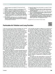
brain and help circulate the cerebrospinal fluid are also motile cilia. Most motile cilia possess nine outer microtubule doublets and a central pair of microtubules (referred to as a ‘‘912’’ configuration) and inner and outer dynein arms that are regularly spaced along the outer doublets and provide the force necessary for ciliary beating (Figure 1). The sperm flagellum is also a motile axonemal structure that has a 912 configuration, although the wave pattern is very different from that of the other motile cilia. Nodal cilia, which are present during embryogenesis and play a key role in the determination of the left–right axis, are also motile but lack a central pair (910), and only a single cilium is found on each cell. ‘‘Primary’’ cilia are nonmotile axonemal structures that lack the central pair and the dynein arms found in motile cilia and are widely distributed throughout the body. Primary cilia are sometimes referred to as ‘‘sensory’’ cilia; however, motile cilia are also known to be sensory, so this nomenclature is somewhat misleading. Examples of primary cilia include those found on the tubular epithelial cells of the kidney (defective in polycystic kidney disease) and the connecting cilia found in the photoreceptor cells of the eye (defective in some retinopathies). Regarding the overlap between primary ciliary dyskinesia (PCD) and other ciliopathies, it would be expected that mutations that affect structures or processes conserved between motile and primary cilia (e.g., basal bodies, microtubules, intraflagellar transport) would result in a disease phenotype reflecting the function of both types of cilia, whereas mutations affecting pathways specific to one type of cilia (e.g., dynein arms in motile cilia) would result in a more limited spectrum of disease. Overlap between PCD and other ciliopathies is clearly demonstrated by mutations in the retinitis pigmentosa GTPase regulator gene (RPGR), which causes PCD and Xlinked retinitis pigmentosa (1). Other studies have suggested a link between PCD and mutations in the oral-facial-digital type I gene (OFD1) and between bronchiectasis and mutations in the polycystic kidney disease genes PKD1 and PKD2 (1, 2). Because a single cilium is assembled from hundreds of proteins, defining the overlap between the many ciliopathies remains a significant challenge. To discern the existence and extent of overlap between the various ciliopathies, investigators need to examine in detail the structure and function of cilia obtained from multiple tissues. To accomplish this, researchers have used a number of different model systems. In this session, ‘‘Cilia and Models for Studying Structure and Function,’’ Dr. Lawrence Ostrowski from The University of North Carolina described some of his recent work using air– liquid interface cultures of human airway epithelium to study the composition of human ciliary axonemes. Dr. Ostrowski’s group is using quantitative proteomics to identify previously unknown proteins in the ciliary axoneme and to study their function. They have also used proteomics to identify proteins missing in cilia isolated from cultures obtained from patients with PCD and are looking for mutations in those proteins. Dr. Susan Dutcher from the Washington University School of Medicine used a comparative genomics approach to identify PCD candidate genes that were not represented in proteomic analysis. Dr. Dutcher also discussed the use of Chlamydomonas
424
PROCEEDINGS OF THE AMERICAN THORACIC SOCIETY VOL 8
2011
Figure 1. Some of the conserved and unique features of motile and primary cilia. Structural features unique to motile cilia are highlighted in red. Numbers indicate structures/processes affected by mutations in the following genes known to cause primary ciliary dyskinesia: (1) DNAH5, DNAH11, DNAI1, DNAI2, and TXNDC3; (2) RSPH4A and RSPH9; (3) CCDC39 and CCDC40; (4) KTU and LRRC50; (5) RPGR; and (6) OFD1 (see Zariwala, Omran, and Ferkol, this issue, pp. 430, for details). IFT 5 intraflagellar transport; PC1-PC2 5 polycystin 1 and 2 complex.
to identify small molecules that may be able to restore motility to PCD cilia. Dr. Cecilia Lo from the University of Pittsburgh closed the session by presenting work using the mouse as a model system for studying ciliopathies. By using a nonspecific mutagen and high throughput screening, Dr. Lo has identified several ciliary proteins, including Dnahc5, as playing a key role in the formation of the developing heart.
PROTEOMIC ANALYSIS OF HUMAN AIRWAY CILIA One approach to increase our understanding of ciliopathies is to use the techniques of proteomics to identify the entire complement of proteins required to assemble a human cilium. By culturing human airway epithelial cells at an air–liquid interface, we have been able to isolate sufficient quantities of human ciliary axonemes for proteomic analysis. Using a combination of several tandem mass spectrometry techniques, we previously identified over 200 axonemal proteins, including many unknown or hypothetical proteins, that are components of the ciliary axoneme (3). To identify additional ciliary proteins and to obtain quantitative data on their abundance, we have more recently begun to apply the technique of LC/MSE to the analysis of ciliary axonemes. This data-independent mass spectrometry technique provides increased sequence coverage, particularly of less abundant proteins, and can provide relative and absolute quantification of each protein present in a complex mixture (4, 5). In our initial studies, total proteins from two independent axonemal preparations were separated by onedimensional SDS-PAGE. The gel lanes were cut into individual bands, subjected to in-gel digestion with trypsin, and analyzed by LC/MSE. These studies identified over 300 proteins, of which approximately 100 have not been previously reported to be part of the ciliary proteome (6). Many of these are uncharacterized hypothetical proteins. As expected, isoforms of a- and b-tubulin were the most abundant proteins (by mass), and the dynein heavy chains DNAH5 and DNAH9 were the next most abundant proteins. When analyzed on a molar basis, a- and b-tubulin remained the most abundant proteins. However, at least 10 proteins were more abundant than DNAH5 and DNAH9, including two hypothetical proteins. In fact, the 10 most abundant hypothetical proteins are estimated to comprise approximately 8% of the ciliary axoneme (by mass). These results demonstrate
that there are still gaps in our knowledge of ciliary structure and highlight the need for additional studies. To begin to investigate the function of the novel proteins identified, siRNA is being used to knock down their expression in cultures of ciliated human airway epithelial cells. Because it is difficult to transfect well differentiated airway cells, integrating viral vectors expressing an shRNA against the protein of interest are used to transduce undifferentiated cultures of primary human airway cells. The vectors also express a drug resistance marker to allow for selection of transduced cells. After selection and expansion, the cells are transferred to an air–liquid interface and differentiate into a ciliated epithelium. Reducing the expression of the most abundant hypothetical protein by approximately 80% resulted in an approximately 60% inhibition of ciliogenesis, compared with cultures transduced with a vector expressing a control shRNA. These results suggest that this hypothetical protein is essential for cilia assembly. We have also used proteomic techniques to identify proteins that are missing in the cilia of airway epithelial cells obtained from patients with PCD who have undergone lung transplants (7). In a recent study, transmission electron micrographs of cilia from a patient with PCD revealed normal inner and outer dynein arms but an apparent absence of several of the central pair projections (Figure 2). Isolation of ciliary axonemes from cultured cells followed by gel electrophoresis and proteomic analysis revealed that the PCD cilia were deficient in the protein hydin (Figure 3). Hydin has previously been identified as a conserved central pair protein (8) with a molecular weight of greater than 575 kD, and in a mouse model, mutation of the hydin gene resulted in hydrocephalus (9). Humans contain two almost identical copies of the hydin gene, one on chromosome 16 containing 86 exons and one with 79 exons on chromosome 1 (10). Because the genes are greater than 99% identical, there are essentially four alleles of each exon present. Therefore, to analyze the hydin gene(s) for the PCD-causing mutation, we first used a combination of cell fusion and selection techniques (conversion technology [11]) to identify mouse/human hybrid cell lines containing only one of the four human hydin alleles. Amplification and sequencing of all 330 exons of hydin (including the coding region and splice junctions) from this patient did not reveal the disease-causing mutation (M. Zariwala, D. Stewart, M. Kesimer, M. Knowles, and L. Ostrowski, unpub-
Ostrowski, Dutcher, and Lo: Cilia and Models for Studying Structure and Function
425
Figure 2. (A) Diagram of a flagellar axoneme illustrating the location of hydin (modified with permission from Reference 37). (B) Electron micrograph of a normal human ciliary axoneme. (C ) Electron micrograph of a ciliary axoneme from the patient with primary ciliary dyskinesia with a central pair defect. The arrows indicate the location of a missing structure in a similar location to the position of hydin in the flagellum.
lished data). Additional candidate genes are being evaluated in this patient. In summary, although many of the basic structural components of the highly conserved ciliary axoneme have been known for many years, proteomic approaches have been useful to identify additional novel proteins that likely play important roles in human cilia. Future studies comparing the proteomes of cilia from different sources (e.g., motile versus primary) will provide additional information on the overlap between PCD and other ciliopathies. By identifying proteins that are missing in cilia from patients with PCD, proteomic analysis can be used to identify candidate genes for the disease-causing mutation. Finally, by comparing different subcomponents of cilia (e.g., membrane, axonemes, basal body) and the number and sites of posttranslational modifications under different conditions (e.g., phosphorylation), proteomic analysis will increase our understanding of the structure and function of this diverse organelle in health and disease.
USING CHLAMYDOMONAS TO STUDY PRIMARY CILIARY DYSKINESIA Cilia and flagella are microtubule-based structures that move by virtue of at least eight biochemically distinct dynein motors. These organelles are conserved from unicellular protists (Giardia to Tetrahymena to Chlamydomonas) to vertebrates. Proteomic studies of flagella isolated from Chlamydomonas suggest there are at least 450 polypeptides present in these organelles (12). Many of these proteins are conserved from Chlamydomonas to human. To identify the proteins involved in ciliary biogenesis and motile functions that are not represented in the proteomic analysis, we undertook a comparative genomics approach that subtracted the proteins present in nonflagellated organisms (Arabidopsis and Saccharomyces) and in C. elegans, which lacks motile cilia, from the shared proteome of the ciliated/flagellated
Figure 3. Analysis of cilia isolated from cultures of a normal individual (left) and a patient with primary ciliary dyskinesia (PCD) (right). Ciliary proteins were separated by SDS-PAGE and silver stained. The highmolecular-weight band missing from the PCD sample was identified as hydin by mass spectrometry.
organisms Chlamydomonas, zebrafish, and human. We identified 200 genes that are present and are candidates for genes involved in PCD (13). This set includes all of the known proteins of the outer and inner dynein arms as well as proteins involved in their transport. Twenty of the 200 proteins are not present in the proteomic dataset. They represent candidates that may be present in low abundance or may be scaffolds or chaperones for ciliary proteins that are not present in the isolated axonemal structure. This high degree of conservation reinforces the idea that Chlamdomonas can serve not only as a source for candidate genes in genetic studies of patients with PCD but can also serve for screening small molecules that rescue motility defects, which may be useful as therapeutics in the future. Our rationale for small molecule screens is influenced by results from genetic screens preformed previously in Chlamydomonas. The loss of radial spokes, which play a role in regulating the dynein arms, results in flagella that are paralyzed or have abnormal beat frequency and waveform. Mutants with abnormal central pair microtubules are paralyzed. Suppressor screens have found that second mutations that alter other substructures of the axoneme can rescue radial spoke and central pair–induced paralysis without restoring the missing substructures. Thus, these mutations can bypass the role of various substructures. At least three examples are known: (1) Mutations in the dynein heavy chain protein (ODA2, the homolog of DNAH5) can restore motility to radial spoke and central pair mutants (14, 15), (2) mutations in the inner dynein arm heavy chain (PF9/IDA1, the homolog of DNAH10) rescue the motility of a central pair mutant (16), and (3) mutations in the nexin links can restore partial motility to radial spoke and central pair defective strains (14, 17). Thus, genetic bypass suppressors with a loss of two substructures of the axoneme show partial rescue of motility, demonstrating that two wrongs can sometimes make a ‘‘right.’’ Defects in dynein arms are the most common cause of PCD, and our lab is looking for genetic suppressors and small molecules that bypass dynein defects. We have used a genetic approach to identify secondary mutations that rescue a mutant with defective inner dynein arms due to a mutation in a phosphatase 2A subunit (18). The inner dynein arm I activity is regulated by phosphorylation and dephosphorylation of one of the intermediate chains (IC138) (19). Mutants that lack this inner dynein arm show medium-velocity swimming and reduced flagellar beat frequency and are defective in phototaxis (16, 20). This PP2A mutant assembles inner dynein arms that appear to be inactive, and, like mutants that lack dynein arms, this mutant shows the same constellation of phenotypes. We have identified 42 independent, unlinked suppressor mutants that rescue the PP2A mutant phenotype, and they fall into at least 11 different loci. Mutations in one of the loci have motility defects of their own, as shown by imaging of the waveforms using an automated method to analyze flagellar motion with high spatial and temporal resolution (Figure 4) (21). This method uses standard brightfield microscopy and high-speed video hardware. It produces
426
PROCEEDINGS OF THE AMERICAN THORACIC SOCIETY VOL 8
2011
Figure 4. Waveforms of wild-type and two allelic suppressors of the PP2A mutant. The suppressors have altered motility in a wild-type background.
a smooth surface that defines the flagellum’s path in space and time. Using this surface representation, we track regions of high curvature to describe dynein activity. Kinematic quantities, such as shear angle, curvature, and bend propagation speed, can be found. For example, wild-type and mutant cells (oda2 and ida3) have markedly different shear amplitudes but similar maximum and minimum curvature values (21). This ability to generate bypass suppressors that restore motility has led us to develop screening methods to identify small molecules that restore motility in collaboration with Dr. Phil Bayly at Washington University. Figure 5 shows four Chlamydomonas strains grown in microtiter plates. Each well in the plate is characterized by the second moment that captures the radial distribution of cells and is translated into a color scheme in the righthand panel. A mutant that cannot move toward a light source (inner dynein arm mutant, ida3; column 1) differs from its parent strain (agg1; column 2) that can perform phototaxis. A mutant with altered waveforms (outer dynein arm mutant, oda2; column 3) is unable to swim as well in viscous medium as the parental strain in column 4. Thus, with this simple screen, it is possible to screen thousands of small molecules to find ones that make the mutants (columns 1 and 3) more closely resemble the controls (columns 2 and 4).
with defects in left–right patterning, such as in heterotaxy when visceral organ situs is randomized. Among the genes recovered in our CHD mutagenesis screen was a gene encoding a motor dynein required for motile cilia function in the airway, Dnahc5 (31). Mutations in the human ortholog DNAH5 are a common cause of PCD (32). Analysis of the Dnahc5 mutants showed that 60% had concordant body situs, presenting as situs solitus (normal; Figure 6A) or situs inversus (mirror symmetric; Figure 6B) without any heart disease, similar to what is seen in patients with PCD with DNAH5 mutations. The situs defects presumably reflect the requirement for motile cilia function for embryonic patterning of the left–right body axis. As in patients with PCD, the airway cilia are immotile and have ultrastructural defects consisting of missing outer dynein arms (31). However, we also observed that 40% of the Dnahc5 mutants had CHD and randomization of visceral organ situs or heterotaxy (Figure 6C). These findings were unexpected because historically CHD and heterotaxy had not been associated with PCD. Because the Dnahc5 mutant is inbred and genetically homogeneous, we can be confident that
LESSONS FROM THE DNAHC5 MOUSE MODEL AND BEYOND Motile and nonmotile (primary) cilia have essential roles to play in a wide range of biological processes (22). Hence, it is no surprise that mutations causing defects involving the cilia have been shown to cause a broad spectrum of human diseases (23). This includes various syndromes with a wide range of developmental anomalies, including Meckel-Gruber syndrome, oralfacialdigital dysplasia, and Bardet-Biedl syndrome. In the course of conducting a high-throughput mouse mutagenesis screen for congenital heart disease (CHD) (24, 25), we unexpectedly found 7 of 11 mutations identified to cause CHDencoded proteins required for the structure and/or function of cilia. Because the mutagenesis screen was entirely phenotype driven using echocardiography for cardiovascular phenotyping, the finding of so many cilia-related mutations suggested that the cilia may play a central role in the pathogenesis of CHD. What might be the role of the cilia in CHD is not immediately apparent, but some of the signaling pathways known to play important roles in cardiovascular development are transduced through the cilia, such as sonic hedgehog (26, 27) and plateletderived growth factor signaling (28). In addition, motile cilia play an essential role during early embryogenesis in breaking bilateral symmetry to establish the left–right body axis (29, 30). Because the heart is the most left–right asymmetric organ in the body and because this asymmetry is essential for efficient oxygenation of blood, it is perhaps not surprising that some of the most complex cases of CHD are observed in conjunction
Figure 5. Development of high-throughput screen for Chlamydomonas flagellar function. (A) Scan of 32 wells of a 96-well plate (U-shape wells) with four Chlamydomonas mutants. Column 1: ida3; agg1. Column 2: agg1. Column 3: oda2. Column 4: Wild-type. agg1 is a mutant that produces negative phototaxis so that cells swim away from the light source. Columns 3 and 4 contain oda2 and wild-type cells with 15% Ficoll to produce higher viscosity, which allows discrimination between the strains. (B) Color-coded images of the same wells that show the second moment of the cell distribution. Drug-induced rescue of ida3; agg1 would move the color code toward blue, which is seen for the agg1 strain in column 2. Drug-induced rescue of oda2 would move the color code to red/orange, which seen in the wild-type cells in column 4.
Ostrowski, Dutcher, and Lo: Cilia and Models for Studying Structure and Function
427
Figure 6. Dnahc5 mutants showing normal situs solitus (A), mirror symmetric situs inversus totalis (B), and randomization of situs or heterotaxy (C ). Arrow denotes cardiac apex, with levocardia (A, C ) and dextracardia (B). H 5 heart; RAA 5 right aortic arch; S 5 stomach. Adapted by permission from Reference 31.
this spectrum of phenotypes (situs solitus, situs inversus, and CHD with heterotaxy) is due to the Dnahc5 mutation only and is not caused by genetic heterogeneity. These findings in the mouse model show definitively that mutation in a gene known to cause PCD in humans (i.e., Dnahc5) can cause complex CHD and heterotaxy. Similar studies performed in collaboration with Dr. L. Ostrowski using a mouse model of PCD caused by deletion of another motor dynein gene (33) yielded similar results. The Dnaic1 knockout mice showed a high incidence of CHD with heterotaxy, as well as situs inversus and situs solitus, confirming that mutations causing PCD can give rise to heterotaxy and CHD (R. Francis, C. Lo, and L. Ostrowski, unpublished observations). The finding of CHD with heterotaxy in the PCD mouse models was at first glance puzzling because clinically patients with PCD have not previously been thought to be at risk for heterotaxy or CHD. One possible explanation is that these anomalies may lead to early prenatal lethality and thus are not represented in the clinical patient population. In support of this is the fact that all of the Dnah5 or Dnaic1 mouse mutants with CHD and heterotaxy die prenatally or at birth. It also should be noted that a newborn mouse is equivalent to an 8- to 10-week gestation human fetus. More direct evidence to address this question came from a retrospective clinical study of over 300 patients with PCD that looked for evidence of CHD and heterotaxy (34). Because heterotaxy and PCD are rare disorders with an incidence of 1 in 10,000 to 15,000, in such a small sample size one would not expect to see any patients with PCD with heterotaxy by chance. Significantly, a 6% incidence of heterotaxy/CHD was observed, indicating a mechanistic link between PCD and CHD/heterotaxy. This likely reflects the dual role of motile cilia in left–right patterning and mucociliary clearance. These findings in the cohort of patients with PCD, together with observations in the mouse PCD models, suggests that PCD-causing mutations can cause PCD and CHD/heterotaxy. These findings also suggest that patients with CHD with heterotaxy may be at risk for PCD, a possibility that has not been fully evaluated. The question of whether patients with CHD with heterotaxy may have PCD is of clinical importance because such patients often have complex structural heart disease and must undergo surgical repair of their heart anomalies. For unknown reasons, these patients have been found to have higher morbidity and mortality. Some develop postsurgical respiratory complications and become ventilator dependent. In a retrospective review of 87 patients with heterotaxy and over 600 other cardiac surgical patients operated on at Children’s National Medical Center in Washington, DC, the patients with heterotaxy were found to
have more postsurgical events, with increased postsurgical mortality and increased risk for respiratory complications when compared to control patients with similar risk-adjusted (RACHS-1) surgical complexity scores (35). In light of the clinical finding that heterotaxy and CHD may present in patients with PCD and the fact that PCD mouse mutants can present with CHD and heterotaxy, it seems compelling to consider whether patients with CHD with heterotaxy may suffer from mucociliary clearance defects like those observed in patients with PCD. Perhaps this could contribute to the respiratory complications and worse outcomes in patients with CHD. To assess this possibility, we have undertaken a prospective recruitment of patients with CHD with heterotaxy to assess for ciliary dysfunction with tests used to evaluate for PCD. This included measuring the nasal nitric oxide level, which is typically low in patients with PCD (36), and performing a nasal scrape to examine ciliary motion in the nasal epithelia with videomicroscopy. These studies showed that 42% of patients with heterotaxy exhibited ciliary dysfunction, as indicated by low nasal nitric oxide and ciliary dysmotilitiy or immotility. These findings suggest that patients with CHD with heterotaxy may indeed be at high risk for ciliary dysfunction similar to those seen in patients with PCD. Studies are underway with exome capture and massively parallel sequencing to look for mutations in the 11 known PCD genes (see Zariwala, Omran, and Ferkol, this issue, pp. 430), as well as other cilia-related genes, to determine whether any of these patients with CHD with heterotaxy may be confirmed to have PCD based on genetic evidence. A clinical outcome study is also being pursued to determine if ciliary dysfunction may be correlated with worse outcomes in patients with CHD with heterotaxy. In summary, studies from the Dnahc5 PCD mouse model have provided unequivocal evidence that mutations in genes causing PCD can cause CHD and heterotaxy. This is consistent with the finding of CHD and heterotaxy among patients with PCD and with our recent observation of airway ciliary dysfunction in patients with CHD with heterotaxy. The challenge now is to determine whether some patients with CHD with heterotaxy may be considered as having a ciliopathy and whether some are bona fide patients with PCD. Because respiratory complications in patients with CHD are typically attributed to the heart disease, it is likely that PCD in patients with CHD may not be diagnosed. The other interesting question to emerge from these studies is why Dnahc5 mutations in mice and in patients can cause all three situs phenotypes (solitus, inversus, and heterotaxy). Because Dnahc5 mutants have cilia in the node but they are paralyzed, having cilia in the node appears to be sufficient at least half the time to generate concordant
428
visceral organ situs (i.e., solitus or inversus). An equally interesting question is why CHD is only observed in Dnahc5 mutants with heterotaxy but not in those with situs solitus or inversus. This suggests that the specification of laterality is intimately integrated with the pathways essential for the regulation of cardiac morphogenesis. As often is the case with the unpredictability of scientific discoveries, our work in the Dnahc5 mutant mouse model has opened an unexpected window into the role of motile cilia in human CHD.
CONCLUSIONS AND FUTURE DIRECTIONS Although our understanding of cilia has increased tremendously in the last decade, researchers are only beginning to understand the complexity of cilia structure and function and the many different roles that are performed by this amazing organelle. The fact that even today many novel and unknown proteins are being discovered in cilia highlights how our understanding of cilia is in some ways rudimentary. Although a complete understanding of cilia is lacking, many major advancements in our knowledge have occurred, especially concerning the role of cilia in the many diseases now referred to as ciliopathies. We are hopeful that these advances in understanding will soon lead to improved treatments. For example, the identification of the causative genes that are mutated in individuals with PCD has opened the door for the design of specific treatments. These may include small molecule correctors, identified by screening techniques in the Chlamydomonas model system described by Dr. Dutcher, or the gene therapy approaches being developed in cultures of differentiated airway cells, as described by Dr. Ostrowski (38, 39). With the continued development of improved proteomic and genomic techniques, it is expected that our knowledge of cilia will continue to advance rapidly along with our ability to diagnose and treat ciliopathies. Author Disclosure: L.E.O. does not have a financial relationship with a commercial entity that has an interest in the subject of this manuscript. S.K.D. received institutional grant support from the Children’s Discovery Institute. She owns stocks/options of Life Technologies and Gilead. C.W.L. does not have a financial relationship with a commercial entity that has an interest in the subject of this manuscript. Acknowledgment: The authors thank the many tissue donors, and Drs. Phil Bayly, and Win Sale for collaborations on the role of dynein arms and motility.
References 1. Leigh MW, Pittman JE, Carson JL, Ferkol TW, Dell SD, Davis SD, Knowles MR, Zariwala MA. Clinical and genetic aspects of primary ciliary dyskinesia/Kartagener syndrome. Genet Med 2009;11:473–487. 2. Driscoll JA, Bhalla S, Liapis H, Ibricevic A, Brody SL. Autosomal dominant polycystic kidney disease is associated with an increased prevalence of radiographic bronchiectasis. Chest 2008;133:1181– 1188. 3. Ostrowski LE, Blackburn K, Radde KM, Moyer MB, Schlatzer DM, Moseley A, Boucher RC. A proteomic analysis of human cilia: identification of novel components. Mol Cell Proteomics 2002;1:451– 465. 4. Silva JC, Gorenstein MV, Li GZ, Vissers JP, Geromanos SJ. Absolute quantification of proteins by LCMSE: a virtue of parallel MS acquisition. Mol Cell Proteomics 2006;5:144–156. 5. Cheng FY, Blackburn K, Lin YM, Goshe MB, Williamson JD. Absolute protein quantification by LC/MS(E) for global analysis of salicylic acid-induced plant protein secretion responses. J Proteome Res 2009; 8:82–93. 6. Blackburn K, Thompson K, Goshe MB, Ostrowski L. Characterizing human respiratory cilia using data-independent LC/MSE provides component level protein stoichiometry measurements. The 58th American Society for Mass Spectrometry Conference on Mass Spectrometry and Allied Topics; Salt Lake City, UT: 2010. 7. Zhang YJ, O’Neal WK, Randell SH, Blackburn K, Moyer MB, Boucher RC, Ostrowski LE. Identification of dynein heavy chain 7 as an inner
PROCEEDINGS OF THE AMERICAN THORACIC SOCIETY VOL 8
8.
9.
10.
11.
12. 13.
14.
15.
16.
17.
18.
19.
20.
21.
22. 23. 24.
25.
26. 27. 28.
29. 30.
31.
2011
arm component of human cilia that is synthesized but not assembled in a case of primary ciliary dyskinesia. J Biol Chem 2002;277:17906–17915. Lechtreck KF, Witman GB. Chlamydomonas reinhardtii hydin is a central pair protein required for flagellar motility. J Cell Biol 2007; 176:473–482. Davy BE, Robinson ML. Congenital hydrocephalus in hy3 mice is caused by a frameshift mutation in Hydin, a large novel gene. Hum Mol Genet 2003;12:1163–1170. Doggett NA, Xie G, Meincke LJ, Sutherland RD, Mundt MO, Berbari NS, Davy BE, Robinson ML, Rudd MK, Weber JL, et al. A 360-kb interchromosomal duplication of the human HYDIN locus. Genomics 2006;88:762–771. Yan H, Papadopoulos N, Marra G, Perrera C, Jiricny J, Boland CR, Lynch HT, Chadwick RB, de la Chapelle A, Berg K, et al: Conversion of diploidy to haploidy. Nature 2000;403:723–724. Pazour GJ, Agrin N, Leszyk J, Witman GB. Proteomic analysis of a eukaryotic cilium. J Cell Biol 2005;170:103–113. Kwan AL, Dutcher SK, Stormo GD. Detecting coevolution of functionally related proteins for automated protein annotation. IEEE Proceedings. 2010;11:99–105. Huang B, Ramanis Z, Luck DJ. Suppressor mutations in Chlamydomonas reveal a regulatory mechanism for Flagellar function. Cell 1982; 28:115–124. Porter ME, Knott JA, Gardner LC, Mitchell DR, Dutcher SK. Mutations in the SUP-PF-1 locus of Chlamydomonas reinhardtii identify a regulatory domain in the beta-dynein heavy chain. J Cell Biol 1994; 126:1495–1507. Porter ME, Power J, Dutcher SK. Extragenic suppressors of paralyzed flagellar mutations in Chlamydomonas reinhardtii identify loci that alter the inner dynein arms. J Cell Biol 1992;118:1163–1176. Heuser T, Raytchev M, Krell J, Porter ME, Nicastro D. The dynein regulatory complex is the nexin link and a major regulatory node in cilia and flagella. J Cell Biol 2009;187:921–933. Elam CA, Wirschell M, Yamamoto R, Fox LA, York K, Kamiya R, Dutcher SK, Sale WS. An axonemal PP2A B-subunit is required for PP2A localization and flagellar motility. Cytoskeleton (Hoboken) 2011;68:363–372. Porter ME, Sale WS. The 9 1 2 axoneme anchors multiple inner arm dyneins and a network of kinases and phosphatases that control motility. J Cell Biol 2000;151:F37–F42. King SJ, Dutcher SK. Phosphoregulation of an inner dynein arm complex in Chlamydomonas reinhardtii is altered in phototactic mutant strains. J Cell Biol 1997;136:177–191. Bayly PV, Lewis BL, Kemp PS, Pless RB, Dutcher SK. Efficient spatiotemporal analysis of the flagellar waveform of Chlamydomonas reinhardtii. Cytoskeleton (Hoboken) 2010;67:56–69. Satir P, Pedersen LB, Christensen ST. The primary cilium at a glance. J Cell Sci 2010;123:499–503. Fliegauf M, Benzing T, Omran H. When cilia go bad: cilia defects and ciliopathies. Nat Rev Mol Cell Biol 2007;8:880–893. Yu Q, Shen Y, Chatterjee B, Siegfried BH, Leatherbury L, Rosenthal J, Lucas JF, Wessels A, Spurney CF, Wu YJ, et al. ENU induced mutations causing congenital cardiovascular anomalies. Development 2004;131:6211–6223. Shen Y, Leatherbury L, Rosenthal J, Yu Q, Pappas MA, Wessels A, Lucas J, Siegfried B, Chatterjee B, Svenson K, et al. Cardiovascular phenotyping of fetal mice by noninvasive high-frequency ultrasound facilitates recovery of ENU-induced mutations causing congenital cardiac and extracardiac defects. Physiol Genomics 2005;24:23–36. Huangfu D, Anderson KV. Cilia and Hedgehog responsiveness in the mouse. Proc Natl Acad Sci USA 2005;102:11325–11330. Anderson KV. Cilia and Hedgehog signaling in the mouse embryo. Harvey Lect 2006;102:103–115. Schneider L, Clement CA, Teilmann SC, Pazour GJ, Hoffmann EK, Satir P, Christensen ST. PDGFRalphaalpha signaling is regulated through the primary cilium in fibroblasts. Curr Biol 2005;15:1861– 1866. Hamada H, Meno C, Watanabe D, Saijoh Y. Establishment of vertebrate left-right asymmetry. Nat Rev Genet 2002;3:103–113. Nonaka S, Shiratori H, Saijoh Y, Hamada H. Determination of left-right patterning of the mouse embryo by artificial nodal flow. Nature 2002; 418:96–99. Tan SY, Rosenthal J, Zhao XQ, Francis RJ, Chatterjee B, Sabol SL, Linask KL, Bracero L, Connelly PS, Daniels MP, et al. Heterotaxy and complex structural heart defects in a mutant mouse model of primary ciliary dyskinesia. J Clin Invest 2007;117:3742–3752.
Ostrowski, Dutcher, and Lo: Cilia and Models for Studying Structure and Function 32. Hornef N, Olbrich H, Horvath J, Zariwala MA, Fliegauf M, Loges NT, Wildhaber J, Noone PG, Kennedy M, Antonarakis SE, et al. DNAH5 mutations are a common cause of primary ciliary dyskinesia with outer dynein arm defects. Am J Respir Crit Care Med 2006;174: 120–126. 33. Ostrowski LE, Yin W, Rogers TD, Busalacchi KB, Chua M, O’Neal WK, Grubb BR. Conditional deletion of dnaic1 in a murine model of primary ciliary dyskinesia causes chronic rhinosinusitis. Am J Respir Cell Mol Biol 2010;43:55–63. 34. Kennedy MP, Omran H, Leigh MW, Dell S, Morgan L, Molina PL, Robinson BV, Minnix SL, Olbrich H, Severin T, et al. Congenital heart disease and other heterotaxic defects in a large cohort of patients with primary ciliary dyskinesia. Circulation 2007;115:2814– 2821.
429
35. Swisher M, Jonas R, Tian X, Lee ES, Lo CW, Leatherbury L. Increased postoperative and respiratory complications in patients with congenital heart disease associated with heterotaxy. J Thorac Cardiovasc Surg 2011;141:637–644, e3. 36. Leigh MW, Zariwala MA, Knowles MR. Primary ciliary dyskinesia: improving the diagnostic approach. Curr Opin Pediatr 2009;21:320–325. 37. Smith EF. Hydin seek: finding a function in ciliary motility. J Cell Biol 2007;176:403–404. 38. Ostrowski LE, Olsen JC. Restoration of ciliary activity to primary ciliary dyskinesia cells using an EIAV-based lentivirus vector. Mol Therapy 2009;17:S30. 39. Ostrowski LE, Yin W, Thompson KE, Patel M, Olsen J. Pilot studies of gene therapy for primary ciliary dyskinesia [abstract]. Am J Respir Crit Care Med 2010;181:A6604.