Classifying EEG and ECoG Signals without Subject Training for Fast BCI. Implementation: Comparison of Non-Paralysed and Completely Paralysed Subjects.
1
Classifying EEG and ECoG Signals without Subject Training for Fast BCI Implementation: Comparison of Non-Paralysed and Completely Paralysed Subjects. N. Jeremy Hill,1 Thomas Navin Lal,1 Michael Schr¨oder,2 Thilo Hinterberger,3 Barbara Wilhelm,3 Femke Nijboer,3 Ursula Mochty,3 Guido Widman,4 Christian Elger,4 Bernhard Sch¨olkopf,1 Andrea K¨ubler3 and Niels Birbaumer3,5 Abstract— We summarize results from a series of related studies that aim to develop a motor-imagery-based brain-computer interface using a single recording session of EEG or ECoG signals for each subject. We apply the same experimental and analytical methods to 11 non-paralysed subjects (8 EEG, 3 ECoG), and to 5 paralysed subjects (4 EEG, 1 ECoG) who had been unable to communicate for some time. While it was relatively easy to obtain classifiable signals quickly from most of the non-paralysed subjects, it proved impossible to classify the signals obtained from the paralysed patients by the same methods. This highlights the fact that though certain BCI paradigms may work well with healthy subjects, this does not necessarily indicate success with the target user group. We outline possible reasons for this failure to transfer.
I. I NTRODUCTION E report the results from experiments in which auto-regressive (AR) models, Support Vector Machine (SVM) classification and Recursive Channel Elimination (RCE) were applied to electroencephalogram (EEG) or electrocorticogram (ECoG) signals in order to implement a Brain-Computer Interface (BCI) with two intentional control states. We conducted four separate motor-imagery studies in which similar task settings and analysis were employed. The end goal is to provide a completely paralysed patient with a means to express a binary decision as reliably as possible, however slowly and regardless of the amount of supervision required.
W
II. S UBJECTS In the studies reported below there were 8 healthy male subjects in their 20s (A–H), 4 patients (I, J, K and L) who had been unable to communicate for some time due to apparent total loss of voluntary motor control, 3 epileptic subjects (M, N and O) with short-term ECoG implants, and one further completely paralysed patient (P) with a long-term ECoG implant. Before becoming paralysed, all the patients had been able to speak German, the language in which the experiments were conducted. The pathology of subjects I, J, K, L and P is briefly described below: The authors gratefully acknowledge the financial support of the MaxPlanck-Gesellschaft, the Deutsche Forschungsgemeinschaft (SFB550/B5 and RO1030/12), the National Institutes of Health (HD30146 and EB00856), the European Community IST program (IST-2002-506778 under the PASCAL Network of Excellence), and the Studienstiftung des Deutschen Volkes (grant awarded to T.N.L.). 1 Max Planck Institute for Biological Cybernetics, T¨ ubingen, Germany. 2 Department of Computer Engineering, Eberhard-Karls University, T¨ ubingen, Germany. 3 Institute of Medical Psychology and Behavioral Neurobiology, Eberhard-Karls University, T¨ubingen, Germany. 4 Department of Epileptology, University of Bonn, Germany. 5 NIH Human Cortical Physiology Unit, Bethesda, USA.
Patient I The patient was a 63-year-old man who had been diagnosed with Amyotrophic Lateral Sclerosis (ALS) 14 years before and had been on artificial ventilation for 12 years. No communication had been possible for 4 years. The patient showed some signs of being able to follow a verbal command to move the eyes left and right—however, this ability did not persist beyond the first few attempts and we can present no statistical evidence of it. Patient J The patient was a 61-year-old man who had had a stroke 10 months before, affecting the motor cortex of the left hemisphere. He was not artificially ventilated, and appeared to fixate faces or objects in the room in a relatively normal manner. However he did not appear to be able to direct eye movements on command or in response to a stimulus, and little or no communication had been possible since the stroke—the only possible signal seemed to be the ability to clench his jaws on command, and (as reported by the patient’s wife) to tighten his lips when he did not want to drink. There were occasional twitches of the hand and tremors of the leg, which seemed to be outside the patient’s voluntary control. Patient K The patient was a 55-year-old man who had suffered a heart attack and postanoxic coma 11 years before and had been on artificial ventilation since then. No communication had been possible since the attack. Blinking and eye movements occurred at close-to-normal frequency, as well as apparently involuntary contractions of the facial muscles, and occasional spasms of pathological laughter. There was no other movement. He showed signs of being able to follow an object with his right eye, and to move the right eye up, down or to the right according to a verbal command, although this ability was not sustained for more than a few consecutive attempts. According to the patient’s wife, even though this behaviour was sometimes possible, he had never been able to associate voluntary eye movement with an “if... then...” contingency to produce a consistent answer to a yes/no question. This raises the possibility that the patient may have general cognitive deficits resulting from his condition. Patient L The patient was a 42-year-old woman who had been diagnosed with chronic Guillan-Barr´e syndrome 13 years before,
2
and had been on artificial ventilation for 12 years. No communication had been possible for 3 years, and she showed no signs of voluntary muscle control. Patient P The patient was a 46-year-old woman who had been diagnosed with ALS 5 years before, and had been artificially ventilated for 3 years. There had been no evidence of voluntary muscle control for 1 year. However, limited communication had been possible via gustatory sensory imagery, which produced reliable changes in salivary pH [1]. With repeated trials of this kind she had been able to answer questions correctly, and to give informed consent to a surgical procedure in which, some weeks prior to the study, she had been implanted with an 8-by-8 array of platinum ECoG electrodes with 10 mm interelectrode spacing, beneath the dura in the left cerebral hemisphere and covering a large area of prefrontal cortex, most of the primary motor strip, and some of the somatosensory strip. Since the procedure, communication had not been possible even by the mouth-pH method. III. M ETHODS A. Experimental Procedure Subjects performed up to 400 trials, typically in a single session (see table), with a few minutes’ break after every 50 (for the non-paralysed subjects) or 20 (for the paralysed subjects). Trials were typically 6 seconds long with a short break (randomized between 3 to 5 seconds) between each. The subject was asked, on each trial, to imagine moving one of two body parts. Our task was to reconstruct what the instruction was on each trial, from motor-imagery-related brain signals. We conducted four separate studies as follows: i EEG recordings from 8 healthy subjects. A visual cue (an arrow to the left or to the right) indicated whether left or right hand motor imagery was to be performed on each trial. An interval of 5 seconds was used for classification, starting 500 msec after cue offset. The setup is described in more detail in [2]. ii EEG recordings from 1 healthy subject and 4 paralysed subjects (see above for pathology of subjects I–L). Since at least three of the patients could not focus or reliably direct their gaze, an auditory cue (a recorded voice saying “left hand” or “right hand”) indicated the motor imagery required on each trial. An interval of 3 seconds was used for classification, starting 500 msec after cue offset (for patients I and J, a slower time-scale was also tested: 7 seconds, beginning 2.5 seconds after cue offset—the results were not substantially different, however). iii ECoG recordings from 3 epileptic patients with shortterm implanted electrode arrays at the Department of Epileptology, University of Bonn. The motor imagery required on each trial was indicated by a visual cue: an arrow for left/right hand, or a picture of a little finger vs. a picture of Einstein sticking out his tongue for the finger/tongue task (the motor imagery task chosen for each patient depended on the exact placement of their electrodes). An interval of 1.5 seconds was used for
classification, starting 500 msec after cue offset. The method and results are described in more detail in [3]. iv ECoG recordings from one completely paralysed ALS patient (subject P) with a long-term implanted electrode array. The motor imagery required on each trial was indicated by a recorded voice saying the German word for “finger” or “tongue”. An interval of 3 seconds was used for classification, starting 500 msec after cue offset. B. Analysis (EEG) The critical task-related modulations were expected to manifest themselves in the amplitude spectra of the signals, as event-related desynchronization (ERD) leads to a decrease in the amplitude of sensorimotor rhythms (SMR) in cortical areas that represent the respective body parts. The general approach is similar to that described in [2], [3] and [4]. Signals were processed on a per-trial basis and classified into two classes—with already completely paralysed patients, speed of communication is a far lower priority than obtaining a means to express reliable binary decisions at all, so we do not aim to implement continuous or multi-class modes of operation. EEG signals (originally sampled at 200 or 256 Hz depending on the equipment used) were lowpass-filtered by a zero-phase-distortion method, with a smooth fall-off from 45 to 50 Hz (the electricity supply is at 50 Hz) and then downsampled at 100 Hz. Each trial was then linearly detrended and then, in a departure from [2], spatial filters were then computed and applied in order to perform blind separation of the EEG sources. The separating matrix was obtained by Independent Components Analysis (ICA) on a random subset of the signal samples from all trials concatenated together (for 400 trials, a 10% subset was found to be sufficient). Spatial filtering is commonly used for ERD classification (the most popular techniques being ICA and the Common Spatial Pattern algorithm) since it leads to significant improvements in performance on EEG data [5]. The algorithm used here was the EEGLAB implementation of Infomax ICA [6]. Each Independent Component was then represented by finding the least-square-error coefficients of a forward-backward autoregressive model of order 4. A linear Support Vector Machine was then trained on the coefficients, with optimization of the regularization parameter by 10-fold cross-validation. The sets of 4 coefficients that corresponded to each independent component were then recursively eliminated using the method first described in [2] and the final number of selected components was determined based on the lowest number of electrodes for which classification error, estimated by 10-fold cross-validation, was within a 95% confidence interval of the minimum, as described in [4]. Offline performance estimates were computed by 2 repeats (with different random seeds) of 10-fold cross-validation. Twotier cross-validation was used wherever necessary: i.e. to avoid overfitting, trials of each outer test fold were excluded from the inner cross-validation procedures used for model selection— this is a standard rule of good practice in machine-learning, as laid out in the context of BCI in [7] and also described in detail in [2]. Test trials were also excluded from the computation of
3
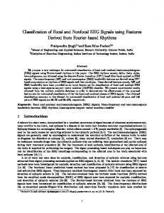
spatial filters, even though class label information is not used by the ICA. C. Analysis (ECoG) Analysis of the ECoG signals, which were sampled at 1 kHz, was identical to the EEG analysis, with the following exceptions (the analysis is described in more detail in [3]): • The signals were not downsampled. • The signals were not temporally filtered for subjects M– O, who were measured in a well-shielded environment. Subject P was tested at home, and the signal was subject to pollution by mains electricity artefacts at 50 and 150 Hz. FIR notch filters were used to remove these. • ICA was not employed—the ECoG signals were used without spatial filtering. • An AR model of order 3 was found to be best for the data. We do not attempt an in-depth comparison of performance in EEG vs. ECoG in the current paper—however, see [5]. IV. R ESULTS Table I reports the results of the four motor-imagery studies. With the exception of D(b), all the entries in the table report the first time that the respective subjects had performed a motor-imagery experiment. The “SMR” column gives a rough indication of the strength of the subject’s sensorimotor rhythm: an amplitude spectrum was computed for each trial and each Independent Component or electrode. The spectra were then averaged across trials, then expressed in log units (dB). For EEG, the final SMR value is the largest peak-to-trough difference in the 7–30 Hz range, among Independent Components judged to be focused close to C3 or C4. For ECoG, the SMR value is simply the largest such peak-to-trough value on the grid. The “performance” column gives an estimate of the percentage of single trials that could be correctly classified offline, plus or minus one standard error. Chance performance is 50% correct. Better-than-chance performance is marked in bold. For 6 out of 8 of the healthy subjects in EEG experiments, classification based on the first recording session is possible at up to 97% accuracy, (average 79% across those 6, or 72% across all 8). For the non-paralysed subjects in ECoG, classification was also possible at around 75% accuracy despite the smaller number of trials. However, for the five completely paralysed patients, irrespective of the amount of background SMR energy and of the number of trials we were able to record, classification was not possible.
inability of the paralysed subjects to communicate at the time of the study, we cannot yet, without extensive additional work, distinguish between a large number of hypotheses at a number of different levels, any of which might explain their inability to yield signals that are classifiable in the same way as those of the non-paralysed subjects. These include: 1) inability to modulate SMR by imagined movement, as a direct neurophysiological consequence of the paralysing motor system pathology; 2) inability to modulate SMR by imagined movement, due to long disuse of the required premotor-motor pathways; 3) difficulty in performing the cognitive task of imagining movement, due to long periods of immobility; 4) general cognitive deficits, resulting in an inability to understand and follow the task instructions; 5) temporary distractedness, lack of alertness, or nonconducive emotional state at the particular time of the experimental session; 6) unwillingness to cooperate in the experiment. “Cognitive deficits” might be expected in some cases as a direct consequence of the original cause of paralysis (for example, extensive lesions due to stroke, or the global effects of anoxia). However, according to one line of reasoning, difficulties may also arise at this level simply from prolonged existence in a paralysed state. A series of studies on rats paralysed with curare (see [8]) shows that operant conditioning becomes impossible when an animal has been unable to affect its environment for an extended period of time. This leads to the hypothesis that a long period without environmental reinforcement of one’s intentions leads to the extinction of the ability to generate intentional states voluntarily—extending this idea to the domain of human cognition, we might call it the extinction of intentional thought. Since falsification of this hypothesis has not been possible thus far, either in the current study or elsewhere, it remains as a challenge to the BCI community. If the hypothesis is correct, it reinforces the idea that BCI training should begin before complete loss of muscle control, or as soon as possible afterwards. It was our intention to take a design that is known to work well with non-paralysed subjects and transfer it to paralysed subjects for direct comparison. However, though a trial-based motor-imagery paradigm without feedback or extensive training is sufficient to achieve good results from normals, it is clear that there are many aspects of such a design that may be unsuitable for paralysed subjects: •
V. D ISCUSSION The results of our motor-imagery studies indicate that it is possible to achieve accurate single-trial classification of EEG and ECoG signals from unpracticed subjects, using automatic classification and feature selection techniques. However, we have only been able to show this for subjects who can actually move their muscles. It is, of course, impossible to prove anything by failing to observe a phenomenon. Given the complete
•
•
•
motor imagery may not be the best neurological phenomenon to choose, because of points (1), (2) and (3) above—it may be better to use an exogenous BCI paradigm, or to look for alternative features that appear to cluster over time into two or more states in the patient’s endogenous EEG/ECoG; the patient may require feedback—with training, this may help overcome problems (2), (3) and perhaps also (4); patients may be unable to keep up with a rapid, regimented trial-based design—a much slower pace, or a slow asynchronous BCI design, may be necessary; multiple sessions, conducted at different times of day, are
4
study i
ii
iii
iv
subject
paralysed
task
signal
channels
trials
sessions
SMR (dB)
performance
notes
A
no
L/R hand
EEG
39
400
1
14.2
65.5 ± 1.6
1
B
no
L/R hand
EEG
39
400
1
6.0
80.9 ± 1.0
1
C
no
L/R hand
EEG
39
400
1
10.1
93.4 ± 0.8
1
D(a)
no
L/R hand
EEG
39
400
1
14.4
96.9 ± 0.7
1,2
E
no
L/R hand
EEG
39
400
1
3.2
75.4 ± 1.7
1
F
no
L/R hand
EEG
39
400
1
6.1
50.4 ± 1.1
1
G
no
L/R hand
EEG
39
400
1
6.2
53.4 ± 1.6
1
H
no
L/R hand
EEG
39
400
1
7.2
61.6 ± 1.9
1
D(b)
no
L/R hand
EEG
64
160
1
11.9
84.7 ± 2.1
2
I
yes
L/R hand
EEG
64
448
2
7.8
50.9 ± 2.0
J
yes
L/R hand
EEG
64
120
1
8.9
46.7 ± 3.1
K
yes
L/R hand
EEG
64
240
1
4.4
49.1 ± 1.9
L
yes
L/R hand
EEG
64
200
1
3.3
45.5 ± 2.0
M
no
L/R hand
ECoG
74
200
1
14.2
75.7 ± 0.4
3
N
no
finger/tongue
ECoG
64
150
1
9.9
73.2 ± 0.7
3
O
no
finger/tongue
ECoG
84
100
1
6.9
76.7 ± 1.3
3
P
yes
finger/tongue
ECoG
64
775
8
10.6
51.2 ± 1.1
Notes: 1 The experiments from subjects A-H were reported by [2]. Quantitative data from F–H were not previously reported since they were unclassifiable. Performance rates reported here are the result of re-analysis of the same data. The use of Independent Components Analysis explains the 5–10% improvement relative to the figures previously published for A–E, and the above-chance performance on previously unclassifiable subject H. 2 Subject D performed two experiments—hence entries for D(a) and D(b). The second occasion was to test the new hardware and software that was to be used with the four paralysed patients I, J, K and L. 3 Results for subjects M, N and O are taken directly with permission from [3]—q.v. for more details. TABLE I: S UMMARY OF R ESULTS
desirable to maximize the chances of avoiding problem (5), and may be necessary in order to allow enough time for feedback training to be effective, and to collect enough data in a slower-paced experiment. It is known that motor-imagery training can be successful in patients if started sufficiently early before complete loss of muscle control: K¨ubler et al. [9] have recently shown that 20 sessions of SMR-modulation training can allow even patients with advanced ALS to exceed the ∼70% level of performance that has been suggested [10] to be the minimum requirement for use in a language support program such as that described by [11]. Thus, there is the potential for such an approach to help the totally paralysed, although as yet there have been no published results to show whether a patient who begins training while able to move can retain the ability to modulate SMR once movement control is lost. The current study, however, compares subjects from the two extreme ends of the spectrum of disability: subjects without any paralysing symptoms against patients who were already unable to communicate when the study started. There has been no report of a patient of the latter type learning to modulate SMR, but we cannot rule out the potential that further work with these five paralysed patients might succeed in training them to do so, or to communicate using some other kind of Brain-Computer Interface. R EFERENCES [1] B. Wilhelm, M. Jordan, and N. Birbaumer, “Communication in LockedIn Syndrome: Effects of imagery on salivary pH,” Neurology, 2005, in press.
[2] T. N. Lal, M. Schr¨oder, T. Hinterberger, J. Weston, M. Bogdan, N. Birbaumer, and B. Sch¨olkopf, “Support Vector channel selection in BCI,” IEEE TBME, vol. 51, no. 6, pp. 1003–1010, June 2004. [3] T. N. Lal, T. Hinterberger, G. Widman, M. Schr¨oder, N. J. Hill, W. Rosenstiel, C. E. Elger, B. Sch¨olkopf, and N. Birbaumer, “Methods towards invasive human brain computer interfaces,” in Advances in Neural Information Processing Systems 17, L. K. Saul, Y. Weiss, and L. Bottou, Eds. Cambridge, MA: MIT Press, 2005, pp. 737–744. [4] T. N. Lal, M. Schr¨oder, N. J. Hill, H. Preissl, T. Hinterberger, J. Mellinger, M. Bogdan, W. Rosenstiel, T. Hofmann, N. Birbaumer, and B. Sch¨olkopf, “A brain computer interface with online feedback based on magnetoencephalography,” L. De Raedt and S. Wrobel, Eds., 2005, pp. 465 – 472. [5] N. J. Hill, T. N. Lal, M. Schr¨oder, T. Hinterberger, G. Widman, C. E. Elger, , B. Sch¨olkopf, and N. Birbaumer, “Classifying event-related desynchronization in EEG, ECoG and MEG signals,” in Towards BrainComputer Interfacing, G. Dornhege, J. del R. Millan, T. Hinterberger, D. J. McFarland, and K.-R. M¨uller, Eds. Cambridge, MA: MIT Press, 2006, in press. [6] S. Makeig, A. J. Bell, T.-P. Jung, and T. J. Sejnowski, “Independent component analysis of electroencephalographic data,” in Advances in Neural Information Processing Systems 8, D. Touretzky, M. Mozer, and M. Hasselmo, Eds. Cambridge, MA: MIT Press, 1996, pp. 145–151. [7] K.-R. M¨uller, M. Krauledat, G. Dornhege, G. Curio, and B. Blankertz, “Machine learning techniques for brain-computer interfaces,” Biomedizinische Technik, vol. 49, no. 1, pp. 11–22, 2004. [8] B. R. Dworkin and N. E. Miller, “Failure to replicate visceral learning in the acute curarized rat preparation,” Behavioral Neuroscience, vol. 100, pp. 299–314, 1986. [9] A. K¨ubler, F. Nijboer, J. Mellinger, T. M. Vaughan, H. Pawelzik, G. Schalk, D. J. McFarland, N. Birbaumer, and J. R. Wolpaw, “Patients with ALS can use sensorimotor rhythms to operate a brain-computer interface,” Neurology, vol. 64, no. 10, pp. 1775–1777, 2005. [10] J. Perelmouter and N. Birbaumer, “A binary spelling interface with random errors.” IEEE TNSRE, vol. 8, pp. 227–232, 2000. [11] J. Perelmouter, B. Kotchoubey, A. K¨ubler, E. Taub, and N. Birbaumer, “Language support program for Thought-Translation-Devices.” Automedica, vol. 18, pp. 67–84, 1999.