CLINICAL AND VACCINE IMMUNOLOGY, Oct. 2008, p. 1580–1589 1556-6811/08/$08.00⫹0 doi:10.1128/CVI.00173-08 Copyright © 2008, American Society for Microbiology. All Rights Reserved.
Vol. 15, No. 10
Clinical and Immunologic Features of an Atypical Intracranial Mycobacterium avium Complex (MAC) Infection Compared with Those of Pulmonary MAC Infections䌤 Mouhannad Sadek,2 Feng Yun Yue,1 Erika Yue Lee,1 Gabor Gyenes,1 R. Brad Jones,1 Victor Hoffstein,2 David G. Munoz,2 Ignatius Fong,2 and Mario Ostrowski1,2* Clinical Sciences Division and Department of Immunology, University of Toronto, Toronto, Ontario, Canada,1 and St. Michael’s Hospital, University of Toronto, Toronto, Ontario, Canada2 Received 15 May 2008/Returned for modification 4 June 2008/Accepted 18 July 2008
Members of the Mycobacterium avium complex (MAC) may cause chronic pulmonary infections in otherwise healthy elderly persons but rarely invade parts of the body outside of the lungs in immunocompetent hosts. We present a case of an isolated intracranial MAC infection in an apparently immunocompetent individual and review previous reports. We studied the T-cell and monocyte responses in healthy volunteers, individuals with a pulmonary MAC infection, and one individual with an isolated intracranial MAC infection. Genomic DNA from the individual with the brain MAC infection was studied for gamma interferon (IFN-␥) receptor mutations. Individuals with localized pulmonary MAC infections showed increased activation of monocytes and enhanced monocyte and T-cell tumor necrosis factor alpha (TNF-␣) production in response to lipopolysaccharide and MAC antigens but defects in T-cell IFN-␥ secretion. The individual with an intracranial MAC infection showed a lack of monocyte activation and deficiencies in both monocyte and T-cell TNF-␣ production and monocyte interleukin-12 (IL-12) production but had preserved T-cell IFN-␥ production. Mutations or deletions in the IFN-␥ receptor were not detected in the individual with the intracranial MAC infection. Our data suggest that distinct immune defects characterize two different manifestations of MAC infection. A relative defect in IFN-␥ production in response to MAC may predispose an individual to localized but partially controlled lung disease, whereas defects leading to reduced IL-12 and TNF-␣ production may allow the dissemination of MAC. Further studies delineating the potential role of TNF-␣ in limiting the spread of MAC outside the lung are warranted. Members of the Mycobacterium avium complex (MAC) are ubiquitous nontuberculous mycobacteria that seldom cause disease in immunocompetent hosts. In human immunodeficiency virus (HIV)-infected individuals who have severe CD4 T-cell depletion, MAC causes disseminated infections. However, MAC can also cause chronic pulmonary infections in elderly persons with apparently normal immune function (34). Characteristically, some older nonsmoking women, generally those with bronchiectasis, may develop right middle lobe infiltrates and a nonproductive cough, termed “Lady Windermere syndrome.” The coordinated function of T cells and monocytes is generally required to resolve mycobacterial infections (10). Few case reports have demonstrated that isolated intracranial MAC infections in the absence of an underlying medical condition account for immunosuppression (7, 8, 32, 40). However, immune defects or genetic predispositions were not adequately described in those reports. Immune defects have previously been reported in elderly persons with pulmonary MAC infections. These have specifically included a level of higher production of interleukin-10 (IL-10) but lower concentrations of gamma interferon (IFN-␥), IL-12, and tumor necrosis factor
alpha (TNF-␣) in peripheral blood mononuclear cells (PBMCs) in response to heat-killed MAC (17, 35, 36, 41). Granuloma formation, which is essential for mycobacterial control, consists of a complex interplay between activated ␣T-cell-receptor-expressing cells that have been primed to respond to mycobacterial antigens and macrophages that have engulfed organisms. Their overall effect is to contain the bacilli and prevent further spread. Signals which aid macrophages in granuloma formation and the clearance of mycobacterial infections are complex and are incompletely understood. These include IFN-␥ and TNF-␣. TNF-␣ is secreted by macrophages and T cells and is able to induce mycobacteriostatic and mycobactericidal activity in both cultured mouse and human macrophages (3, 14). Recently, TNF-␣ has been demonstrated to recruit T cells to the granuloma site, thus maintaining the granuloma (12). IFN-␥ is a macrophage-activating factor secreted by T-helper cells that leads to multiple cellular pathways within the macrophage, including the production of superoxide, TNF-␣, IL-12, IL-1, and IL-6 (4). IL-12 is thought to potentiate IFN-␥ secretion by T cells via a positive-feedback mechanism and thus contributes to the control of mycobacterial infections (2, 15). Defects in either IFN-␥ or TNF-␣ signaling are associated with poorly defined granuloma formation and the escape of the mycobacteria (37). Granulomas also induce intense inflammatory responses in the presence of persisting mycobacteria, which can lead to tissue damage and clinical manifestations of mycobacterial disease. In this regard, IL-10, a counterregulatory cytokine, prevents T-cell prolifera-
* Corresponding author. Mailing address: Clinical Sciences Division, Rm. 6271, 1 King’s College Circle, University of Toronto, Toronto, Ontario M5S 1A8, Canada. Phone: (416) 946-5805. Fax: (416) 978-8765. E-mail:
[email protected]. 䌤 Published ahead of print on 13 August 2008. 1580
VOL. 15, 2008
PULMONARY AND INTRACRANIAL MAC
1581
tion and inhibits T-cell responses (19), thus alleviating excess inflammation. It is also, however, responsible for the induction of anergy and suppression of the immune response to mycobacteria (5), likely by inhibition of TNF-␣ expression (9). The aims of this study were to (i) examine in depth the immune response of an index patient with an intracranial MAC infection who had an apparently normal immune function and (ii) compare the cytokine profiles of the index case to those of individuals with pulmonary MAC infections and healthy controls. Our findings have elucidated potential immune mechanisms important against atypical mycobacterial organisms in humans.
antibodies to CD3 and CD28 (0.1 g/ml and 1 g/ml, respectively; BD Biosciences) at 37°C under a 5% CO2 atmosphere for 5 days. The supernatants were collected and stored at ⫺20°C. The concentrations of IL-12 (after 24 h) and IFN-␥ (after 5 days) in the supernatants were measured by enzyme-linked immunosorbent assay, according to the manufacturer’s instructions (R&D Systems, Minneapolis, MN). Sequencing of IFN-␥R1 and IFN-␥R2 genes. DNA was extracted from PBMCs from the index patient (Puregene kit; Qiagen). For the detection of partial IFN-␥ receptor 1 (IFN-␥R1) deficiency, exon 3 and exon 6 of the IFN-␥R1 gene were amplified by PCR and sequenced as described previously (26). For the detection of mutations in IFN-␥R2, exon 3 of IFN-␥R2 was amplified and sequenced as described previously (26). Statistical analysis. Data were compared by Student’s t test (two tailed) with SPSS software (Chicago, IL).
MATERIALS AND METHODS
RESULTS
Study population. Written informed consent was obtained from the participants in accordance with the guidelines for the conduct of clinical research at the University of Toronto and St. Michael’s Hospital, and the protocol was approved by the review board of St. Michael’s Hospital. (i) Patients. Blood was obtained from four patients (age range, 60 to 63 years) with a diagnosis of pulmonary or intracranial disease due to MAC. A diagnosis was made on the basis of the guidelines recommended by the American Thoracic Society (18). In all cases of pulmonary MAC, sputum culture results were positive, chest radiographs were abnormal, the disease was confined to the lung, and the patients were symptomatic. They were excluded if they had (i) evidence of immunological compromise (e.g., HIV infection) or (ii) if they were taking immunosuppressive medication. (ii) Healthy control subjects. Blood was obtained from four healthy control subjects (age range, 28 to 46 years). Volunteers were excluded if they had a history of respiratory illness or were taking any immunosuppressive medication. Isolation of PBMCs. PBMCs were isolated from blood at the time of diagnosis by differential centrifugation over Ficoll-Paque (Amersham Phamacia Biotech), and the samples were stored in a nitrogen freezer (⫺150°C) for future use. Antigens used. MAC antigen was produced as an aqueous solution of a sonicated heat-killed culture of an M. avium intracellulare isolate derived from a patient from the same geographical area as the study subjects and was obtained from the provincial public health laboratory (Ontario Public Health Laboratories). Various dilutions of MAC antigen (1/10, 1/50, 1/100, 1/200, 1/500, and 1/1,000) were tested in PBMC cultures, and the levels of production of cytokines from monocytes was found to be the highest at the 1/500 dilution, which was set as our standard. Lipopolysaccharide (LPS; Sigma, Oakville, Ontario, Canada) was used at 1 g/ml to stimulate monocytes, and anti-CD3 (BD Biosciences) was used at 0.1 g/ml to stimulate T cells. Flow cytometry and intracellular cytokine determination. To assess the monocytes ex vivo, freshly isolated PBMCs were stained with CD14 ex vivo and run on a FACSCalibur flow cytometer. Monocytes were gated according to their forward and side scatters and assessed for CD14 expression. In addition, the PBMCs were cultured in the presence of medium or antigen (LPS or MAC antigen) for 6 h and then assessed for CD14 expression by flow cytometry. The procedure for intracellular staining of cytokines in T cells and monocytes from PBMC samples was performed according to the protocols of BD Biosciences (San Diego, CA). Briefly, PBMCs were incubated with one of four antigens, (i) medium alone, (ii) MAC antigen, (iii) LPS, and (iv) anti-CD3, for 6 h in the presence of 10 g/ml brefeldin A and 1 g/ml anti-CD49d and anti-CD28 antibodies for costimulation (BD Biosciences). Samples were stained with a panel of conjugated antibodies after they were permeabilized (BD Biosciences). The following antibodies were used: IFN-␥–fluorescein isothiocyanate, IL-10–phycoerthyrin, CD3-peridinin chlorophyll protein complex, CD14-peridinin chlorophyll protein complex, and TNF-␣–antigen-presenting cells. The cells were then washed and resuspended in 1% paraformadehyde–phosphate-buffered saline and were then analyzed on the following day on a FACSCalibur flow cytometer (BD Biosciences). Data were acquired by the use of CellQuest software (BD Biosciences) and were analyzed with the FloJo program (Tree Star, San Carlos, CA). A total of 100,000 events were acquired per sample. Measurement of extracellular cytokine concentration. PBMCs (1 ⫻ 106) were cultured in triplicate in 48-well plates in RPMI 1640 supplemented with 10% heat-inactivated fetal bovine serum, 2 mM L-glutamine, 25 mM HEPES, and antibiotics (Invitrogen Life Tech, Carlsbad, CA) under the following conditions: (i) with medium alone, (ii) with MAC antigen, (iii) with LPS, or (iv) with
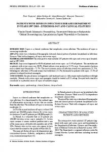
Case report and previous literature. A 63-year-old man presented with a history of 4 months of headaches and wordfinding difficulties but no constitutional symptoms. His medical history was significant for a history of pulmonary sarcoidosis, of which he had no respiratory symptoms at the time of presentation. He was not on any oral medications, had not recently traveled, and had no infectious exposures. The physical examination was unremarkable. His total white cell count was 9,000 cells/mm3, and his lymphocyte count was 1,170 cells/mm3 (CD4 count, 220 cells/mm3; CD8 count, 157 cells/mm3). A test for HIV and the Mantoux skin test were negative. A contrast head computed tomography revealed a ring enhancing lesion in the left frontal lobe (Fig. 1a). A left frontal craniotomy was performed and revealed purulent-looking necrotic tissue. Microscopic examination revealed multiple foci of spindle cell pseudotumor formation (Fig. 1b). Ziehl-Neelsen staining demonstrated a large number of acid-fast bacilli (Fig. 1c). There was no evidence of an intrapulmonary MAC infection either clinically or radiologically. He was started on clarithromycin and ethambutol. After approximately 1 year of treatment, he showed resolution of all symptoms, and imaging demonstrated abscess resolution with residual scarring. The clinical characteristics of previously reported cases and our case of isolated intracranial MAC infection in the absence of HIV infection are depicted in Table 1. Of note is that three of five cases had a history of sarcoidosis. Three of the four previously reported cases had a history of intermittent steroid treatment, and the current case had a history of local inhaled steroid use but had not used inhaled steroids during at least the 6 months prior to presentation. At least three of the five cases had preexisting CD4 lymphocytopenia. Treatment included ethambutol and a macrolide for most cases, and the outcomes were variable, with poor responses reported in two of the five cases. Detailed immune studies were reported for only one case, who showed a reduced ability to produce TNF-␣ and IFN-␥ in response to mitogen stimulation. Study population. In order to identify the immune defects that might be associated with the extrapulmonary dissemination of MAC, as observed in our index patient, we studied three individuals with localized pulmonary MAC infection and four healthy volunteers. Monocyte activation pattern. The activation of CD14 monocytes has previously been shown to be associated with downregulation of the CD14 receptor, which produces a CD14dim phenotype. CD14dim monocytes are thought to be major pro-
1582
SADEK ET AL.
CLIN. VACCINE IMMUNOL.
FIG. 1. A contrast-enhanced head computed tomography of index patient revealed a ring enhancing lesion in the left frontal lobe (a). Microscopic examination of the brain biopsy specimen revealed multiple foci of spindle cell pseudotumor formation, consistent with a poorly developed granulomatous response (b). Ziehl-Neelsen stains of the biopsy specimen demonstrate a large number of acid-fast bacilli (c).
ducers of cytokines, and the levels of CD14dim monocytes have previously been shown to be elevated in septic patients (13). In addition, the percentage of CD14dim (activated) monocytes has been shown to increase after stimulation with sepsis-specific antigens, such as LPS, staphylococcal enterotoxin B, peptidoglycan, and even TNF-␣ (38). Ex vivo PBMCs were isolated, and the percentage of CD14dim monocytes was measured as a marker of baseline inflammatory responses. Patients with localized pulmonary MAC infections had a higher percentage of CD14dim monocytes circulating in their peripheral blood than the controls and the individual with a brain MAC infection, even after stimulation with MAC antigen in culture (Table 2). LPS and MAC antigens activated monocytes in all subjects with pulmonary MAC and all controls, as assessed by CD14 downregulation, but this effect was less pronounced in our index patient with an intracranial MAC infection (Table 2).
Monocyte responses. Monocytes were examined for their ability to produce cytokines in response to LPS (a Toll-like receptor 4 [TLR4] agonist) and to MAC antigens (a TLR2 and TLR4 agonist) (Fig. 2a and b). Monocytes from the individuals with pulmonary MAC infections produced significantly more TNF-␣ and significantly less IL-10 in response to LPS or MAC antigen than the healthy controls (for LPS, 86.9 ⫾ 14.5% TNF-␣ and 9.3 ⫾ 5.6% IL-10 versus 37.6 ⫾ 15.3% TNF-␣ and 39.6 ⫾ 1.9% IL-10, respectively [P ⬍ 0.01]; for MAC antigen, 75.3 ⫾ 15.5% TNF-␣ and 8.2 ⫾ 4.6% IL-10 versus 40.1 ⫾ 14.0% TNF-␣ and 24.5 ⫾ 4.2% IL-10, respectively [P ⬍ 0.05]). Monocytes from our patient with intracranial MAC infection showed levels of TNF-␣ expression similar to those for the healthy controls in response to LPS but reduced levels of TNF-␣ production in response to MAC antigen. In addition, monocytes from this individual produced small amounts of
1583 PULMONARY AND INTRACRANIAL MAC VOL. 15, 2008
Authors (reference no.) or patient Uldry et al. (40)
Dickerman et al. (7)
Sex
Presentation
4 mo
2 mo
4 yr
Symptom duration prior to treatment
Antibiotic treatment
Treatment duration (mo)
Response to antimycobacterial therapy
Poor response, death
Immunology study results
11
Underlying disease
Ciprofloxacin, amikacin, ethambutol, ansamycin, and clofazimine
6
Diagnostic method
Not done
Rifampin, ethambutol, clarithromycin
Not available
None
CD4⫹ lymphocytopenia, low extracellular INF␥ and TNF-␣ levels compared to those for healthy controls
Rifampin, ethambutol, clarithromycin
Brain biopsy
Brain biopsy
Sarcoidosis (treated with steroids until 3 yr prior to symptoms)
CD4⫹ lymphocytopenia (90 ⫻ 106 cells/liter)
Not done See Results
11
Brain biopsy
Brain biopsy
SLE (treated with prednisone and azathioprine) Sarcoidosis
Resolution of neurological symptoms
Sarcoidosis (intermittent treatment with steroids)
Not done
Brain biopsy
Ethambutol, clarithromycin
Poor response; required repeat surgical debridement, with good outcome Condition stabilized after multiple surgical debridement, with a good outcome
Not done
Not done
Leukocytes, 45.3 ⫻ 106/ liter; lymphocytes, 80%; protein concn, 2.76 g/liter; glucose concn, 3.1 mM; culture negative Not done
CSF examination
TABLE 1. Clinicopathologic profile of cases of isolated intracranial central nervous system mycobacterial infection in the absence of HIV infectiona Age (yr) Headaches, nausea, vomiting, ataxia
Yr presented
Seizure, ataxia
F
M
31
38
1985
1996
M
Enlarging scalp mass
38
F
1999
50 M
Morrison et al. (32)
2000 63
Headaches, slurred speech, nystagmus, ataxia
Di Patre et al. (8) 2005
Headaches, word-finding difficulties
Abbreviations: CSF, cerebrospinal fluid; F, female; M, male; SLE, systemic lupus erythematosus.
Index patient (this study) a
1584
SADEK ET AL.
CLIN. VACCINE IMMUNOL.
TABLE 2. CD14dim profiles ex vivo and after stimulation % CD14
dim
monocytes
Infection group
Ex vivo and unstimulated
After LPS stimulation
After MAC antigen stimulation
Pulmonary MAC Brain MAC Controls
55.1 ⫾ 7.8a 20.1 25.8 ⫾ 5.8
71.1 ⫾ 12.5 44.1 69.9 ⫾ 9.5b
83.1 ⫾ 5.1a,b 46 62.0 ⫾ 10.8b
a P ⬍ 0.01 for patients with lung MAC infection compared with the results for the healthy controls. b P ⬍ 0.01 compared with the results obtained in the unstimulated condition.
IL-10 to LPS and MAC compared to the amounts produced by the monocytes from the healthy controls. We also measured the relative contributions of cytokine expression within the CD14bright and CD14dim fractions in each sample and found that TNF-␣ was equally distributed in both fractions, whereas the majority of IL-10 produced in the healthy controls was found in the CD14dim subset (data not
shown) under both LPS and MAC antigen stimulation conditions. Overall, monocytes isolated from the individuals with pulmonary MAC infections produced more TNF-␣ than the healthy controls and the patient with intracranial MAC infection and less IL-10 than the healthy controls, whether it was in response to LPS or MAC antigen. Monocytes from the index patient with intracranial MAC infection appeared to have a relative defect in TNF-␣ production in response to MAC antigen, even compared to that for the controls, and this defect was not associated with any increased production of IL-10. In addition, the CD14dim monocytes from the healthy controls produced significant amounts of IL-10 compared to the amounts produced by the patients with pulmonary and intracranial MAC infections. T-cell response to CD3/CD28. In order to test for general defects in T-cell function, the cytokine response to incubation with CD3/CD28 costimulation was measured (Fig. 3a). T cells from individuals with pulmonary MAC infection showed sig-
FIG. 2. Production of TNF-␣ and IL-10 by antigen-stimulated CD14 monocytes. PBMCs from patients with pulmonary MAC infections (n ⫽ 3), an intracranial MAC infection (n ⫽ 1), and healthy controls (n ⫽ 4) were incubated ex vivo with LPS (a) or heat-killed MAC antigen (b) for 6 h in the presence of brefeldin A. The cells were then harvested, stained for CD14, and then permeabilized and stained for cytokines. Intracellular cytokine expression within CD14 gated cells was subsequently measured by flow cytometry, as described in the text. Mean values ⫾ standards deviations are shown. *, P ⬍ 0.05 for patients with pulmonary MAC infections compared with the results for the healthy controls; **, P ⬍ 0.01 for patients with pulmonary MAC infections compared with the results for the healthy controls.
VOL. 15, 2008
PULMONARY AND INTRACRANIAL MAC
1585
1586
CLIN. VACCINE IMMUNOL.
SADEK ET AL.
nificantly reduced IFN-␥ responses compared to those for the healthy controls but showed TNF-␣ responses similar to those for the healthy controls (for TNF-␣, 2.6 ⫾ 0.8% and 2.3 ⫾ 0.9%, respectively; for IFN-␥, 0.5 ⫾ 0.2% and 3.0 ⫾ 0.5%, respectively [P ⬍ 0.01]). T cells from our patient with intracranial MAC infection showed reduced IFN-␥ and TNF-␣ responses compared to those of T cells from the healthy controls. The IL-10 responses were comparable between the MAC-infected individuals and the healthy controls. Thus, compared to the healthy controls, T cells from individuals with localized MAC infections display a reduced capacity to produce IFN-␥ in response to T-cell stimulation via the CD3 receptor, and T cells from the patient with the intracranial MAC infection showed a reduced capacity to produce both TNF-␣ and IFN-␥ when they were stimulated with CD3. T-cell responses to MAC antigen. In order to test for specific T-cell dysfunction in response to MAC infection, the response to incubation with isolated MAC antigen was measured. The results of a representative experiment are shown in Fig. 3b, and summary data are depicted in Fig. 3c. T cells from individuals with localized pulmonary MAC infections showed significantly enhanced TNF-␣ production compared to that for the healthy controls (15.2 ⫾ 3.6% and 2.5 ⫾ 3.1%, respectively [P ⬍ 0.01]) and a more modest but not significant (P ⫽ 0.14) increase in the level of IFN-␥ production compared to that for the healthy controls. The individual with brain MAC infection showed TNF-␣ responses to MAC similar to those for the healthy controls, but the TNF-␣ levels were at least threefold lower than those seen in the patients with pulmonary MAC infection, that is, in the context of an infection. Conversely, the IFN-␥ responses to MAC in this individual were greater than those of the healthy controls and the patients with pulmonary MAC infections (Fig. 3b). There was no evidence of enhanced IL-10 production in response to MAC antigen in any individual. PBMC extracellular cytokine production in response to MAC. It has previously been reported that individuals with pulmonary MAC infection may have T cells capable of intracellular IFN-␥ production but have an inability to secrete them (35). In order to test for a relative deficiency in the ability to secrete cytokines in response to MAC antigen, PMBCs were incubated with MAC antigen and an enzyme-linked immunosorbent assay was performed to measure IFN-␥ and IL-12 levels (Fig. 4a and b, respectively). For IFN-␥, PBMCs from the individual with brain MAC infections produced large amounts in response to MAC antigen, whereas the group with localized pulmonary MAC did not show any enhanced IFN-␥ secretion compared to that for the healthy controls. Conversely, the index patient with brain MAC infection had a deficiency in IL-12 production in response to MAC antigen and LPS compared to the responses of the healthy controls,
whereas individuals with pulmonary MAC infections produced larger amounts of IL-12 than the healthy controls. Sequencing of exons 3 and 6 of IFN-␥R1 and exon 3 of IFN-␥R2. Given that the index patient secreted high levels of IFN-␥ in response to MAC antigen, we hypothesized that a defect in the receptors for IFN-␥, i.e., IFN-␥R1 and IFN-␥R2, could explain defective immune control of mycobacteria. Partial IFN-␥R1 and IFN-␥R2 deficiency has been associated with late-onset mycobacterial infections (23). The most frequently described mutations have included nucleotide substitutions at positions 187 (C to T) and 260 (T to C) in exon 3 or deletions in exon 6 (position 818) for IFN-␥R2 deficiency and mutations at position 114 (C to T) of exon 3 for IFN-␥R1 deficiency (23, 29, 30). Sequencing of exons 3 and 6 of IFN-␥R1 and exon 3 of IFN-␥R2 in the index patient revealed none of these previously described mutations or deletions. DISCUSSION Isolated intracranial MAC infections are rare in the absence of obvious immunosuppression, such as HIV infection (20). Evaluation of our case with those reported previously revealed a number of features associated with intracranial MAC infections. These include the presence of sarcoidosis, intermittent steroid use, and CD4 lymphocytopenia. We noted a striking association between the reported cases of intracranial MAC and sarcoidosis. Whether some cases of sarcoidosis represent occult MAC pulmonary infections prior to the development of intracranial infection will require further study. We contrasted the immunologic features of our case with those of a more common manifestation of MAC infection, that is, localized pulmonary MAC infection. Adequate monocyte/ macrophage function is critical for the containment of mycobacterial infections. Patients with localized pulmonary MAC infections have a higher percentage of CD14dim monocytes, reflecting a greater degree of monocyte activation in vivo. This suggests an activated immune response at the baseline, which may be due to the chronic infectious process. We did not see this baseline level of activation in healthy controls, and the lack of monocyte activation in our index patient with an intracranial MAC infection suggests a defect in monocyte activation in the presence of MAC antigen. Despite the presence of ongoing monocyte activation in patients with localized pulmonary MAC infections, these individuals are unable to clear the infection from their lungs. We show that localized pulmonary MAC infections are associated with the production of high levels of TNF-␣ in response to MAC but low levels of production of IFN-␥ relative to those in healthy controls. There have been conflicting reports about the pathophysiology of localized pul-
FIG. 3. Production of TNF-␣, IFN-␥, and IL-10 by antigen-stimulated T cells. T cells from patients with pulmonary MAC infections (n ⫽ 3), an intracranial MAC infection (n ⫽ 1), and healthy controls (n ⫽ 4) were incubated with antibodies to CD3 and CD28 (a) or heat-killed MAC antigen (b and c) for 6 h in the presence of brefeldin A. The cells were harvested and stained, and cytokine expression on gated CD3-positive cells was subsequently measured by flow cytometry, as described in the text. Representative data from the index patient, a healthy volunteer, and an individual with a localized pulmonary MAC infection are shown in panel b. In panel b, the values above the boxes indicate the percentage of CD3 staining cells expressing TNF-␣ In panels a and c, mean values ⫾ standards deviation are shown. *, P ⬍ 0.01 for patients with pulmonary MAC infections compared with the results for the healthy controls; **, P ⬍ 0.05 for patients with pulmonary MAC infections compared with the results for the healthy controls.
VOL. 15, 2008
PULMONARY AND INTRACRANIAL MAC
1587
FIG. 4. Individuals with localized lung MAC infections may have an IFN-␥ secretion defect in response to MAC antigen, and the patient with an intracranial MAC infection displays IL-12 deficiency in response to MAC antigen and LPS. The extracellular production of IFN-␥ (a) and IL-12 (b) by MAC antigen-stimulated PBMCs was measured. PBMCs were isolated from patients with pulmonary MAC infections, a patient with an intracranial MAC infection, and healthy controls and were cultured with heat-killed MAC antigen and/or LPS. The cytokine concentrations within the supernatants were measured by enzyme-linked immunosorbent assay. Mean values ⫾ standard deviations are shown.
monary MAC infections. Previous reports support structural abnormalities of the thorax that predispose elderly patients to such infections (27, 28). However, more recent data have documented IFN-␥ deficiency as an underlying etiology (17, 36). Other reports have documented a defect in IFN-␥ secretion as the main factor (35, 41). IFN-␥ is important in antimycobacterial host defense via macrophage activation and granuloma formation. A defect in the synthesis and, possibly, secretion of IFN-␥ predisposes individuals to persistent nontuberculous mycobacterial infections and may explain the poor response to treatment and frequent relapses in these patients (25, 34). Our data also show that in response to MAC antigens, patients with pulmonary MAC infections displayed a relative IFN-␥ deficiency. Comparison of the intracellular levels to the extracellular levels of IFN-␥ in patients with pulmonary MAC infections also suggests that a secretory defect leading to relatively low levels of extracellular IFN-␥ may also be a contributing factor. Studies have shown clinical improvement with the administration of IFN-␥ in patients with pulmonary MAC infections (21), and this supports a secretory dysfunction rather than a receptor abnormality. The mechanism of this IFN-␥ secretion
defect has yet to be characterized. The presence however, of a potent TNF-␣ response suggests that TNF-␣ may be important in limiting the spread of a localized infection as well as contributing to overall macrophage activation. Our patient with an intracranial MAC infection showed a selective TNF-␣ and IL-12 defect of T cells and monocytes in response to MAC antigen but high levels of IFN-␥. This cytokine profile is suggestive of a partial IFN-␥ receptor deficiency state. The lack of IL-12 production in response to LPS or MAC antigen, despite the induction of high levels of IFN-␥, is in keeping with a failure of signaling through the IFN-␥ receptor, since IFN-␥ primes monocytes/macrophages to produce IL-12 (39). Previous work with patients with IFN-␥ receptor deficiency showed that such mutations lead to the loss of augmentation of TNF-␣ production by PBMCs (24), with the result being disseminated MAC infections (11, 22). We did not, however, detect the commonly described mutations in the IFN␥R1 and IFN-␥R2 genes in this individual. Further studies examining mutations elsewhere in these genes or in other genes such as stat-5 (downstream from IFN-␥ signaling) or IL-12 will likely be required to further elucidate a genetic basis for the index patient’s immune abnormalities. Of note is a
1588
SADEK ET AL.
recent report which also could not find any evidence of partial deficiencies of IFN-␥R1 and IFN-␥R2 in association with localized nontuberculous mycobacterial lung disease (26). TNF-␣ deficiency leads to susceptibility to infections by M. tuberculosis and the observed lack of granuloma formation (1), the latter of which was reflected in our index patient. The lack of TNF-␣ induction also suggests that this cytokine plays a central role in macrophage activation and the prevention of the spread of MAC to other tissues in humans. In this regard, previous reports have indicated that humans with tuberculosis were treated with TNF-␣-blocking agents, such as etanercept or infliximab (16). It is thus quite possible that the increasing use of such agents will lead to more cases involving extrapulmonary MAC infections. Boussiotos et al. previously showed that in patients with Mycobacterium tuberculosis infections, the IL-10 production induced by M. tuberculosis induces a relative immune suppression that prevents control of the organism (5). We did not find support for enhanced IL-10 expression as a mechanism for mycobacterial persistence either in patients with localized pulmonary MAC or in the patient with the intracranial MAC infection. Of interest, however, was that in the healthy controls, activated monocytes produced large amounts of IL-10, suggesting that monocytes may have a regulatory function in the absence of an active infectious process. The association of sarcoidosis with our case and those reported previously is interesting and requires further study. The immune profile of our case differed from that usually described in patients with sarcoidosis; that is, the enhanced production of T cells producing IFN-␥ and proinflammatory cytokines, such as TNF-␣, is generally reported in patients with sarcoidosis (33). Recently, Carlisle et al. (6) demonstrated that 12/30 individuals with sarcoidosis had detectable IFN-␥ responses by enzyme-linked immunospot assay to two or more mycobacterial antigens in their PBMCs, even though they were negative for purified protein derivative. It was unclear whether these individuals indeed had persistent mycobacterial infections which were responsible for their symptoms. However, such findings warrant further studies to determine whether a subset of cases of sarcoidosis are due to persistent mycobacteria or insufficient immune responses to the organism and if such persistence gives the potential for dissemination to other organs. In addition, a recent report showed that in patients with sarcoidosis, there is an amplification of T regulatory cells in the lungs, which is associated with a local proliferative defect of T cells in response to antigen (31). Whether such a defect predisposes individuals to invasive mycobacterial infections will also require further study. In summary, we highlight the immunologic features of two forms of MAC infection. We find that individuals with the more common, localized pulmonary MAC infections have a relative deficiency of IFN-␥ production that leads to a localized inability to eradicate the mycobacteria. In our case of an intracranial MAC infection, we find defects in both TNF-␣ and IL-12 production. Further studies are warranted to investigate the role of defects in the production of these cytokines in humans in the development of MAC-related diseases. ACKNOWLEDGMENTS We thank our patients for contributing their time to this study.
CLIN. VACCINE IMMUNOL. Financial support for this study was obtained from the Canadian Institutes of Health Research (CIHR) and the Ontario HIV Treatment Network (OHTN). M.O. is a career scientist for the CIHR and OHTN. M.O. receives unrestricted research grants from Sanofi-Pasteur and Pfizer. REFERENCES 1. Algood, H. M., P. L. Lin, D. Yankura, A. Jones, J. Chan, and J. L. Flynn. 2004. TNF influences chemokine expression of macrophages in vitro and that of CD11b⫹ cells in vivo during Mycobacterium tuberculosis infection. J. Immunol. 172:6846–6857. 2. Altare, F., A. Durandy, D. Lammas, J. F. Emile, S. Lamhamedi, F. Le Deist, P. Drysdale, E. Jouanguy, R. Doffinger, F. Bernaudin, O. Jeppsson, J. A. Gollob, E. Meinl, A. W. Segal, A. Fischer, D. Kumararatne, and J. L. Casanova. 1998. Impairment of mycobacterial immunity in human interleukin-12 receptor deficiency. Science 280:1432–1435. 3. Bean, A. G., D. R. Roach, H. Briscoe, M. P. France, H. Korner, J. D. Sedgwick, and W. J. Britton. 1999. Structural deficiencies in granuloma formation in TNF gene-targeted mice underlie the heightened susceptibility to aerosol Mycobacterium tuberculosis infection, which is not compensated for by lymphotoxin. J. Immunol. 162:3504–3511. 4. Boehm, U., T. Klamp, M. Groot, and J. C. Howard. 1997. Cellular responses to interferon-gamma. Annu. Rev. Immunol. 15:749–795. 5. Boussiotis, V. A., E. Y. Tsai, E. J. Yunis, S. Thim, J. C. Delgado, C. C. Dascher, A. Berezovskaya, D. Rousset, J. M. Reynes, and A. E. Goldfeld. 2000. IL-10-producing T cells suppress immune responses in anergic tuberculosis patients. J. Clin. Investig. 105:1317–1325. 6. Carlisle, J., W. Evans, R. Hajizadeh, M. Nadaf, B. Shepherd, R. D. Ott, K. Richter, and W. Drake. 2007. Multiple Mycobacterium antigens induce interferon-gamma production from sarcoidosis peripheral blood mononuclear cells. Clin. Exp. Immunol. 150:460–468. 7. Dickerman, R. D., Q. E. Stevens, R. Rak, S. E. Dorman, S. M. Holland, and T. T. Nguyen. 2003. Isolated intracranial infection with Mycobacterium avium complex. J. Neurosurg. Sci. 47:101–105. 8. Di Patre, P. L., W. Radziszewski, N. A. Martin, A. Brooks, and H. V. Vinters. 2000. A meningioma-mimicking tumor caused by Mycobacterium avium complex in an immunocompromised patient. Am. J. Surg. Pathol. 24:136– 139. 9. Donnelly, R. P., S. L. Freeman, and M. P. Hayes. 1995. Inhibition of IL-10 expression by IFN-gamma up-regulates transcription of TNF-alpha in human monocytes. J. Immunol. 155:1420–1427. 10. Dorman, S. E., and S. M. Holland. 1998. Mutation in the signal-transducing chain of the interferon-gamma receptor and susceptibility to mycobacterial infection. J. Clin. Investig. 101:2364–2369. 11. Dorman, S. E., C. Picard, D. Lammas, K. Heyne, J. T. van Dissel, R. Baretto, S. D. Rosenzweig, M. Newport, M. Levin, J. Roesler, D. Kumararatne, J. L. Casanova, and S. M. Holland. 2004. Clinical features of dominant and recessive interferon gamma receptor 1 deficiencies. Lancet 364:2113–2121. 12. Egen, J. G., A. G. Rothfuchs, C. G. Feng, N. Winter, A. Sher, and R. N. Germain. 2008. Macrophage and T cell dynamics during the development and disintegration of mycobacterial granulomas. Immunity 28:271–284. 13. Fingerle, G., A. Pforte, B. Passlick, M. Blumenstein, M. Strobel, and H. W. Ziegler-Heitbrock. 1993. The novel subset of CD14⫹/CD16⫹ blood monocytes is expanded in sepsis patients. Blood 82:3170–3176. 14. Flynn, J. L., M. M. Goldstein, J. Chan, K. J. Triebold, K. Pfeffer, C. J. Lowenstein, R. Schreiber, T. W. Mak, and B. R. Bloom. 1995. Tumor necrosis factor-alpha is required in the protective immune response against Mycobacterium tuberculosis in mice. Immunity 2:561–572. 15. Frucht, D. M., and S. M. Holland. 1996. Defective monocyte costimulation for IFN-gamma production in familial disseminated Mycobacterium avium complex infection: abnormal IL-12 regulation. J. Immunol. 157:411–416. 16. Gomez-Reino, J. J., L. Carmona, and M. Angel Descalzo. 2007. Risk of tuberculosis in patients treated with tumor necrosis factor antagonists due to incomplete prevention of reactivation of latent infection. Arthritis Rheum. 57:756–761. 17. Greinert, U., M. Schlaak, S. Rusch-Gerdes, H. D. Flad, and M. Ernst. 2000. Low in vitro production of interferon-gamma and tumor necrosis factoralpha in HIV-seronegative patients with pulmonary disease caused by nontuberculous mycobacteria. J. Clin. Immunol. 20:445–452. 18. Griffith, D. E., T. Aksamit, B. A. Brown-Elliott, A. Catanzaro, C. Daley, F. Gordin, S. M. Holland, R. Horsburgh, G. Huitt, M. F. Iademarco, M. Iseman, K. Olivier, S. Ruoss, C. F. von Reyn, R. J. Wallace, Jr., and K. Winthrop. 2007. An official ATS/IDSA statement: diagnosis, treatment, and prevention of nontuberculous mycobacterial diseases. Am. J. Respir. Crit. Care Med. 175:367–416. 19. Groux, H., M. Bigler, J. E. de Vries, and M. G. Roncarolo. 1996. Interleukin-10 induces a long-term antigen-specific anergic state in human CD4⫹ T cells. J. Exp. Med. 184:19–29. 20. Gyure, K. A., R. A. Prayson, M. L. Estes, and G. S. Hall. 1995. Symptomatic Mycobacterium avium complex infection of the central nervous system. A
VOL. 15, 2008
21. 22.
23.
24.
25.
26.
27. 28.
29.
30.
case report and review of the literature. Arch. Pathol. Lab. Med. 119:836– 839. Hallstrand, T. S., H. D. Ochs, Q. Zhu, and W. C. Liles. 2004. Inhaled IFN-gamma for persistent nontuberculous mycobacterial pulmonary disease due to functional IFN-gamma deficiency. Eur. Respir. J. 24:367–370. Han, J. Y., S. D. Rosenzweig, J. A. Church, S. M. Holland, and L. A. Ross. 2004. Variable presentation of disseminated nontuberculous mycobacterial infections in a family with an interferon-gamma receptor mutation. Clin. Infect. Dis. 39:868–870. Haverkamp, M. H., J. T. van Dissel, and S. M. Holland. 2006. Human host genetic factors in nontuberculous mycobacterial infection: lessons from single gene disorders affecting innate and adaptive immunity and lessons from molecular defects in interferon-gamma-dependent signaling. Microbes Infect. 8:1157–1166. Holland, S. M., S. E. Dorman, A. Kwon, I. F. Pitha-Rowe, D. M. Frucht, S. M. Gerstberger, G. J. Noel, P. Vesterhus, M. R. Brown, and T. A. Fleisher. 1998. Abnormal regulation of interferon-gamma, interleukin-12, and tumor necrosis factor-alpha in human interferon-gamma receptor 1 deficiency. J. Infect. Dis. 178:1095–1104. Huang, J. H., P. N. Kao, V. Adi, and S. J. Ruoss. 1999. Mycobacterium avium-intracellulare pulmonary infection in HIV-negative patients without preexisting lung disease: diagnostic and management limitations. Chest 115: 1033–1040. Hwang, J. H., W. J. Koh, E. J. Kim, E. H. Kang, G. Y. Suh, M. P. Chung, H. Kim, and O. J. Kwon. 2006. Partial interferon-gamma receptor deficiency and non-tuberculous mycobacterial lung disease. Tuberculosis (Edinburgh) 86:382–385. Iseman, M. D. 1996. That’s no lady. Chest 109:1411. Iseman, M. D., D. L. Buschman, and L. M. Ackerson. 1991. Pectus excavatum and scoliosis. Thoracic anomalies associated with pulmonary disease caused by Mycobacterium avium complex. Am. Rev. Respir. Dis. 144:914– 916. Jouanguy, E., S. Lamhamedi-Cherradi, F. Altare, M. C. Fondaneche, D. Tuerlinckx, S. Blanche, J. F. Emile, J. L. Gaillard, R. Schreiber, M. Levin, A. Fischer, C. Hivroz, and J. L. Casanova. 1997. Partial interferon-gamma receptor 1 deficiency in a child with tuberculoid bacillus Calmette-Guerin infection and a sibling with clinical tuberculosis. J. Clin. Investig. 100:2658– 2664. Jouanguy, E., S. Lamhamedi-Cherradi, D. Lammas, S. E. Dorman, M. C. Fondaneche, S. Dupuis, R. Doffinger, F. Altare, J. Girdlestone, J. F. Emile,
PULMONARY AND INTRACRANIAL MAC
31.
32.
33. 34.
35.
36.
37. 38.
39. 40.
41.
1589
H. Ducoulombier, D. Edgar, J. Clarke, V. A. Oxelius, M. Brai, V. Novelli, K. Heyne, A. Fischer, S. M. Holland, D. S. Kumararatne, R. D. Schreiber, and J. L. Casanova. 1999. A human IFNGR1 small deletion hotspot associated with dominant susceptibility to mycobacterial infection. Nat. Genet. 21:370– 378. Miyara, M., Z. Amoura, C. Parizot, C. Badoual, K. Dorgham, S. Trad, M. Kambouchner, D. Valeyre, C. Chapelon-Abric, P. Debre, J. C. Piette, and G. Gorochov. 2006. The immune paradox of sarcoidosis and regulatory T cells. J. Exp. Med. 203:359–370. Morrison, A., K. A. Gyure, J. Stone, K. Wong, P. McEvoy, K. Koeller, and H. Mena. 1999. Mycobacterial spindle cell pseudotumor of the brain: a case report and review of the literature. Am. J. Surg. Pathol. 23:1294–1299. Newman, L. S., C. S. Rose, and L. A. Maier. 1997. Sarcoidosis. N. Engl. J. Med. 336:1224–1234. Prince, D. S., D. D. Peterson, R. M. Steiner, J. E. Gottlieb, R. Scott, H. L. Israel, W. G. Figueroa, and J. E. Fish. 1989. Infection with Mycobacterium avium complex in patients without predisposing conditions. N. Engl. J. Med. 321:863–868. Safdar, A., D. Armstrong, and H. W. Murray. 2003. A novel defect in interferon-gamma secretion in patients with refractory nontuberculous pulmonary mycobacteriosis. Ann. Intern. Med. 138:521. Safdar, A., D. A. White, D. Stover, D. Armstrong, and H. W. Murray. 2002. Profound interferon gamma deficiency in patients with chronic pulmonary nontuberculous mycobacteriosis. Am. J. Med. 113:756–759. Saunders, B. M., and W. J. Britton. 2007. Life and death in the granuloma: immunopathology of tuberculosis. Immunol. Cell Biol. 85:103–111. Skinner, N. A., C. M. MacIsaac, J. A. Hamilton, and K. Visvanathan. 2005. Regulation of Toll-like receptor (TLR)2 and TLR4 on CD14dimCD16⫹ monocytes in response to sepsis-related antigens. Clin. Exp. Immunol. 141: 270–278. Trinchieri, G., and F. Gerosa. 1996. Immunoregulation by interleukin-12. J. Leukoc. Biol. 59:505–511. Uldry, P. A., J. Bogousslavsky, F. Regli, J. P. Chave, and V. Beer. 1992. Chronic Mycobacterium avium complex infection of the central nervous system in a nonimmunosuppressed woman. Eur. Neurol. 32:285–288. Vankayalapati, R., B. Wizel, B. Samten, D. E. Griffith, H. Shams, M. R. Galland, C. F. Von Reyn, W. M. Girard, R. J. Wallace, Jr., and P. F. Barnes. 2001. Cytokine profiles in immunocompetent persons infected with Mycobacterium avium complex. J. Infect. Dis. 183:478–484.