J Pediatr Rev. 2015 January; 3(1):e194.
DOI: 10.5812/jpr.194 Review Article
Published online 2015 January 20.
Clinical Features and Management of Cartilage-Hair Hypoplasia: A Narrative Review Kobra Shiasi Arani
1,*
1Research Center for Biochemistry and Nutrition in Metabolic Disorders, Kashan University of Medical Sciences, Kashan, IR Iran
*Corresponding author: Kobra Shiasi Arani, Research Center for Biochemistry and Nutrition in Metabolic Disorders, Kashan University of Medical Sciences, Kashan, IR Iran. Tel: +983155580190 Fax: +98-3155548900, E-mail:
[email protected]
Received: November 2, 2014; Accepted: December 13, 2014
Context: Cartilage-hair hypoplasia is a rare hereditary cause of short stature. The aim of this study was to familiarize physicians with this rare but important disease. Evidence Acquisition: This article is a narrative review of the scientific literature to inform about clinical features and management of Cartilage-hair hypoplasia. A systematic search identified 127 papers include original and review articles and case reports. Results: Cartilage-Hair Hypoplasia characterized by short-limb dwarfism associated with metaphyseal chondrodysplasia. The inheritance is autosomal recessive. Other findings include hair hypoplasia, anemia, immunodeficiency, propensity to infections, gastrointestinal disorders (Hirschsprung disease, anal stenosis, esophageal atresia and malabsorption), defective spermatogenesis, increased risk of malignancies and higher rate of mortality. Immunodeficiency in cartilage-hair hypoplasia may be an isolated B-cell or isolated T-cell immunodeficiency or combined B and T-cell immunodeficiency; however, severe combined immunodeficiency is rare. There is no known treatment for hair hypoplasia. Growth hormone was used with conflicting results for short stature in children with Cartilagehair hypoplasia. Skeletal problems must be managed with physiotherapy and appropriate orthopedic interventions. Hirschsprung disease, anal stenosis and esophageal atresia should be surgically corrected. Patients with severe hypoplastic anemia require repeated transfusions. Bone marrow transplantation may be required for patients with severe combined immunodeficiency or severe persistent hypoplastic anemia. Treatment with G-CSF is useful for neutropenia. Patients should be monitored closely for developing malignancy such as skin neoplasms, lymphomas and leukemias. Conclusions: Cartilage-hair hypoplasia is an important hereditary disease with different medical aspects. The high rate of consanguineous marriages in Iran necessitates considering CHH in any child with severe short stature or other clinical features of disorder. Keywords: Cartilage Hair Hypoplasia; Hirschsprung Disease; Immunologic Deficiency Syndromes; Osteochondrodysplasias
1. Context Cartilage-hair hypoplasia (CHH) or McKusick type metaphyseal chondrodysplasia (OMIM disease number 250250) originally described by Victor McKusick in 1965 in Amish children. McKusick noticed an association between short-limbed dwarfism and sparseness of hair in old Amish persons and later reported 77 cases (1). The inheritance is autosomal recessive. The hallmark of disease is short-limb dwarfism associated with metaphyseal chondrodysplasia. Other findings include hair hypoplasia, anemia, immunodeficiency, propensity to infections, GI dysfunction (Hirschsprung disease), defective spermatogenesis and increased risk of malignancies. Later the disease has been described in non-Amish persons in the United States, Europe and Mexico (2, 3). The Amish is a religious group living in Lancaster County, Philadelphia. Inbreeding within the community has resulted in two rare forms of autosomal recessive dwarfism (CHH and Ellis Van Creveld dwarfism) occurring with unusual frequency. In the Amish population, the incidence was estimated at 1 per 1340 person and a carrier frequency of 1 per 19 (4). In other
study on the Amish population, the incidence was estimated as 1-2: 1000 and a carrier frequency of 1: 10 (1). Another accumulation of the disease has been reported in Finland; the incidence was estimated as 1: 23000 live births and the carrier rate of 1:76 (5). The incidence in other populations is low (French, Dutch, British, Danes, Germans, Italians, Spanish, Polish and Mexicans) (5). The frequency is equal in male and female (according to autosomal recessive inheritance). We previously reported the first case of CHH in Iran (6). There is no recent review article about clinical features and management of CHH as searched in the literature. The aim of this study was to familiarize physicians with this rare but important disease.
2. Evidence Asquisition
This article is a narrative review of the scientific literature to inform about clinical features and management of Cartilage-hair hypoplasia. A systematic search identified 127 papers include original and review articles and case reports. Nine review articles were found, last of them published three years ago (7).
Copyright © 2015, Mazandaran University of Medical Sciences. This is an open-access article distributed under the terms of the Creative Commons Attribution-NonCommercial 4.0 International License (http://creativecommons.org/licenses/by-nc/4.0/) which permits copy and redistribute the material just in noncommercial usages, provided the original work is properly cited.
Shiasi Arani K
3. Results Cartilage-hair hypoplasia is caused by a mutation in the RMRP gene (the ribonuclease mitochondrial RNA processing gene), mapped to 9p12 (4, 8). RMRP gene encodes the untranslated RNA component of RNase mitochondrial RNA processing complex (RNase MRP) that has two functions; cleavage of RNA in mitochondrial DNA synthesis and nucleolar cleaving of preribosomal RNA (prerRNA). RMRP is required for cell growth. Clinical features relatively depend on RNase MRP function. The severity of skeletal dysplasia correlates with the rRNA cleavage activity, whereas significant reduced mRNA cleavage activity is essential for immunodeficiency (9).
3.1. Clinical Features 3.1.1. Short Stature
The hallmark of disease is short-limb dwarfism associated with metaphyseal chondrodysplasia seen in all patients. Prenatal growth failure is characteristic. Shortness of limbs or stature or both was noticed in 76% of affected neonates, reached to 98% by the age of one year in 108 Finnish patients (birth length was below -2.0 SD in 70% of patients) (10, 11). Affected newborns have short and pudgy hands, and redundant skin folds around the neck and extremities. The pubertal maturation is normal; however, pubertal growth spurt is weak or absent resulting in progressive growth failure. Relative length of the humerus, radius, ulna, tibia and fibula decreases rapidly in early childhood and again at puberty. Relatively short and broad phalanges of the hands are observed. The disproportionate short stature is due to a long trunk but short limbs. Proportionate short stature has been reported in some patients (11-13). Mean relative weight is above the normal mean and is further increased at puberty; most adult patients are obese (10). Average height in adulthood is 122.5 cm for females and 131.1 cm for males ranged from 103.7 cm to 149.0 cm (10, 14). In other studies, final height of 107-157 cm (40-60 inch) was reported (adult heights ranged from -11.4 to -5.2 SD) (11, 14). Head size is within the normal reference range at all ages (10).
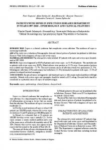
3.1.2. Hair Hypoplasia Hair hypoplasia is characteristic in CHH patients; fine, sparse and light colored hair of the scalp, eyebrows and eyelashes is evident at birth and throughout the life and the hair lacks a central pigmented core with an abnormally small caliber on microscopic examination (Figure 1). Body hair is also affected; hair may darken with age. The hair was normal in six patients (7%) of 108 Finnish patients (1, 10).
3.1.3. Joints and Skeleton Skeletal features include increased lumbar lordosis, ligamentous laxity, mild platyspondylia and mild scoliosis 2
Figure 1. An Iranian Patient; Hair of the Scalp, Eyebrows and Eyelashes is Light, Fine and Sparse
(that may cause arthralgic pains in knees, ankles or lumbar spine), bowing of the lower limbs and chest deformi-
ty (flaring of lower rib cage, narrow thorax, prominent sternum, Harrison’s grooves). Bow legs may necessitate corrective osteotomy (in 14% of the Finnish patients) (11). In 108 Finnish patients, increased ligamentous laxity was present in 95%, limited extension of the elbows in 92%, increased lumbar lordosis in 85%, thoracal deformity in 68%, genu varum in 63% and scoliosis in 21% of the patients (11).
3.1.4. Gastrointestinal Disorders There are several reports of Hirschsprung's disease in patients with CHH (1, 11, 15-22). Eight patients from 108 Finnish patients (7%) had Hirschsprung's disease and one patient had anal stenosis and one esophageal atresia (11). Primary malabsorption was not observed in any of 108 Finnish patients, although, gastrointestinal infection was confirmed in two patients with symptoms of malabsorption (11). In the report of Makitie et al. (1965) one of the 77 Amish CHH patients had celiac disease and in five other patients malabsorption was suspected (11). Therefore, primary malabsorption is only exceptionally associated with CHH; mimicking symptoms may result from an underlying gastrointestinal infection. Fryns et al. reported seven CHH patients with splenomegaly, hypersplenism and portal hypertension (23).
3.1.5. Anaemia Hypoplastic anemia is observed in most patients, usually as mild macrocytic or normocytic anemia in early childhood and often disappears by the age of 2-3 years. However, some patients showed severe anemia, which was permanent in more than 50% of patients (24). Macrocytic anemia reported in CHH patients is unrelated to vitamin B12 or folate deficiency (13, 15, 24-26). Anemia ocJ Pediatr Rev. 2015;3(1):e194
Shiasi Arani K curred in 79% of 108 Finnish patients, which was severe in 14 patients (16%) (hemoglobin value 3-7.5 g/dL) (11). Macrocytosis may be seen without anemia. The reticulocyte index was low considering the hemoglobin value (11). Routine bone marrow examination is not recommended. Williams et al. reported 12 CHH patients with severe anemia (defined as hemoglobin < 3.0 g/dL or a history of repeated blood transfusions). In 11 patients, anemia developed before five months of age; one patient developed aplastic anemia at 2.5 years of age, 10 patients had normocytic anemia, but two had macrocytic anemia, five patients had thrombocytosis. Three patients expired at ages 2 weeks, 3 months and 6 months, respectively. Spontaneous recovery was seen only in two patients, a 13-year-old girl and a 40-year-old man (24). Anemia is probably related to growth hormone parameter. Makitie et al. in a prospective study of 21 patients with CHH showed that hemoglobin level in these patients was correlated with height, insulin-like growth factor I and IGF-binding protein-3. Fetal hemoglobin was increased, parallel to decreased hemoglobin (27).
3.1.6. Autoimmune Hemolytic Anemia
At first Ashby et al. reported an 11-month-old CHH girl with purpura and thrombocytopenia, five days after a mild upper respiratory tract infection. The patient later developed autoimmune hemolytic anemia, jaundice and hepatosplenomegaly. The anemia had good response to corticosteroid therapy in addition with irradiated blood transfusion (28). Rider et al. reported an Amish CHH boy with autoimmune hemolytic anemia as a complication of BMT (29).
3.1.7. Granulomatous Inflammation
Moshous et al. described granulomatous inflammation as a new feature in patients with CHH. The patients had epithelioid cell granulomatous inflammation in the visceral organs and skin. TNF-α antagonists therapy had some therapeutic effects in their patients, but one patient had fatal progressive leukoencephalopathy (30). Also, skin granulomatous lesion with severe T cell immunodeficiency described as initial presentation in otherwise healthy child with short stature (31).
3.1.8. Impaired Spermatogenesis
Only one report is present about spermatogenesis in CHH patients. Makitie et al. evaluated eleven adult CHH males (21-49 years), all patients had abnormal semen analysis characterized by low sperm count, decreased motility and morphological changes. The testicular size was small in some patients, but the serum levels of gonadotropins, inhibin B and testosterone were normal. Neither patient complained of infertility or erectile dysfunction (32).
3.2. Radiographic Features
The long bones are short and thick for age with irregular J Pediatr Rev. 2015;3(1):e194
metaphyseal borders of the growth plates. The metaphyseal ends are widened, scalloped and irregularly sclerotic, often with cystic areas; the epiphyses are less affected (13). The metaphyseal changes are most evident in the knees and ankles; the hips are only mildly affected. Delayed ossification and trabeculation of the long bones are also characteristic findings on X-rays (1, 33). Flaring and irregularity of the ribs at costochondral junction and long fibula were reported; anterolateral chest deformity (like Harrison grooves) may be seen (1, 5). These radiographic features develop by the age of 6-9 months and are diagnostic. At adulthood with the closure of the epiphyseal plates, metaphyseal irregularities disappear but the ends remain somewhat flared and angulated. Hand and wrist X-rays may show rachitic changes and may be relatively normal. Makitie et al. analyzed 149 skeletal radiographic surveys of 82 Finnish CHH patients; skeletal age delay was observed in 14% of patients. One-fourth of patients had mild scoliosis (5). The spine shows few abnormalities, the vertebral bodies are usually normal and caudal widening of the interpediculate distances (though less obvious than normal) is present in most patients. Lumbar lordosis is increased (11). Glass et al. reported radiologic changes in four CHH children under the age of two years, they reported angulation of the entire sternum, a sign not previously described in CHH (in a 2-week-old girl who had short ribs with anterior flaring). They also reported widening of the atlantoaxial space in a 5-month-old girl (33). Bonafe et al. suggested a diagnostic tell-tale sign for CHH as cone-shaped epiphyses in the phalanges associated with metaphyseal chondrodysplasia (34).
3.2.1. Immune Deficiency Immune dysregulation in CHH has a wide spectrum of clinical manifestations including variable grades of immunodeficiency, autoimmune complications and malignancies. The immunodeficiency in CHH was first suspected because of attacks of severe varicella infection among the Amish CHH patients (1). Later, deficiency of cell-mediated immunity was confirmed, which showed decreased delayed hypersensitivity, mild to moderate lymphopenia and impaired in vitro responsiveness of lymphocytes to PHA and decreased delayed hypersensitivity (35-38). In a large series of 141 patients with primary immunodeficiency diseases due to defects in lymphocytes, only one patient had cartilage-hair hypoplasia (39). The immunodeficiency in CHH may be an isolated T-cell or B-cell immunodeficiency, or rarely combined B-cell and T-cell immunodeficiency (SCID). However, severity of immunodeficiency is variable and many CHH patients may live healthy.
3.2.2. Susceptibility to Infections Susceptibility to infections in most patients is limited. The infection problems are most frequent in early childhood but may persist to adult age. Patients with cartilage3
Shiasi Arani K hair hypoplasia are at higher risk of infections with opportunistic microorganisms, especially severe varicella infection, which is fatal in a few cases. McKusick et al. reported six cases of fatal varicella pneumonia in Amish patients (1). In one study, approximately 56% of 108 Finnish patients had increased susceptibility to infections (11). In another Finnish study, increased rate of infections was reported in 31% of 35 CHH patients during the preceding year of study, fifteen patients (43%) had a history of increased infections during the first years of life (40). The patients occasionally have infections with pathogens such as cytomegalovirus, poliovirus, vaccinia, P carinii and Candida species commonly observed in T-cell immunodeficiency. Increased susceptibility to bacterial sinopulmonary infections is also reported (40). Repeated pulmonary infection predisposes patients to bronchiectasis. Patients with bronchiectasis had more severe growth failure and more prevalence of humoral immunodeficiency than general patients with CHH (41). Diffuse dilated lymphoplasmacytic bronchiolitis with chronic obstructive symptoms were reported in three CHH children due to Haemophilus influenza or Streptococcus pneumonia infection with good response to long-term clarithromycin therapy (42).
3.2.3. Cellular Immunity Defective cellular immunity is characterized by mild to moderate lymphopenia, decreased delayed hypersensitivity and impaired in vitro responsiveness of lymphocytes to mitogen stimulation. Lymphopenia was present in 62% of 88 patients (1). Approximately 88% of 108 Finnish patients had defective cellular immunity (11). Makitie et al. analyzed lymphocyte subpopulations and proliferative responses in mitogen stimulation in 35 Finnish patients (12 males and 23 females). Low number of CD4+ cells was present in 57% of patients (which led to a decreased total count of T-lymphocytes in 52% and a subnormal CD4+/CD8+ cell ratio in 32%) and 36% had lymphopenia. B-lymphocyte count was normal. Natural killer cell count was higher than normal in 6/15 (40%) of patients. Lymphocyte stimulation indices (concanavalin A, phytohaemagglutinin and pokeweed mitogen) were subnormal. Total lymphocytes, T-lymphocytes, CD4+ cells and pokeweed mitogen stimulation indexes were inversely correlated with rate of infection. Six patients of 35 Finnish CHH patients (6%) died because of primary infections. SO common indexes of cellular immune function poorly predict the clinical outcome of patients. They suggested to follow up all CHH patients carefully because of the risk of serious infections and malignancies (40). Trojak et al. evaluated 18 old Amish CHH patients and nine unaffected sibs. None of the subjects had a history suggestive of persistent immune dysfunction. Patients with CHH had significantly lower lymphocyte mitogenic and allogeneic cell stimulation responses compared with unaffected sibs and unrelated control subjects (similar to those reported in Finnish CHH patients) (37). 4
Lux et al. reported two CHH patients with recurrent respiratory-tract infections and severe varicella infection. Serum immunoglobulin levels were normal or elevated and patients were able to synthesize antibodies to a wide variety of viral and bacterial antigens. One child had chronic neutropenia secondary to a failure of myeloid maturation. Both children had diminished delayed skin hypersensitivity, persistent lymphopenia and diminished in vitro responsiveness of their lymphocytes. One child had delayed rejection of a skin allograft (35). Polmar and Pierce described marked impairment of T-cell function due to an intrinsic defect in cell proliferation in patients with CHH. Since defective proliferation was showed in B cells and fibroblasts, they concluded that the defect in cell proliferation is generalized. Accordingly, proliferation-independent natural killer activity was normal (15).
3.2.4. Humoral Immunity Humoral immunity may be normal or impaired in CHH and contributes to increased susceptibility to infections. In one report of 16 patients, none of them had subnormal levels of IgG, IgA or IgM. Most patients had normal number of B-cells (40). However in another study of 25 patients with CHH (five patients with recurrent infections), seven (35%) had defective humoral immunity. One patient had severe hypogammaglobulinemia and three had multiple IgG subclass deficiencies. Three patients had IgA deficiency. IgG4 was low in most patients. Increased rate of infections was associated with supranormal IgG and IgG1 and subnormal IgA, IgG2, or IgG4 concentrations (29). Kawasaki et al. reported a one-year-old boy patient with recurrent upper respiratory infections. He had impaired cellular immunity and a selective IgG2 deficiency (43). Matesic et al. reported a 39-year-old man with CHH with a history of recurrent respiratory infections, allergies and chronic cough. He experienced chronic sinusitis, recurrent pneumonias and central bronchiectasis. He had moderate lymphopenia with decreased counts in all subpopulations except for natural killer cells. Mitogen-induced blastogenesis was diminished. Natural killer cell and neutrophil function were normal. Their patient had a profound humoral deficiency with remarkably low IgG2, IgG3 and IgG4 subclass and a high-normal total IgG level. His response to pneumococcal polysaccharide was extremely limited (44).
3.2.5. Neutropenia
Neutropenia is common in patients with CHH, which occurred in 24% of 88 patients (25). The main mechanism is maturation arrest, but autoimmune neutropenia is also reported. The severity of neutropenia and anemia usually decreases with time; cyclic neutropenia was seen in patients with CHH (11, 23, 35).
3.2.6. Severe Combined Immunodeficiency
Cartilage-hair hypoplasia is a rare cause of severe combined immunodeficiency (SCID). Children with cartilageJ Pediatr Rev. 2015;3(1):e194
Shiasi Arani K hair hypoplasia and SCID have a greater susceptibility to overwhelming and opportunistic infections and graft versus host disease (43). Guggenheim et al. reported three CHH patients with SCID. All three patients had profound lymphopenia, profoundly depressed mitogenic responses to PHA and markedly reduced number of CD3T cells. The number of B lymphocytes was normal; natural killer (NK) cells were normal in two patients and reduced in one patient. The humoral immune system was affected in these patients. One patient had no detectable IgM and IgA, but normal IgG. The second patient had no IgA and low normal Immunoglobulin and IgM levels and the third patient had panhypogammaglobulinemia. These patients were effectively treated by bone marrow transplantation and were alive and well 5-20 years after BMT (45). Kainulainen et al. reported two patients with CHH with a prolonged viral infection due to combined T cell and B cell immunodeficiency; interestingly hypogammaglobulinemia disappeared during the first years of life (46).
3.2.7. Increased Risk of Cancer Patients with CHH are at increased risk of cancer. Reported cancers include skin neoplasm (the most common is basal cell carcinoma), non-Hodgkin and Hodgkin lymphoma, leukemia, intestinal lymphosarcoma, malignant testicular tumor, epithelial carcinoma of the vocal cord, bile duct carcinoma and ocular cancer (11, 47, 48). Makitie et al. reported results of a cohort study on 122 Finnish patients with CHH identified through two epidemiologic surveys in 1974 and 1986. Their parents and healthy siblings were also included. This cohort underwent follow-up for cancer incidence to the end of 1995. The standardized incidence ratio (SIR) of cancer among patients with CHH was 6.9 (95% CI 2.3 to 16), mainly attributable to non-Hodgkin’s lymphoma and basal cell carcinoma. The cancer incidence in their parents or siblings was not different from the average cancer incidence in the Finnish population (49). Taskinen et al. published the result of a cohort study of 123 Finnish patients with CHH (51 males), which were followed regarding malignancy. Mean follow-up time was 19.2 years. The number of identified cancers in patients with CHH was compared with expected number of cancer in general population. In CHH group, 14 patients with cancer were diagnosed (expected number 2.0; SIR 7.0, CI 3.8-12). The most frequent cancer was non-Hodgkin lymphoma followed by squamous cell carcinoma, Hodgkin lymphoma and leukemia. Nine of 14 cancers (64%) were diagnosed in patients younger than 45 years. In addition, 10 patients had basal cell carcinoma of the skin (standardize incidence ratio, 33.2) (50).
3.3. Mortality
Makitie et al. performed a cohort of 120 patients with CHH (50 men and 70 women) to assess mortality from 1971 to 1995. The mean length of follow-up was 11.4 years. During the follow-up, seven disease related deaths were J Pediatr Rev. 2015;3(1):e194
observed, three deaths due to pneumonia (aged 3 weeks, 2 and 8 years); two deaths due to other infections; septicemia by Candida albicans (aged one year) and encephalitis (aged 13 years) and finally two deaths due to non-Hodgkin’s lymphoma (aged 22 and 45 years). The disease mortality in ages 0-14 was 21-fold the mortality of the Finnish population of the same age (51).
3.4. Management 3.4.1. Short Stature
Few and conflicting results are available regarding the efficacy of treatment with growth hormone (GH) in children with CHH (52-54). A 3-year-old boy was treated with GH for seven years and underwent a leg-lengthening surgical procedure; height standard deviations (SD) improved from -4.2 to -2.1 (53). In another report, GH was used to treat four patients. The duration of GH therapy was 2-6.5 years. Slight improvement of growth rate was reported during the first year of treatment, varying from 0.2-0.8 SD, but the growth was not sustained, and no increase in final height was achieved (54). Surgical bone lengthening may be considered. However, the risk of infection in patients with CHH is increased and extra attention to prevention and treatment of infections is necessary.
3.4.2. Hair Hypoplasia There is no known treatment for hair hypoplasia in these patients.
3.4.3. Joints and Skeleton Skeletal problems must be managed with physiotherapy and appropriate orthopedic interventions. Bone marrow transplantation had no effect on chondrodysplasia (45).
3.4.5. Gastrointestinal Disorders Hirschsprung’s disease, anal stenosis and oesophageal atresia should be surgically corrected. Malabsorption, diarrhea and failure to thrive require symptomatic treatment.
3.4.6. Anemia Patients with severe hypoplastic anemia (6% of patients) require repeated transfusions. Few patients may need lifelong regular transfusions or bone marrow transplantation (24). Chronic blood transfusions can lead to iron overload and requirement for chelation therapy. There is a report suggesting well toleration of deferoxamine, deferiprone and deferasirox in patients with CHH. Chelation therapy would prepare patients for hematopoietic stem cell transplantation (55). Bone marrow cultures in six patients with CHH showed reduced or absence of ery5
Shiasi Arani K throid colony formation, which was not affected by GH treatment in vivo or by GH or IGF-I in vitro (27).
3.4.7. Vaccination In patients with impaired cellular immunity, immunization with live vaccines is contraindicated (9). The use of varicella vaccine for prophylaxis should be avoided (56). However, an attenuated live polio vaccine might have risks of severe disease in a patient with CHH (57). Reactivation of varicella-zoster virus from latency causes zoster and is common among recipients of hematopoietic cell transplants, e.g. those performed for Hodgkin and non-Hodgkin lymphomas. Hata et al. found that inactivated varicella vaccine given before hematopoietic cell transplantation and during the first 90 days thereafter reduced the risk of zoster. This finding had possible relevance to the use of inactivated varicella vaccine in CHH (58).
3.4.8. Immunodeficiency Treatment of immunodeficiency depends on the presence and type of immunodeficiency. In CHH patients with isolated T-cell immunodeficiency, varicella is the most common infection that may severe and lifethreatening. Prophylaxis with acyclovir, varicella-zoster immune globulin (VZIG), or both can be administered in patients exposed to varicella; acyclovir is also recommended in the treatment of varicella infections (57). Leukocyte interferon may be useful in CHH children with varicella, because of benefit in immunosuppressed children with cancer (59). For patients with humoral immunodeficiency and recurrent bacterial infections, antibody replacement therapy is indicated (9). Treatment with granulocyte colony-stimulating factor (G-CSF) is useful for neutropenia in patients with cartilage-hair hypoplasia (44). Ammann et al. reported a 6-year-old patient with CHH and chronic neutropenia who had recurrent lower respiratory tract infections with good response to longterm treatment with granulocyte-colony stimulating factor (G-CSF). Growth was not affected by treatment (60). In patients with severe T-cell or combined immunodeficiency, BMT has been recommended (45). There are multiple reports indicating successes of BMT for cartilage-hair hypoplasia (45, 61). The need for BMT in patients with CHH is low as most individuals have mild to moderate immune deficiency. However, in patients with severe immunodeficiency BMT should be considered before the development of severe infections or malignancy. Bordon et al. reported the largest experience about BMT in patients with CHH. They performed allogeneic hematopoietic stem cell transplantation (HSCT) for 16 patients with CHH (13 patients at early childhood (a mean of 2.5 years) and 3 adolescent patients). Ten patients (62.5%) were followed for a median of seven years. In all survivors, autoimmunity was resolved and T-lymphocyte function and numbers were normalized (61). 6
3.4.9. Malignancies Patients with CHH should be monitored closely for developing malignancy such as skin neoplasms, lymphomas and leukemias.
4. Conclusions
Cartilage-hair hypoplasia is a rare but important hereditary disease with different medical aspects. The high rate of consanguineous marriages in Iran necessitates considering CHH in any child with severe short stature associated with hair hypoplasia and metaphyseal chondrodysplasia. These patients are at increased risk of anemia, immunodeficiency, propensity to infections, GI dysfunction (Hirschsprung’s disease) and malignancies. Each problem must be well recognized and appropriately managed.
Acknowledgements
I am thankful to Dr Ghasemi seyed Emadoddin for aid to diagnosis first case of cartilage hair hypoplasia in Iran.
References 1. 2. 3. 4.
5. 6. 7.
8. 9.
10. 11. 12. 13.
14. 15.
McKusick VA, Eldridge R, Hostetler JA, Ruangwit U, Egeland JA. DWARFISM IN THE AMISH. II. CARTILAGE-HAIR HYPOPLASIA. Bull Johns Hopkins Hosp. 1965;116:285–326. Makitie O. Cartilage-hair hypoplasia in Finland: epidemiological and genetic aspects of 107 patients. J Med Genet. 1992;29(9):652–5. Hermanns P, Tran A, Munivez E, Carter S, Zabel B, Lee B, Leroy JG, et al. RMRP mutations in cartilage-hair hypoplasia. Am J Med Genet A. 2006;140(19):2121–30. Ridanpaa M, van Eenennaam H, Pelin K, Chadwick R, Johnson C, Yuan B, vanVenrooij W, Pruijn G, Salmela R, Rockas S, Mäkitie O, Kaitila I, de la Chapelle A, et al. Mutations in the RNA component of RNase MRP cause a pleiotropic human disease, cartilage-hair hypoplasia. Cell. 2001;104(2):195–203. Mäkitie O, Marttinen E, Kaitila I. Skeletal growth in cartilage-hair hypoplasia. A radiological study of 82 patients. Pediatr Radiol. 1992;22(6):434–9. Shiasi Arani K. Cartilage Hair Hypoplasia: First report from Iran. Med J Islam Repub Iran. 2013;27(3):157–60. Kwan A, Manning MA, Zollars LK, Hoyme HE. Marked variability in the radiographic features of cartilage-hair hypoplasia: case report and review of the literature. Am J Med Genet A. 2012;158A(11):2911–6. Reicherter K, Veeramani AI, Jagadeesh S. Cartilage-hair hypoplasia caused by novel compound heterozygous RMRP mutations. Indian Pediatr. 2011;48(7):559–61. Thiel CT. Cartilage-Hair Hypoplasia - Anauxetic Dysplasia Spectrum Disorders. In: Pagon RA, Adam MP, Ardinger HH, Bird TD, Dolan CR, Fong CT, Smith RJH, Stephens K, et al editors. GeneReviews(R).. Seattle WA: University of Washington, Seattle; 1993. Makitie O, Perheentupa J, Kaitila I. Growth in cartilage-hair hypoplasia. Pediatr Res. 1992;31(2):176–80. Mäkitie O, Kaitila I. Cartilage-hair hypoplasia--clinical manifestations in 108 Finnish patients. Eur J Pediatr. 1993;152(3):211–7. Savage MO. Metaphyseal dysplasia in siblings: a variant of cartilage-hair hypoplasia. Proc R Soc Med. 1972;65(8):727. van der Burgt I, Haraldsson A, Oosterwijk JC, van Essen AJ, Weemaes C, Hamel B. Cartilage hair hypoplasia, metaphyseal chondrodysplasia type McKusick: description of seven patients and review of the literature. Am J Med Genet. 1991;41(3):371–80. Makitie O, Kaitila I. Growth in diastrophic dysplasia. J Pediatr. 1997;130(4):641–6. Polmar SH, Pierce GF. Cartilage hair hypoplasia: immunological
J Pediatr Rev. 2015;3(1):e194
Shiasi Arani K
16. 17.
18. 19. 20. 21. 22. 23.
24. 25. 26. 27.
28. 29.
30.
31.
32. 33. 34.
35.
36.
aspects and their clinical implications. Clin Immunol Immunopathol. 1986;40(1):87–93. Lischka A, Frisch H, Weissenbacher G. [Radiologic changes in metaphyseal chondrodystrophy of the McKusick type (cartilagehair hypoplasia)]. Monatsschr Kinderheilkd. 1984;132(7):550–3. Wilson WG, Aylsworth AS, Folds JD, Whisnant JK. Cartilage-hair hypoplasia (metaphyseal chondrodysplasia, type McKusick) with combined immune deficiency: variable expression and development of immunologic functions in sibs. Birth Defects Orig Artic Ser. 1978;14(6A):117–29. Harris RE, Baehner RL, Gleiser S, Weaver DD, Hodes ME. Cartilagehair hypoplasia, defective T-cell function, and Diamond-Blackfan anemia in an Amish child. Am J Med Genet. 1981;8(3):291–7. Roberts PA, Mann TP, Rubin J. Hirschsprung's disease associated with a variant form of achondroplasia, in sister and brother. Proc R Soc Med. 1969;62(4):329. Fauchier C, Regy JM, Combe P. [Familial dystrophic nanism with Hirschsprung's disease]. Ann Pediatr (Paris). 1969;16(8):496–502. Boothby CB, Bower BD. Cartilage hair hypoplasia. Arch Dis Child. 1973;48(11):918–21. le Merrer M, Briard ML, Chauvet ML, Maroteaux P. [Autosomal recessive metaphyseal chondrodysplasia and Hirschsprung's disease]. Ann Pediatr (Paris). 1991;38(1):27–30. Fryns JP. Hypersplenism and portal hypertension with vena porta thrombosis in cartilage-hair hypoplasia (metaphyseal chondrodysplasia, McKusick type, MIM *250250). Genet Couns. 2000;11(3):277–8. Williams MS, Ettinger RS, Hermanns P, Lee B, Carlsson G, Taskinen M, Mäkitie O, et al. The natural history of severe anemia in cartilage-hair hypoplasia. Am J Med Genet A. 2005;138(1):35–40. Mäkitie O, Rajantie J, Kaitila I. Anaemia and macrocytosis--unrecognized features in cartilage-hair hypoplasia. Acta Paediatr. 1992;81(12):1026–9. Sacrez R, Levy JM, Godar G, Castanier J. [Blackfan-diamond anemia associated with multiple malformations]. Med Infant (Paris). 1965;72(7):493–9. Makitie O, Juvonen E, Dunkel L, Kaitila I, Siimes MA. Anemia in children with cartilage-hair hypoplasia is related to body growth and to the insulin-like growth factor system. J Clin Endocrinol Metab. 2000;85(2):563–8. Ashby GH, Evans DI. Cartilage hair hypoplasia with thrombocytopenic purpura, autoimmune haemolytic anaemia and cellmediated immunodeficiency. J R Soc Med. 1986;79(2):113–4. Rider NL, Morton DH, Puffenberger E, Hendrickson CL, Robinson DL, Strauss KA. Immunologic and clinical features of 25 Amish patients with RMRP 70 A-->G cartilage hair hypoplasia. Clin Immunol. 2009;131(1):119–28. Moshous D, Meyts I, Fraitag S, Janssen CE, Debre M, Suarez F, Toelen J, De Boeck K, Roskams T, Deschildre A, Picard C, Bodemer C, Wouters C, Fischer A, et al. Granulomatous inflammation in cartilage-hair hypoplasia: risks and benefits of anti-TNF-alpha mAbs. J Allergy Clin Immunol. 2011;128(4):847–53. McCann LJ, McPartland J, Barge D, Strain L, Bourn D, Calonje E, Verbov J, Riordan A, Kokai G, Bacon CM, Wright M, Abinun M, et al. Phenotypic variations of cartilage hair hypoplasia: granulomatous skin inflammation and severe T cell immunodeficiency as initial clinical presentation in otherwise well child with short stature. J Clin Immunol. 2014;34(1):42–8. Mäkitie OM, Tapanainen PJ, Dunkel L, Siimes MA. Impaired spermatogenesis: an unrecognized feature of cartilage-hair hypoplasia. Ann Med. 2001;33(3):201–5. Glass RB, Tifft CJ. Radiologic changes in infancy in McKusick cartilage hair hypoplasia. Am J Med Genet. 1999;86(4):312–5. Bonafe L, Schmitt K, Eich G, Giedion A, Superti-Furga A. RMRP gene sequence analysis confirms a cartilage-hair hypoplasia variant with only skeletal manifestations and reveals a high density of single-nucleotide polymorphisms. Clin Genet. 2002;61(2):146–51. Lux SE, Johnston RJ, August CS, Say B, Penchaszadeh VB, Rosen FS, McKusick VA, et al. Chronic neutropenia and abnormal cellular immunity in cartilage-hair hypoplasia. N Engl J Med. 1970;282(5):231–6. Ranki A, Perheentupa J, Andersson LC, Hayry P. In vitro T- and B-cell reactivity in cartilage hair hypoplasia. Clin Exp Immunol.
J Pediatr Rev. 2015;3(1):e194
37.
38. 39. 40. 41. 42. 43. 44. 45.
46. 47. 48. 49. 50. 51.
52. 53.
54. 55. 56.
57.
58.
59.
1978;32(2):352–60. Trojak JE, Polmar SH, Winkelstein JA, Hsu S, Francomano C, Pierce GF, Scillian JJ, Gale AN, McKusick VA, et al. Immunologic studies of cartilage-hair hypoplasia in the Amish. Johns Hopkins Med J. 1981;148(4):157–64. Virolainen M, Savilahti E, Kaitila I, Perheentupa J. Cellular and humoral immmunity in cartilage-hair hypoplasia. Pediatr Res. 1978;12(10):961–6. Buckley RH. Primary immunodeficiency diseases due to defects in lymphocytes. N Engl J Med. 2000;343(18):1313–24. Mäkitie O, Kaitila I, Savilahti E. Susceptibility to infections and in vitro immune functions in cartilage-hair hypoplasia. Eur J Pediatr. 1998;157(10):816–20. Toiviainen-Salo S, Kajosaari M, Piilonen A, Makitie O. Patients with cartilage-hair hypoplasia have an increased risk for bronchiectasis. J Pediatr. 2008;152(3):422–8. Bailly-Botuha C, Jaubert F, Taam RA, Galmiche L, Picard C, Bellon G, de Blic J, et al. Diffuse lymphoplasmacytic bronchiolitis in cartilage-hair hypoplasia. J Pediatr. 2008;152(3):429–33. Kawasaki H, Kohdera U, Taniuchi S, Kobayashi Y. Cartilage-hair hypoplasia associated with IgG2 deficiency. Acta Paediatr Jpn. 1995;37(6):703–5. Matesic D, Hagan JB. Cartilage-hair hypoplasia. Mayo Clin Proc. 2007;82(6):655. Fryns JP. Hypersplenism and portal hypertension with vena porta thrombosis in cartilage-hair hypoplasia (metaphyseal chondrodysplasia, McKusick type, MIM *250250). Genet Couns. 2000;11(3):277–8. Guggenheim R, Somech R, Grunebaum E, Atkinson A, Roifman CM. Bone marrow transplantation for cartilage-hair-hypoplasia. Bone Marrow Transplant. 2006;38(11):751–6. Kainulainen L, Lassila O, Ruuskanen O. Cartilage-hair hypoplasia: follow-up of immunodeficiency in two patients. J Clin Immunol. 2014;34(2):256–9. Castori M, Morrone A, Kanitakis J, Grammatico P. Genetic skin diseases predisposing to basal cell carcinoma. Eur J Dermatol. 2012;22(3):299–309. Taskinen M, Makitie O. [Cartilage-hair hypoplasia--much more than growth problem]. Duodecim. 2011;127(3):273–9. Makitie O, Pukkala E, Teppo L, Kaitila I. Increased incidence of cancer in patients with cartilage-hair hypoplasia. J Pediatr. 1999;134(3):315–8. Taskinen M, Ranki A, Pukkala E, Jeskanen L, Kaitila I, Makitie O. Extended follow-up of the Finnish cartilage-hair hypoplasia cohort confirms high incidence of non-Hodgkin lymphoma and basal cell carcinoma. Am J Med Genet A. 2008;146A(18):2370–5. Makitie O, Pukkala E, Kaitila I. Increased mortality in cartilagehair hypoplasia. Arch Dis Child. 2001;84(1):65–7. Obara-Moszynska M, Wielanowska W, Rojek A, Wolnik-Brzozowska D, Niedziela M. Treatment of cartilage-hair hypoplasia with recombinant human growth hormone. Pediatr Int. 2013;55(6):e162– 4. Harada D, Yamanaka Y, Ueda K, Shimizu J, Inoue M, Seino Y, Tanaka H, et al. An effective case of growth hormone treatment on cartilage-hair hypoplasia. Bone. 2005;36(2):317–22. Bocca G, Weemaes CM, van der Burgt I, Otten BJ. Growth hormone treatment in cartilage-hair hypoplasia: effects on growth and the immune system. J Pediatr Endocrinol Metab. 2004;17(1):47–54. Taskinen M, Toiviainen-Salo S, Lohi J, Vuolukka P, Grasbeck M, Makitie O. Hypoplastic Anemia in Cartilage-Hair HypoplasiaBalancing between Iron Overload and Chelation. J Pediatr. 2013;162(4):844–9. Rubin LG, Levin MJ, Ljungman P, Davies EG, Avery R, Tomblyn M, Bousvaros A, Dhanireddy S, Sung L, Keyserling H, Kang I, Infectious Diseases Society of A, et al. 2013 IDSA clinical practice guideline for vaccination of the immunocompromised host. Clin Infect Dis. 2014;58(3):309–18. Hardy I, Gershon AA, Steinberg SP, LaRussa P. The incidence of zoster after immunization with live attenuated varicella vaccine. A study in children with leukemia. Varicella Vaccine Collaborative Study Group. N Engl J Med. 1991;325(22):1545–50. Hata A, Asanuma H, Rinki M, Sharp M, Wong RM, Blume K, Arvin AM, et al. Use of an inactivated varicella vaccine in recipients of
7
Shiasi Arani K 60. 61.
8
hematopoietic-cell transplants. N Engl J Med. 2002;347(1):26–34. Arvin AM, Kushner JH, Feldman S, Baehner RL, Hammond D, Merigan TC. Human leukocyte interferon for the treatment of varicella in children with cancer. N Engl J Med. 1982;306(13):761–5. Ammann RA, Duppenthaler A, Bux J, Aebi C. Granulocyte colonystimulating factor-responsive chronic neutropenia in cartilagehair hypoplasia. J Pediatr Hematol Oncol. 2004;26(6):379–81.
62.
Bordon V, Gennery AR, Slatter MA, Vandecruys E, Laureys G, Veys P, Qasim W, Friedrich W, Wulfraat NM, Scherer F, Cant AJ, Fischer A, Cavazzana-Calvo M, Bredius RG, Notarangelo LD, Mazzolari E, Neven B, Gungor T, Inborn Error Working Party of the European Bone Marrow Transplantation G, et al. Clinical and immunologic outcome of patients with cartilage hair hypoplasia after hematopoietic stem cell transplantation. Blood. 2010;116(1):27–35.
J Pediatr Rev. 2015;3(1):e194