. 301 .
Chinese Journal of Traumatology 2008; 11(5):301-305
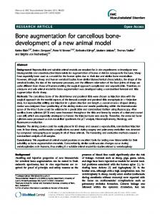
Clinical observation of particulate cancellous bone impaction grafting in combination with total hip arthroplasty for acetabular reconstruction LIU Xian-zhe 刘先哲, YANG Shu-hua 杨述华*, XU Wei-hua 许伟华, LIU Guo-hui 刘国辉, YANG Cao 杨操, LI Jin 李进, YE Zhe-wei 叶哲伟, LIU Yong刘勇 and ZHANG Yu-kun 张宇坤 Objective: To investigate the effect of particulate cancellous bone impaction grafting in combination with total hip arthroplasty (THA) for acetabular reconstruction in patients with posttraumatic arthritis and bone loss after acetabular fractures. Methods: Totally 15 consecutive cases with unilateral acetabular fracture were treated with bone impaction grafting in combination with THA in our department. There were 10 males and 5 females with mean age of 48.2 years (ranging from 36 to 73 years). Eight cases had the fracture at left hips, 7 at right hips. The average age at injury was 28 years (ranging from 18 to 68 years). The mean follow-up period was 4.3 years (ranging from 2 to 7 years). Results: Compared with mean 42 points (ranging from 10 to 62) of the preoperative Harris score, the survival cases at the final follow-up had mean 84 points (ranging from 58 to 98). One patient had mild pain in the hip. No revision of the acetabular or femoral component was undertaken during
the follow-up. Normal rotational centre of most hips was recovered except 2 cases in which it was 0.8 mm higher than that in opposite side. All of them had a stable radiographic appearance. Progressive radiolucent lines were observed in I, III zones in 2 cases. One patient had a nonprogressive radiolucent line in zone III. The cup prosthesis was obviously displaced (6 mm) in one patient, but had not been revised. Conclusion: Particulate cancellous bone impaction grafting in combination with THA as a biological solution is an attractive procedure for acetabular reconstruction in patients with posttraumatic arthritis and bone loss after acetabular fracture, which can not only restore acetabular bone stock but also repair normal hip anatomy and its function. Key words: Reconstructive surgical procedures; Arthroplasty, replacement, hip; Bone transplantation Chin J Traumatol 2008; 11(5):301-305
A
cetabular fracture is a kind of complicated fracture caused by high energy injury, which can lead to the permanent disability. The outcomes of conservative treatment and open-reduction internal fixation are not satisfactory. Traumatic arthritis, femoral head necrosis and ectopic ossification may occur in advanced stage and influence patients’ joint function. So it is necessary to perform total hip arthroplasty (THA).1, 2 However, traumatic arthritis is always accompanied by bone loss and deformity of acetabular bone so that it can effect the clinical outcome of THA and result in the failure of surgery.3-5 Particulate morselized bone impacDepartment of Orthopedics, Union Hospital, Tongji Medical College, Huazhong University of Science and Technology, Wuhan 430022, China (Liu XZ, Yang SH, Xu WH, Liu GH, Yang C, Li J, Ye ZW, Liu Y and Zhang YK) *Corresponding author: Tel:86-27-85351689, E-mail:
[email protected]
tion grafting combined with THA for acetabular reconstruction is benefitial for acetabular recovery, meanwhile it can restore anatomical and functional activity by filling the defect of bone. 6 This article retrospectively analyzed 15 cases of particulate morselized bone impaction grafting in combination with THA to discuss operative procedures and therapeutical effect.
METHODS General data From 1998 to 2006, we had performed particulate cancellous bone impaction grafting in combination with THA in 15 patients with posttraumatic arthritis secondary to acetabular fracture, including 10 males and 5 females, with mean age of 28 years (ranging from 18 to 68 years) at the time of injury and 48.2 years (ranging from 36 to 73 years) at the time of operation. Three cases were conservatively treated and the other 12 were
. 302 .
Chinese Journal of Traumatology 2008; 11(5):301-305
treated by open-reduction internal fixation. There were 12 cases of traffic accident injury and 3 falling injury. According to American orthopaedic association standard, 7 there were 3 cases of typeⅠ, 6 typeⅡ, 5 typeⅢ and 1 type V. Ten patients used bone cement femoral prosthesis and the other 5 adopted uncemented femoral prosthesis. Surgical technique Lateral operative approach was applied in all patients in order to thoroughly expose acetabulum and proximal part of femur. Anterior superior iliac spine was exposed to broaden the incision and show anterior structure of acetabular joint. During operation, it was necessary to identify greater and lesser trochanters, tendons of gluteus maximus, inferior boarder of gluteus medius and gluteus minimus to avoid damaging sciatic nerve. Eliminate surrounding scar tissue completely, loosen contractual soft tissue and scrape proliferative fibrous tissue on acetabular wall by curette. Surgeons should pay attention to the ligamentum transversum on lower boarder of acetabulum, which is an important marker of reconstruction in acetabular biomechanics.
Fig.2. Diagram of particulate cancellous bone impaction grafting in combination with THA for acetabular reconstruction.
Fig. 3. A. The acetanulum had severe fracture and dislocation
All particulate cancellous bone was autogeneic (Fig.1). We cut femoral head in 9 cases, iliac bone in 1 case and both parts of bone in 5 cases. Remove the cartilage on the surface of femoral head, divide it to 4 pieces and make them into 1 mm3 blocks by rongeur forceps. During operation, check the defect of acetabulum carefully and use hammer and compressor to impact spaces after washing. At the last time of impaction grafting, the diameter of compressors or models should be 4 mm longer than that of acetabular cup (Fig. 2). Three cases with obvious defect of acetabular cups were fixed by antibiotic cement and titanium net. The acetabulum was fixed biologically in other 12 cases (Fig. 3. A and B).
Fig.1. Particulate cancellous bone for impaction grafting.
before operation.
Fig.3. B. The patient had good bone healing and recovery of normal anatomic structure 6 weeks after operation.
Postoperative management Dependent on their own conditions, the patients were allowed to do some activities after surgery. Generally speaking, at 24 hours after operation, patients could begin passive exercise. Six weeks later, they could get out of bed to have partial weight loading exercise and the amount of exercise should be gradually increased. Oral anticoagulation drugs were administrated routinely after operation to prevent deep venous thrombosis in lower extremities.
. 303 .
Chinese Journal of Traumatology 2008; 11(5):301-305
Follow-up All 15 patients had been followed up. The therapeutic effect were evaluated in the 3rd, 6th, 12th month and every 12 months afterwards, including clinical evaluation and radiographic evaluations. Clinical evaluation contained Harris score, pain evaluation, dependence on ambulatory aid and patients’ satisfaction. Any circumstance that needed the revision of acetabular prosthesis were regarded as failure in clinical evaluation. We observed the growing of particulate bone according to anterior posterior X-ray film and Conn’s theory of proliferating pattern of bone graft.8 With the triangle principle described by DeLee & Charnley,9 we could measure the width between acetabular cup and bone graft. It was defined as radiographic failure if positive lucent band appeared in three areas of acetabulum or prosthesis moved no less than 5 mm in any direction.10
RESULTS One patient was dead 3 years later, but the death was not related to operation. Other patients were satisfied with the clinical effect of operation. Compared with average 42 points (ranging from 10 to 62) of preoperative Harris score, the survived patients had a mean HHS of 84 points (ranging from 58 to 98) at the final follow-up. Only one patient complained about joint pain and felt relieved after taking non-steroidal anti-inflammatory drugs. No revision was carried out during the period of follow-up. In postoperative radiologic evaluation, there was the same bone density between bone graft and host bone tissue with formation of continuous trabecula. Area I and III were progressively widened in 2 cases and lucent band in area III was discovered in 1 case 6 months after surgery without aggravation. Seven years after operation, a patient was found with prosthesis of acetabular cup displaced for about 6 mm, progressively widened lucent bands in area I & III and Harris score of 95 points. Although this patient should be defined as failure in radiographic evaluation, the operation of revision was not essential. In most cases, the hip joints’ rotation centers became normal except 2 cases, in which the center was 0.8 mm higher than that in opposite side. We observed the complications as follows: ectopic
ossification (n=3), sciatic nerve dysfunction (n=1), bone dislocation (n=1) and deep venous thrombosis of lower extremity (n=1). After decompression of surrounding swelling tissue, the nerve function had been restored.
DISCUSSION Open-reduction internal fixation is the standard method to treat acetabular displacement, which may be associated with traumatic arthritis, necrosis of femoral head and ectopic ossification in advanced stage. Traumatic arthritis can lead to the defect and malformation of acetabulum. So we chose THA to reserve the function of joint but the therapeutic effect was not significant because of bone defect and arthritis. In this circumstance, we must pay much attention to acetabular defects and do the reconstruction well so that the normal anatomic structure and functional activity can be restored. There are many ways of acetabulum reconstruction. In biomechanics, when we do bone grafting in the location of defects, the speed of recovery in internal part is much slower than that in external part and sometimes it is constricted in superficial contact surface.11, 12 It may be attributed to down-regulation of antigenic substances in process of bone healing. Particulate cancellous bone impaction grafting in combination with THA can optimally repair the defect of acetabulum, reconstruct hip joint and gain long-term stability. It has been reported that the patients with rheumatoid arthritis associated with acetabular defects can have better long-term therapeutic effect if they undergo bone grafting.13, 14 The patients who have acetabular fracture internal fixation secondary to bone defect obtain satisfactory long-term results in the follow-up and the rates of complications and revision are low. Particulate cancellous bone impaction grafting is a biological reconstruction method for treatment of the acetabular defect. Firstly, particulate cancellous bone is so small that it can fit the tiny gap between area of defect and bone bed, which is benefitial for fusion of host bone and transplanted bone. Secondly, impacted particulate cancellous bone becomes much more consistent and its surface is attached to cement well. It compresses cement into spaces of particulate cancellous bone to enhance the initial stability of prosthesis and neotrebacula growing between cement and graft.15,16 There exists disputes on whether cement can grow into cancellous bone. Lots of experiments show that it can
. 304 .
achieve integration finally. Roffman et al.17 found that there was newly-formed bone growing into bone graft and surface of cement. Schimmel16 has found that transplanted bone could be attached closely to surrounding bone bed 3 weeks after operation and the pannus could penetrate into particulate cancellouse bone to form new trabecula according to a series of researches on goats’ acetabulum models. Using cement for fixation was not suitable because sclerosed bone might inhabit its combination.18 In cases that particulate cancellous bone impaction grafting in combination with cement-type acetabular cup reconstruction were adopted, there was the same bone density between bone graft and host bone tissue with formation of continuous trabecula. Areas I and III were progressively widened in 2 cases and lucent band in area III was observed without aggravation in 1 case. The hip joints’ rotation centers became normal except 2 cases in which it was 0.8 mm between two side centers. Seven years after operation, a patient had prosthesis of acetabular cup displaced for about 6 mm, progressively widened lucent bands in area I & III and Harris score of 95 points. Although this patient should be defined as failure case in radiographic evaluation, his clinical symptom was not obvious. Radiographic follow-up showed that most cases had stable changes in radiology and this kind of operation approach was feasible and effective. No patients had acetabular revision except 1 case of death 3 years after operation. Other patients were satisfied with the clinical effect of operation. Compared with average 42 points (ranging from 10 to 62 points) of the preoperative Harris score, the surviving cases at the final follow-up had mean 84 points (ranging from 58 to 98 points). Only one patient complained about joint pain. Selection of femoral prosthesis is dependent on patients’ age and bone condition. Generally speaking, we chose uncemented prosthesis for young patients and vice versa. Romness and Lewallen19 reported that during mean 7.5-year follow-up, 55 patients received THA with cement prosthesis after acetabular fracture, with 52.9% loosening rate and 13.7% revision rate. Pritchett and Bortel20 reported that the results of THA in 19 cases of acetabular central dislocation were satisfactory. Harris score increased from 42 to 84 points and no patients had revision and any complications like infection, ectopic ossification and dislocation. Weber et al.21 has reported that the clinical results of THA in 66 cases of acetabular fracture secondary to traumatic arthritis were
Chinese Journal of Traumatology 2008; 11(5):301-305
satisfiactory. There was no revision case in our research, which might be due to short period of follow-up. However, bone impaction grafting also played an important role. Rate of revision surgery is related to extent of acetabular defect, anatomic abnormality and prompt operation. In the early stage of acetabular fracture, openreduction internal fixation can cause dissection of surrounding soft tissue, resulting in bone necrosis, disunion, ectopic ossification, proliferation of scar tissue and blockage of implants. It may affect reoperation, even cause the failure of surgery.22 In our opinion, we can carry out THA for patients at the early stage of injury and its combination with particulate cancellous bone impaction grafting can have better therapeutic effect in process of acetabular reconstruction. There are many factors related to reoperation for acetabular fracture secondary to traumatic arthritis accompanied by bone defect. So we must be familiar with preparations of particulate cancellous bone and surgical skills when performing the procedures of bone impaction grafting combined with THA to reconstruct acetabulum. In this study, we chose autogeneic femoral head or ilium. Different from femur reconstruction, 1 mm3 to 0.4 mm3 autogeneic blocks were preferred. At the last time of bone impaction grafting, the radius of compressors or models must be 4 mm longer than that of acetabular cup. Our study demonstrates that as a biological method to reconstruct acetabulum, particulate cancellous bone grafting combined with THA for treatment of acetabulum fracture is promising. However, it is suggested that the operation should be performed by experienced doctors.
REFERENCES 1. Matta JM. Fractures of the acetabulum: accuracy of reduction and clinical results in patients managed operatively within three weeks after the injury. J Bone Joint Surg Am 1996; 78(11): 1632-1645. 2. Ragnarsson B, Mjoberg B. Arthrosis after surgically treated acetabular fractures. A retrospective study of 60 cases. Acta Orthop Scand 1992; 63(5): 511-514. 3. Mears DC, Velyvis JH. Primary total hip arthroplasty after acetabular fracture. Instr Course Lect 2001; 50: 335-354.
. 305 .
Chinese Journal of Traumatology 2008; 11(5):301-305
4. Berry DJ, Halasy M. Uncemented acetabular components
14. Schreurs BW, Thien TM, de Waal Malefijt MC, et al.
for arthritis after acetabular fracture. Clin Orthop Relat Res 2002;
Acetabular revision with impacted morselized cancellous bone
(405):164-167.
graft and a cemented cup in patients with rheumatoid arthritis:
5. Tile M, Jimenez ML, Borkhoff C. Delayed total hip arthroplasty following acetabular fracture. In Tile M, Helfet DL, Kelhem JF (eds). Fracture of the pelvis and acetabulum. 3rd ed. Philadephia: Lippincott, Williams and Wilkins, 2003; 786-794. 6. Slooff TJ, Huiskes R, van Horn J, et al. Bone grafting in total hip replacement for acetabular protrusion. Acta Orthop Scand 1984; 55(6): 593-596. 7. D’ Antonio JA, Capello WN, Borden LS, et al. classification and management of acetabular abnormalities in total hip arthroplasty. Clin Orthop Relat Res 1989;(243): 126-137.
three to fourteen year follow-up. J Bone Joint Surg Am 2003;85A(4): 647-652. 15. Schreurs BW, Slooff TJ, Gardeniers JW, et al. Acetabular reconstruction with bone impaction grafting and a cemented cup: 20 years’ experience. Clin Orthop Relat Res 2001;( 393):202-215. 16. Schimmel JW, Buma P, Versleyen D, et al. Acetabular reconstruction with impacted morselized cancellous allografts in cemented hip arthroplasty: a histological and biomechanical study on the goat. J Arthroplasty 1998; 13(4): 438-448. 17. Roffman M, Silberman M, Mendes D. Incorporation of
8. Conn RA, Peterson LFA, Stauffer RN, et al. Management
bone graft covered with methylmethacrylate onto acetabular wall.
of acetabular deficiency: long-term results of bone-grafting the
An experimental study. Acta Orthop Scand 1983; 54(4):580-586.
acetabulum in total hip arthroplasty. Orthop Trans 1985; 9: 451-452.
18. Kershaw CJ, Atkins RM, Dodd CA, et al. Revision total
9. DeLee JG, Charnley J. Radiological demarcation of ce-
hip arthroplasty for aseptic failure. A review of 276 cases. J Bone
mented sockets in total hip replacement. Clin Orthop Relat Res 1976;(121): 20-32. 10. Schreurs BW, Zengerink M, Welten ML, et al. Bone impaction grafting and a cemented cup after acetabular fracture at 318 years. Clin Orthop Relat Res 2005; (437): 145-151. 11. Enneking WF, Mindell ER. Observations on massive retrieved human allografts. J Bone Joint Surg Am 1991; 73(8): 11231142. 12. Gross AE, Lavoie MV, McDermott P, et al. The use of allograft bone in revision of total hip arthroplasty. Clin Orthop Relat Res 1985;(197): 115-122. 13. Rosenberg WW, Schreurs BW, de Waal Malegijt MC, et al. Impacted morselized bone grafting and cemented primary total
Joint Surg Br 1991;73(4):564-568. 19. Romness DW, Lewallen DG. Total hip arthroplasty after fracture of acetabulum. Long-term results. J Bone Joint Surg Br 1990;72(5):761-764. 20. Pritchett JW, Bortel DT. Total hip replacement after central fracture dislocation of the acetabulum. Orthop Rev 1991; 20 (7): 607-610. 21. Weber M, Berry DJ, Harmsen WS. Total hip arthroplasty after operative treatment of an acetabular fracture. J Bone Joint Surg Am 1998; 80(9): 1295-1305. 22. Bellabarba C, Berger RA, Bentley CD, et al. Cementless acetabular reconstruction after acetabular fracture. J Bone Joint Surg Am 2001; 83-A(6): 868-876.
hip arthroplasty for acetabular protrusion in patients with rheumatoid arthritis: an 8-18 year follow-up study of 36 hips. Acta Orthop Scand 2000;71(2):143-146.
(Received November 27, 2007) Edited by LIU Jun-lan