Arq Neuropsiquiatr 2004;62(3-B):808-814
CLINICAL PRESENTATION, TREATMENT AND OUTCOME OF PATIENTS WITH CEREBRAL METASTASES The University of São Paulo series Fernanda Andrade1, Paulo Henrique Aguiar2, Ricardo Bragança de Vasconcellos Fontes1,2, Edison Nakagawa2, Joel Augusto Teixeira2, Flavio Key Miura2, Guilherme Lepski2, Suely Kazue Nagahashi Marie2, Raul Marino Jr2 ABSTRACT - Introduction: Secondary neoplasias are the most common tumors affecting the central nervous system and several clinical aspects of this disease are still controversial. Method: Forty-seven consecutive patients with the diagnosis of cerebral metastases (CM) were retrospectively studied at the Clinical Hospital of São Paulo University Medical School. Mean age was 53.9 years and 25 patients were female. Results: The most frequent primary sites were breast, lung and skin. Symptoms were related to increased intracranial pressure (ICP) in 48.9%, focal neurological events in 27.7% and both in 17.0%. Single brain metastases were found in 57.4% of those cases, the frontal lobe being most frequently affected. Surgical treatment was performed in 68.1%, radiotherapy in 40.4% and chemotherapy in 17.0%. Conclusion: After statistical analysis, there was a trend towards prolonged survival of female patients, patients with ICP symptoms and the surgical group. Data from different centers are essential to establish the best management of CM. KEY WORDS: CNS tumors, secondary neoplasias, cerebral metastases.
Apresentação clínica, tratamento e desfecho de pacientes com metástases cerebrais: a experiência da Universidade de São Paulo RESUMO - Introdução: As neoplasias secundárias são o principal grupo de tumores que afetam o sistema nervoso central. Diversos aspectos da evolução e tratamento desta doença são controversos. Método: Quarenta e sete pacientes com metástase cerebral foram estudados retrospectivamente no Hospital das Clinicas da Faculdade de Medicina da Universidade de São Paulo. A idade média foi 53,9 anos e 25 pacientes eram do sexo feminino. Resultados: Os sítios primários mais freqüentes foram mama, pulmão e pele. Os sintomas apresentados foram relacionados à hipertensão intracraniana (HIC) em 48,9%, sintomas focais em 27,7% e ambos em 17,0%. Metástases únicas foram encontradas em 57,8% dos casos. O tratamento foi cirurgia em 68,1% dos casos, radioterapia em 40,4% e quimioterapia em 17,0%. Conclusão: Após análise estatística, foram encontradas tendências a maior sobrevida nos pacientes do sexo feminino, no grupo de pacientes que apresentava sintomas de HIC e no grupo cirúrgico. Dados de diferentes centros são essenciais para o estabelecer a melhor forma de tratamento para as mestástases cerebrais. PALAVRAS-CHAVE: tumores SNC, metástases cerebrais, neoplasias secundárias.
The most common group of neoplastic diseases affecting the central nervous system (CNS) is composed of secondary neoplasias, representing a significant cause of morbidity and mortality. The proportion of all cancer patients who develop cerebral metastases (CM) at some time during the course of their disease lies between 20 and 30%1. This frequency is further increased in patient series including only certain neoplastic diseases such as mela-
noma, lung and breast cancer or series which underwent magnetic resonnance to detect CM, reaching 50% in an autopsy series of patients with lung cancer2-5. Even though the skull and the meninges may be affected by metastases, the most common site of tumour implantation is the cerebral parenchyma itself. This is due to the hematogenic seeding of tumour emboli from distant body sources. Thus, it is not surprising that the cerebral hemisphe-
Division of Neurosurgery, Hospital das Clínicas (HC), São Paulo University Medical School (FMUSP) São Paulo SP, Brazil: 1Neurosurgery League, FMUSP; 2Department of Neurology and Neurosurgery, FMUSP. Received 15 September 2003, received in final form 9 March 2004. Accepted 4 May 2004. Dra. Fernanda Andrade - Rua Rio Grande 678/54 - 04018-001 São Paulo SP - Brasil. E-mail:
[email protected]
Arq Neuropsiquiatr 2004;62(3-B)
res are the most commonly affected (80%), followed by the cerebellum (15%) and brain stem (5%), reflecting division of blood supply to the brain6. The usual clinical picture is similar to any other expansive lesion of the CNS, and often includes symptoms of increased intracranial pressure (ICP) such as headache, nausea, vomiting and seizures, or localized symptoms such as motor and sensitive deficits, ataxia, aphasia and simple partial seizures7. Symptoms are commonly progressive and may appear before, during or after the detection of the primary neoplasm site. Symptom onset, progression and severity depend on several characteristics of the metastatic lesion, such as number, anatomic site, size, growth rate and histology8. Being composed of diverse neoplastic diseases, each with its own characteristics, the clinical management of CM is still controversial on many aspects. Neurosurgeons continue to debate as to the progression of both the primary and the secondary lesions can be predicted by some form of clinical index or characteristic, so that the treatment of CM could be tailored for each patient. Overly aggressive treatments are often questionable, since the primary neoplastic disease may not be controllable. These aspects are further complicated by the fact that CM present regional and demographic peculiarities which extrapolate the variations in the individual frequencies of the different neoplasms, thus justifying both extensive and regional clinical studies. Therefore, the objective of this study is to report and analyze the clinical data of patients diagnosed with CM and followed by the Brain Tumor Group at the Hospital das Clinicas da Faculdade de Medicina da Universidade de Sao Paulo (HC-FMUSP) over the period between November 1999 and May 2002, emphasizing the clinical presentation of the disease and its evolution after treatment. METHOD Forty-seven consecutive patients diagnosed with CM and followed by the Brain Tumor Group at the HC-FMUSP from November 1999 until May 2002 were retrospectively studied. Medical recorders were searched for information about age, gender, primary tumor site, clinical presentation, evolution and treatment, number and location of the metastases. Demographic data of the 47 patients are shown in Table 1. These data were submitted to statistical analysis employing SPSS 10.0 (SPSS Inc., Chicago, IL, USA). The log-rank test was used to detect statistically significant differences while the Kaplan-Meier test was utilized to correlate clinical information with patient survival9. Statistical significance was set at 0.05 level.
809
Table 1. Patient data. Patients
47
Mean age (range)
53.9 (22-79)
Gender Female (F)
25 (53.2%)
Male (M)
22 (45.8%)
RESULTS The most frequent primary neoplasm was breast cancer (23.4%) followed by lung (19.1%) and melanoma (8.5%). Breast cancer represented 44% of the female cases, as opposed to lung cancer, which was the most frequent primary neoplasm among men (31.8%). Tumors with unknown primary site accounted for 17% of our cases (8 patients); all had the histological diagnosis of undifferentiated adenocarcinoma except for one which had been diagnosed as undifferentiated small cell neoplasm (Table 2). The breast was the most frequent primary site in patients under 45 years of age, while neoplasms with undetermined primary site and lung neoplasms were found with the same frequency (22.7%) in patients aged 45-65 years. Additionally, older patients presented a high frequency of primary lung cancer (23.1%) followed by primary gastrointestinal (GI) tumors. Breast cancer was a significant cause of cerebral metastases across all age groups (Table 3).
Table 2. Individual frequency of primary neoplasms. Primary site
Female
Male
Total
(n=25)
(n=22)
(n=47)
Breast
11 (44.0%)
Lung
2 (8.0%)
7 (31.8%)
9 (19.1%)
Undetermined
2 (8.0%)
6 (27.3%)
8 (17.0%)
Melanoma
1 (4.0%)
3 (13.6%)
4 (8.5%)
GI tumors
1 (4.0%)
3 (13.6%)
4 (8.5%)
Lymphoma
2 (8.0%)
1 (4.5%)
3 (6.4%)
Ovary
2 (8.0%)
Kidney
1 (4.0%)
Seminoma
11 (23.4%)
2 (4.3%) 1 (4.5%)
2 (4.3%)
1 (4.5%)
1 (2.1%)
Uterus
1 (4.0%)
1 (2.1%)
Thyroid
1 (4.0%)
1 (2.1%)
Sarcoma
1 (4.0%)
1 (2.1%)
810
Arq Neuropsiquiatr 2004;62(3-B)
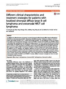
The most common symptoms were related to increased ICP (48.9%), especially headache (39.1%) and generalized seizures (30.4%). In contrast, focal neurological events such as motor deficits or simple partial seizures were present in 27.7% of our patients. Symptoms of both localized lesions and increased ICP were reported by 17.0% of our patients and 6.4% (3 patients) related that a bulging mass was their first symptom. Mean time between symptom onset and the first medical evaluation was 5.8 months. Focal manifestations led patients to seek medical attention sooner than increased ICP symptoms (p < 0.05). More than 75% of the patients with focal symptoms sought medical care in less than one month, as opposed to only 5% of those with increased ICP symptoms. Single brain metastases comprised 57.4% (27 patients) of our cases (Fig 1A/B). One patient (2.1%) had an extra-cranial metastasis only. The frontal lobe was the most affected site (33.3%) followed by the cerebellum (14.8%), the parietal lobe (11.1%), the temporal lobe (3.7%) and the thalamus (3.7%). Furthermore, diffuse single lesions (those involving more than one lobe) were responsible for 33.3% of these cases. Multiple metastases were found in 19 patients (40.4 %) (Fig 1 C/D). Two lesions were identified in 42.1% of these cases, three in 21.1% and more than three in 36.8%. In addition, symptoms of increased ICP (alone or with focal neurological events) were more frequent in patients with multiple metastases (78.9% versus 59.3% of the pa-
tients with a single tumor), though statistical significance was not reached (p = 0.07). No correlation could be found between symptom (focal or increased ICP) and the affected cerebral lobe (p = 0.1). Division of our cases according to supra- or infratentorial location revealed interesting correlations. Most patients had supratentorial metastases (33 patients - 70.2%), while infratentorial and multiple (at least one supratentorial and one infratentorial tumor) accounted for 12.8% and 17.0% respectively. Focal and increased ICP symptoms were both reported by 40.6% (13 patients) of the patients with supratentorial masses. All patients with infratentorial metastases reported increased ICP symptoms and 50% also presented some form of cerebellar syndrome. None of these patients related only focal symptoms. This correlation between supra/infratentorial location and the presented symptoms proved to be statistically significant (p=0.01). One case presented only extracranial symptoms (2.1%).
Table 3. Primary site distribution according to age. Primary Site
0 - 45 y
45 - 65 y
65y and above
Breast
3 (25.0%)
4 (18.2%)
4 (30.8%)
Lung
1 (8.3%)
5 (22.7%)
3 (23.1%)
Undetermined
2 (16.7%)
5 (22.7%)
1 (7.7%)
Melanoma
2 (16.7%)
2 (9.1%)
GI tumors Lymphoma
1 (4.5%) 2 (16.7%)
3 (23.1%)
1 (4.5%)
Ovary
1 (4.5%)
1 (7.7%)
Kidney
1 (4.5%)
1 (7.7%)
Seminoma
frontal mass. This mass is a metastatic deposit from a GI tumor in a male patient with focal symptoms. (B) Cerebral comput-
1 (8.3%)
Uterus
1 (4.5%)
Thyroid
1 (4.5%)
Sarcoma Total (n=47)
ed tomography after contrast injection revealing a hyperdense homogeneous left frontal mass from an ovary metastatic tumor. Patient had ICP symptoms. (C/D) Axial T1 weighted MRI after contrast injection showing two hyperintense heterogeneous mass (left fontal and occipital lobes) from a lung metastatic
1 (8.3%) 12 (25.5%)
Fig 1. Single brain metastases. (A) Axial T1 weighted MRI after contrast injection showing a hyperintense homogeneous left
22 (46.8%)
13 (27.7%)
tumor. Patient had symptoms of ICP.
Arq Neuropsiquiatr 2004;62(3-B)
811
Table 4. Treatment information. Number
Location
Other sites of metastases?
Single
Multiple
Extracranial
Supratentorial
Infratentorial
Both
Extracranial
Yes
No
20 62.5%
11 34.4%
1 3.1%
24 75.0%
5 15.5%
2 6.3%
1 3.1%
5 15.6%
27 84.4%
Surgery Yes N=32 No
7
8
0
8
1
6
1
3
12
46.7%
53.3%
-
53.3%
6.7%
40.0%
6.7%
20.0%
80.0%
Yes N=19
9 47.7%
10 52.6%
0 -
14 73.7%
0 -
5 26.3%
0 -
5 26.3%
14 73.7%
No N=28
18 64.3%
9 32.1%
1 3.6%
18 64.3%
6 21,4%
3 10.7%
1 3.6%
3 10.7%
25 89.3%
Yes N=8
4 50.0%
4 50.0%
0 -
5 62.5%
0 -
3 37.5%
0 -
3 37.5%
5 62.5%
No N=39
23 59.0%
15 38.5%
1 2.6%
27 69.2%
6 15.4%
5 12.8%
1 2.6%
5 12.8%
32 87.2%
Yes N=4
4 100.0%
0 -
0 -
2 50.0%
2 50.0%
0 -
0 -
0 -
4 100%
No N=43
23 53.4%
19 44.2%
1 2.3%
30 69.8%
4 9.3%
8 18.6%
1 2.3%
8 18.6%
35 81.4%
27
19
1
32
6
8
1
8
39
57.4%
40.4%
2.1%
68.1%
12.8%
17%
2.1%
17.0%
83.0%
N=15 RDT
Chemo
Other
Total N=47
RDT, radiation therapy; Chemo, chemotherapy.
There was a strong correlation (p < 0.001) between the number of metastases and supra- or infratentorial location. Single tumors were predominantly supratentorial (81.5%) while multiple metastases were exclusively supratentorial in 52.6%, while being exclusively infratentorial in 5.3% and both supra- and infratentorial in 42.1%. No correlation was identified between the number of metastases and primary tumor site (p = 0.8). Breast and lung cancer metastases were multiple in 63.6% and 44.4% of cases, respectively, and GI, thyroid, kidney tumors and seminoma cases did present only single metastases. Clinical details according to treatment option are given in Table 4. The treatment category named “Other” in this table refers to 4 patients who underwent steroid therapy only. Radiosurgery was em-
ployed in two operated patients after tumor recurrence. Two patients with metastases in different lobes were operated on (one case with bilateral frontal metastases and one with left cerebellar and parietal tumors). Three other patients had systemic disease with local symptoms (one case each of lymphoma, breast and thyroid cancer). No statistical significance was found between surgical treatment and the number of metastases (p = 0.4), however supratentorial location was found to be a significant factor (p = 0.014). Sixteen patients (34%) were still being followed as of November 2002. All surviving patients at that time had a Karnofsky Performance Status over 70. The maximum follow-up period was 36 months with a mean survival time after treatment of 8.3 months (Fig 2A). Mean survival time after surgical
812
Arq Neuropsiquiatr 2004;62(3-B)
Fig 2. (A) Overall survival. (B/C/D) Kaplan-Meier survival curve (B) according to treatment option (p = 0.2), (C) according to the presence of systemic disease activity (p = 0.68) and (D) for neurological symptoms (p= 0.08).
Fig 3. Kaplan-Meier survival curve (A) according to patient gender (p = 0.055), (B) for patient age (p = 0.78), (C) for metastases localization in the brain (p = 0.30) and (D) according to the number of metastases (p = 0.42).
Arq Neuropsiquiatr 2004;62(3-B)
treatment was longer than after other treatment options (14.0 versus 5.0 months), but statistical significance was not reached (p = 0.20). Six-month survival rates after surgery and other treatments were 66.7 and 25%, respectively (Fig 2B). Survival according to the presence of systemic disease activity is shown in Figure 2C. The evidence of extracranial disease did not statistically affect survival time (p = 0.68). Patients with focal symptoms had worse survival rates than those with ICP and extra-encephalic symptoms, though this was not statistically significant (p = 0.08, Fig 2D). There is a trend towards prolonged survival of female patients (p = 0.055 - Fig 3A). Additionally, no association was noted between the patient age (p = 0.78), location (p = 0.30) and the number (p = 0.42) of metastases and survival (Fig 3B, 3C and 3D) DISCUSSION Cerebral metastases are the most prevalent of CNS neoplasms, affecting almost 20% of all cancer patients8. Among intracranial diseases, CM are second only to cerebrovascular disease as the leading cause of mortality7,10. Most studies report that CM tend to involve male patients more frequently, but this seldom reaches statistical significance. In our series, there is a slight female prevalence, probably owing to the large number of breast neoplasm cases when compared to other patient series11. Despite single metastases being the most common, the most frequently reported symptom was ICP. This is largely due to the nature of tumor growth and its surrounding edema. Focal symptoms were more frequent in supratentorial lesions confined to one lobe only, but a direct relationship between symptoms and the number of metastases could not be established. Most patients of this series (68.1%) underwent surgical treatment. Our group adopts classical indications for surgical treatment, taking into account not only the number of metastases, but also the presence of systemic disease, patient age and symptoms, as well as the tumor site (supra- or infratentorial)11-17. Lagerwaard, et al. report systemic tumor activity as an independent, high-impact prognostic factor, and secondary prognostic factors as patient age, number of metastases and histological diagnosis of the primary tumor11. Sixty-two percent of the operated patients in our series had a single cerebral lesion, while 34.4% had more than one brain lesion and 3.1% had only extra-encephalic symptoms.
813
Only five of the 32 operated patients had uncontrolled systemic disease or were treating it. All five patients were under 65 years of age and three of them had single supratentorial metastases. The remaining two cases had two metastases each, but both could be removed during the same craniotomy. Surgical treatment was justified in these cases because of their age, evident signs of neurological deterioration and the likelihood that complete resection might improve the survival time12-15,17. Another nine cases with controlled systemic disease and multiple metastases were also operated on. All nine of these patients were under 65 years and the removal of all lesions could be performed via the same craniotomy. Neurological deterioration and controlled systemic disease also played an important role in the decision-making process for these cases. In the study of Bindal et al., the survival of patients with controlled systemic disease and multiple CM (which were all removed though one craniotomy) was compared to the survival of those with single metastases15. Both groups were submitted to surgical treatment and survival rates were similar. Furthermore, complete removal of multiple foci was associated with improved survival rates than suboptimal removal of single metastases. In line with Bindal et al. recommendations, patients in our series who had both multiple CM and were more than 65 years old were not submitted to surgical treatment15. Even though mean survival time after surgery was longer than after other treatments, statistical significance was not reached. Similarly, mean survival time of patients with controlled systemic disease (11 months), though longer, did not reach statistical significance when compared to patients with uncontrolled systemic disease (8 months) (p=0.68). Other secondary factors such as location and number of metastases, age and histological diagnosis were not associated with increased survival in our series. The only clinical factors which did suggest a direct association with increased mean survival time were patient gender and symptoms at onset. The patient gender link is possibly a result of selection bias, since our series possesses more breast neoplasm cases than most series. Those with extra-encephalic or increased ICP symptoms had a higher mean survival time than those with focal symptoms. Focal symptoms may be linked to a more rapidly deteriorating neurological condition, which can be apparent by a shorter time interval between symptom onset and the seek-
814
Arq Neuropsiquiatr 2004;62(3-B)
ing of medical attention (p < 0.05). In the present series the data about Karnofsky’s performance scales and/or ECOG (Eastern Cooperative Oncology Group) were not complete; however they have been long recognized as independent, high-impact prognostic factors in the clinical progression of brain metastases11. On the other hand, the results about the onset symptoms might refine medical decision-making and become a prognostic factor comparable to those scales in the future. Further investigations into the data presented here should be undertaken before our results can be introduced into clinical practice. In the age of evidence-based medicine, the average neurosurgeon is faced with a myriad of guidelines and recommendations which are often conflicting. Therefore, critical judgement is necessary to analyze the foundations upon which scientific papers are based. Frequently, guidelines are established based on inadequate research designs, which can lead to catastrophic results. Establishing clinical recommendations based on retrospective, non-randomized studies such as the present study can often be difficult. This study design has several inherent deficiencies, including selection bias. Treatment options were considered in the light of numerous factors, which may in turn have altered final outcome. On the other hand, it remains doubtful as to whether any form of prospective, randomized study could be conducted in a scientifically correct and ethical manner in the special case of brain metastases. Perhaps the most useful features of our study are the clinical characterization of this disease in Brazil and the association of presenting clinical signs and symptoms with other disease characteristics (such as metastases location and number) and final outcome. The analysis of treatment results and their relation to final outco-
me should, however, be examined thoroughly and compared to both clinical studies and neurosurgeons’ individual experience. REFERENCES 1. Aronson SM, Garcia JH, Aronson BE. Metastatic neoplasms of the brain: their frequency in relation to age. Cancer 1964;17:558-563. 2. le Chevalier T, Smith FP, Caille P, et al. Sites of primary malignancies in patients presenting with cerebral metastases: a review of 120 cases. Cancer 1985;56:880-882. 3. Schellinger PD, Meinck HM, Thron A. Diagnostic accuracy of MRI compared to CT in patients with brain metastases. J Neurooncol 1999;44:275-281. 4. Hochstenbag MMH, Twijnstra A, Wilmink JT, et al. Asymptomatic brain metastases in small cell lung cancer: MR-imaging is useful at initial diagnosis. J Neurooncol 2000;48:243-248. 5. Hirsch FR, Paulson OB, Hansen HH, Vraa-Jensen J. Intracranial metastases in small cell carcinoma of lung: correlation of clinical and autopsy findings. Cancer 1982;50:2433-2437. 6. Subramanian A, Harris A, Piggott K, Shieff C, Bradford R. Metastasis to and from the central nervous system: the “relative protected site”. Lancet Oncol 2002;3:498-507. 7. Victor M, Ropper AH. Intracranial neoplasms and paraneoplastic disorders. In Adams and Victor’s. Principles of neurology. Ed 5. McGrawHill, 2001;676-733. 8. Crusius P. Metástases intracranianas. In Siqueira MG, Novaes V (eds.). Tumores intracranianos - biologia, diagnóstico e tratamento, Rio de Janeiro: Editora Revinter, 1999. 9. Kaplan EL, Meier P. Nonparametric estimation from incomplete observations. J Am Stat Assoc 1958;53:475-482. 10. Antunes AC, Coutinho MF, Coutinho LM. Hemorrhage in intracranial metastases of melanoma: report of two cases Arq Neuropsiquiatr 1979;37:180-184. 11. Lagerwaard FJ, Levendag PC, Nowak PJCM, et al. Identification of prognostic factors in patients with brain metastases: a rewiew of 1292 patients. Int J Radat Oncol Biol Phys 1999;43:795-803. 12. Vecht CJ, Haaxma-Reiche H, Noordiijk EM,et al. Treatment of single brain metastasis: radiotherapy alone or combined with neurosurgery? Ann Neurol 1993;33:583-590. 13. Galicich JH, Sundareasan N, Arbit E, et al. Surgical treatment of single brain metastasis: factor associated with survival. Cancer 1980;45:381-386. 14. Patchell RA, Tibbs PA, Walsh JW, et al. A radomized trial of surgery in the treatment of single metatases to the brain. N Engl J Med 1990;322:494-500. 15. Bindal RJ, Sawaya R, Leavens ME, Lee JJ. Surgical treatment of multiple brain metastases. J Neurosurg 1993;79:210-216. 16. Lang FF, Wildrick DM, Sawaya R. Management of cerebral metastases: the role of surgery. Cancer Control 1998;5:124-129. 17. Araujo JF, Sperlescu A, Melacci EB, Balbo RJ. Brain metastasis of malinant melanoma: analysis of 13 cases. Arq Neuropsiquiatr 1997;55:292-297.