Introduction. Penetrating head injuries are rare in civilian practice, accounting for 0.4% of all head injuries (1), but are commoner in those affected by conflict.
J Royal Naval Medical Service 2013, Vol 99.2
55
Clinical Penetrating brain injury: a case of survival following blast fragmentation injuries to the head J McKinlay, JE Smith Abstract We present a case of penetrating head injuries caused by blast fragmentation, along with other serious injuries (including to the arms, face and neck), where a good recovery was made despite an Injury Severity Score (ISS) of 75. We suggest that survival and outcome are reliant on several factors and cannot be predicted from ISS, velocity of penetrating injury or presenting Glasgow Coma Scale (GCS) alone.
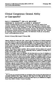
Introduction Penetrating head injuries are rare in civilian practice, accounting for 0.4% of all head injuries (1), but are commoner in those affected by conflict. Predictors of outcome (including discharge from hospital) usually rely on initial GCS (2), but other factors such as penetration across the midline have also been employed to predict outcome. A case of penetrating brain injury is presented that reminds us of the possibility of good neurological outcome from some types of penetrating head injury, despite attracting a maximum ISS of 75. Case study A 23 year old Royal Marine was brought to the emergency department (ED) at the Role 3 Medical Treatment Facility at Camp Bastion, Helmand Province, Afghanistan. He presented via helicopter retrieval (following an integrated trauma system pathway) approximately 30 minutes after sustaining severe head, face and extremity injuries from blast fragmentation following an explosion. He was noted to be conscious, with a patent airway, palpable radial pulse and a GCS of 15 (he was recorded as alert, talking and obeying commands) during transfer. On arrival in the ED his GCS had dropped to 9 (E4V3M2), and he underwent rapid sequence induction of anaesthesia and intubation prior to computed tomography (CT) scan (from head to hips) to define his injuries. This showed a penetrating injury to the brain via the right greater wing of the sphenoid bone, with the tract crossing the midline via the right frontal lobe, passing close to the head of the right caudate nucleus, through the lateral ventricles and left parietal lobe with an 8x6mm metallic fragment lying predominantly in the left parietal lobe. There was evidence of haemorrhage and small bony fragments along the tract with haemorrhage in the lateral ventricles, sub-arachnoid haemorrhage and small volume pneumocephalus (see Figure 1). No other injuries requiring immediate surgery were identified; the patient
remained sedated and was transferred to a neurosurgical facility (in Kandahar) where an intracranial pressure monitor was placed prior to repatriation to definitive care in the UK. His injuries were consistent with an ISS of 75 (New Injury Severity Score, NISS, was also 75). Following transfer back to the UK he spent 34 days in hospital (including 19 days on an intensive care unit) before discharge home to undertake outpatient rehabilitation. His GCS was 15 on discharge with some residual right-sided weakness. Discussion Penetrating brain injury may be caused by high and low velocity gunshot wound, blast fragmentation, or other nonballistic weapons such as knives and machetes. The kinetic energy (= ½×mass×velocity2) of an object that is delivered to the tissues following penetrating injury determines the type of injury inflicted. At lower velocities, injury results from direct disruption and laceration of tissues, while at higher velocities, tissue cavitation is the main cause of tissue damage. The highest velocities tend to have the worst associated damage (3). In this case the injuries were caused by blast fragments, which can be either of high or low velocity Figure 1. CT head scan showing penetrating injury with entry via the right greater wing of the sphenoid bone, the tract crossing the midline via the right frontal lobe, and an 8x6mm metallic fragment lying in the left parietal lobe.
56 Clinical
depending on their size and proximity to the explosive. Resuscitation measures aiming to minimise secondary brain injury are the mainstay of management, with prevention or early correction of hypoxia and hypotension improving outcome (4). There is scant evidence to guide formal prediction of outcome in different penetrating mechanisms of head injury. Traditionally, outcome prediction in penetrating brain injury has not differentiated the specific ballistic mechanism affecting the patient, and relies on initial GCS and other factors such as whether the missile crosses the midline (5-7). In this case, due to the nature of the patient’s injuries (both intracranial and other injuries) it was feared that outcome would be poor, and his survival and subsequent discharge could not have been foreseen from the usual outcome prediction measures. The GCS is one of the most commonly used scores for reliable assessment of the degree of coma in patients with craniocerebral injuries (8). The scale can assess brain function and damage, and give an indication of patient prognosis, particularly the motor score. Assessment of the overall severity of multiple system injuries is fundamental for appropriate treatment. One of the most important and frequently used measures of multiple injuries is the ISS (9). One series of patients (both military and civilian) with penetrating head injury showed that patients sustaining severe injuries following high velocity missile injury to the head have high early mortality (5), although the authors here discuss gunshot wound(s) alone rather than injuries from blast fragmentation. However, patients surviving to participate in an inpatient rehabilitation program had the potential for good functional improvement. Another study (6) suggested that the summed War Head Injury Score (WHIS) is a better predictor of mortality than either the GCS score or the ISS alone, due to the fact that battle casualties often have multiple injuries. Polin et al. performed
a multivariate analysis and prediction of outcome following penetrating head injury by high velocity gunshot wound (7). They looked at predictive factors from several studies and concluded that presenting GCS was a powerful predictor of outcome and that admission coagulopathy, the number of midline planes crossed by the projectile, and the volume of contused brain may be equally important. A prospective study of 61 patients following low velocity shrapnel injuries to the brain showed that non-operative management was favourable in terms of outcome in patients without significant haematoma on CT, or CSF leak via compound skull fracture (10). They also concluded that good outcome was evident in patients who initially had a GCS less than or equal to 8, in whom there was normal pupillary reaction and single lobe involvement with no midline shift on CT. The practical implication of this case study is that GCS on presentation may be a better predictor of survivability in patients with blast fragment penetrating brain injury than ISS alone. The prognosis for different mechanisms of penetrating brain injury may differ, and outcome prediction without thorough clinical evaluation is not reliable. Cavitation from high velocity gunshot wound within the cranial cavity may cause neurological disruption far in excess of a lower velocity fragment, and it is suggested that this may need to be considered when assessing prognosis in patients with penetrating head injury. An ISS of 75 is usually associated with unsurvivable injury (11). This case illustrates that ISS and patient outcome in terms of quality of life may not always be simply predicted, and cases should be dealt with on an individual basis with specific consideration to mechanism of injury.
References 1. Paiva WS, Monaco B, Prudente M et al. Surgical treatment of a transorbital penetrating brain injury. Clin Ophthalmol 2010;4(1):1103-1105.
7. Polin RS, Shaffrey ME, Phillips CD et al. Multivariate analysis and prediction of outcome following penetrating head injury. Neurosurg Clin N Am 1995;6(4):689-699.
2. Roberts P. 2004 The British Military Pocket Book. Crown Copyright. Chapter 16. AC 12552.
8. Teasdale G, Jennett B. Assessment of coma and impaired consciousness: A practical scale. Lancet 1974;2:81–83.
3. Dawodu S. Traumatic Brain Injury: Definition, Epidemiology, Pathophysiology. http://emedicine.medscape.com/ [retrieved Feb 6, 2007].
9. Baker SP, O’Neil B, Haddon Jr W et al. The Injury Severity Score: A method for describing patients with multiple injuries and evaluating emergency care. J Trauma 1974;14:187-196.
4. Coats TJ, Kirk CJC, Dawson M. Outcome after severe head injury treated by an integrated trauma system. J Accid Emerg Med 1999;16:182-18. 5. Zafonte RD, Wood DL, Harrison-Felix CL et al. Penetrating head injury: a prospective study of outcomes. Neurol Res 2001;23(2-3):219-226. 6. Sustic S. War head injury score: An outcome prediction model in war casualties with acute penetrating head injury. Mil Med 2001;166(4):331-334.
Acknowledgement The Academic Department of Military Emergency Medicine (ADMEM) is thanked for collecting and collating the appropriate data for this paper.
10. Wani AA, Ramzan AU, Nizami FA et al. Conservative management of bomb shrapnel injuries to the brain. Neurosurg Q 2012;22:94-98. 11. Copes WS, Champion HR, Sacco WJ, Lawnick MM, Keast SL, Bain LW. The Injury Severity Score Revisited. J Trauma 1988; 28(1) 69-77.
Authors Surgeon Lieutenant Commander J McKinlay1, Surgeon Commander JE Smith2,3 Affiliations 1 Specialty Trainee in Emergency Medicine, Emergency Department, Royal Infirmary of Edinburgh; 2,3 Consultant in Emergency Medicine, Emergency Department, Derriford Hospital, Plymouth, UK and Academic Department of Military Emergency Medicine, Royal Centre for Defence Medicine, Birmingham, UK. Corresponding Author: Surgeon Lieutenant Commander J McKinlay RN, Emergency Department, Royal Infirmary of Edinburgh, Edinburgh EH164SA