persed middle repetitive DNA sequence from Drosophila melanogaster that is concentrated on the euchromatic portion of the X chromosome.
Proc. Natl. Acad. Sci. USA Vol. 84, pp. 2843-2847, May 1987 Genetics
Cloning and characterization of a dispersed, multicopy, X chromosome sequence in Drosophila melanogaster (middle repetitive DNA/X chromosome structure/DNA sequence homology)
GAIL L. WARING AND JANE C. POLLACK Biology Department, Marquette University, Milwaukee, WI 53233
Communicated by Joseph G. Gall, December 24, 1986 (received for review May 12, 1986)
ABSTRACT We have isolated and characterized a dispersed middle repetitive DNA sequence from Drosophila melanogaster that is concentrated on the euchromatic portion of the X chromosome. In situ hybridization of the repeat unit to salivary gland chromosomes shows the sequence is distributed among approximately 10 major and 20 minor X chromosomal sites. Based on DNA sequence analysis of homologous sequences from three different cytogenetic regions, the 372base-pair repeat unit appears to be (A+T)-rich and noncoding and shows strong sequence conservation among units from different chromosomal regions. The nature and distribution of this sequence are suggestive of the hypothetical X chromosome DNA sequences thought to be involved in the primary establishment of sex determination and dosage compensation in Drosophila.
The Drosophila genome consists of three kinetic components, highly repetitive, middle repetitive, and unique DNA (1). The middle repetitive fraction constitutes -417% of the total genomic DNA and includes the tandemly repeated rRNA, 5S RNA, and histone genes. Most of the middle repetitive DNA in Drosophila melanogaster, however, is not tandemly repeated but rather is dispersed over the entire genome (2). It is composed of at least 50-100 different sequence families, each of which generally consists of 10-100 closely related sequences (2). The number of sequences within a family seems to be conserved in different strains of the species, but their chromosomal positions are highly variable. In marked contrast, in this paper we describe a dispersed middle repetitive family with repeats at chromosomal positions that are conserved between strains. More importantly, the repeat units are concentrated in the euchromatic portion of the X chromosome. A functional link between family members is suggested by the extensive sequence homology found between repeats from different chromosomal regions. The intriguing possibility that these sequences may play a role in the primary establishment of sex determination and dosage compensation in Drosophila is considered.
smaller quantities, DNA was prepared from phage lysates with RNase, NaDodSO4, and phenol. Subclones were generated by inserting gel-purified restriction fragments into the appropriate sites of the pGem-1 and pGem-2 transcription vectors (Promega Biotec, Madison, WI). Recombinant plasmids were used to transfect Escherichia coli HB101. To confirm their identity, alkaline minipreparations of plasmid subclones (6) were digested with restriction endonucleases. The resultant fragments were separated on agarose gels, transferred to nitrocellulose paper, and probed with RNA transcripts prepared from p112-2.3R DNA or its derivatives (see Fig. 2). Large-scale plasmid preparations were made by modification of the method outlined by Promega Biotec. Fly or embryonic DNA was prepared by homogenizing 300-500 flies or 1 ml of embryos in 5 ml of buffer (0.1 M NaCl/30 mM TrisHCl, pH 8.0/10 mM EDTA/7.7 mM 2-mercaptoethanol) containing 0.5% Triton X-100. Following low-speed centrifugation (3000 rpm, 15 min) the pellet was resuspended in extraction buffer (30 mM Tris4HCl, pH 8.0/100 mM EDTA, 0.5 mg of proteinase K per ml) made 1% in sarkosyl. After digestion overnight at 37°C, DNA was purified by CsCl gradient centrifugation. Small quantities of embryonic and salivary gland DNA were prepared from sonicated tissues treated with RNase, proteinase K, and
phenol/chloroform. Labeling Nucleic Acids. Nick-translation of DNA was carried out as described by Maniatis et al. (6). For highspecific-activity RNA probes, DNA cloned into pGem transcription vectors was linearized and transcribed with SP6 or T7 RNA polymerase as described by the supplier (Promega Biotec). Transcripts were labeled by including 12 /LM [32P]GTP in the transcription reaction mixture. DNA Sequence Analysis. Drosophila DNA fragments cloned into pGem transcription vectors were sequenced according to the manufacturer's protocols (Promega Biotec). Briefly, linearized DNA was denatured, reannealed in the presence of primers, and sequenced with DNA polymerase I (Klenow fragment) by the dideoxy chain-termination method (7). Reaction mixtures were analyzed on 4% and 8% acrylamide gels containing 8 M urea. In Situ Hybridization to Polytene Chromosomes. Polytene chromosome squashes were prepared as described by Bonner and Pardue (8). Hybridization with biotinylated probes was carried out according to Enzo Biochemicals (New York). DNA was nick-translated using Bio-11-dUTP (Bethesda Research Laboratories) and single-stranded DNA transcripts were synthesized using Bio-11-UTP (Bethesda Research Laboratories). Signal detection was achieved by using a streptavidin biotinylated peroxidase complex (Enzo Biochemicals).
MATERIALS AND METHODS Preparation of Nucleic Acids. The Maniatis X bacteriophage library of D. melanogaster genomic DNA (3) was screened by in situ plaque hybridization (4) under low stringency hybridization [4x SET (0.6 M NaCl/8 mM EDTA/0.12 M Tris HCl, pH 8), 43% formamide, 37°C] and wash conditions (0.3 M NaCl/30 mM sodium citrate, pH 7, 37°C) with the 32P-labeled single-stranded RNA probe described in the text. Phage from single plaques were purified from liquid cultures by centrifugation in CsCl block gradients (5); phage DNA was purified with NaDodSO4, proteinase K, and phenol. For
RESULTS The publication costs of this article were defrayed in part by page charge payment. This article must therefore be hereby marked "advertisement" in accordance with 18 U.S.C. §1734 solely to indicate this fact.
Isolation of Dispersed X Chromosome-Linked Sequences Homologous to 7F Region Sequences. In previous studies on 2843
2844
Proc. Natl. Acad. Sci. USA 84
Genetics: Waring and Pollack a
b c
d
e
f
h i
g
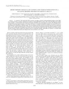
clones from regions other than 7F, we used single-stranded RNA probes and low stringency hybridization and wash conditions. A 2.3-kb SP6 transcript made in vitro from p112-2.3R was used to screen "=50,000 plaques. Based on our prior experiences, the probe hybridized to about 20 times the number of plaques (125) expected for a single-copy sequence. Fifteen positive plaques were picked at random for further analysis. EcoRI restriction digests of the phage DNAs indicated that we had isolated nine new independent nonoverlapping clones. Southern blot analysis (11) showed that all nine phage contained sequences homologous to p112-2.3R (Fig. 1). Though several phages showed more than one homologous fragment, five of the phages shared a 400-base-pair (bp) hybridizable fragment. To determine the cytogenetic location of the cloned sequences, the phage DNAs were nick-translatOd using biotinylated dUTP and hybridized in situ to salivary gland chromosomes. All of the phage gave a primary hybridization signal on the X chromosome (Fig. 2). In addition, several of the phage gave secondary signals at 7E and 8B. Since the primary hybridization signals were always on the X chromosome, these results strongly suggested that sequences homologous to p112-2.3R were dispersed but restricted to the X chromosome. Defining the Cloned Repeat Unit. To more closely define the extent of the homologous region, double digests of p112-2.3R were electrophoresed, transferred to nitrocellulose, and hybridized with homologous sequences subcloned from three different cytogenetic regions (llA, 13B, ilEF). Sequences subcloned for probes included a 2.3-kb EcoRI fragment from X25, a 0.4-kb EcoRI fragment from X12 (pX-12R, Fig. 2), and a 3.5-kb Pst I fragment from X30 (pX30P, Fig. 2) that included all of the 7F homologous sequences contained in this genomic clone. Hae III-EcoRI double digests of p112-2.3 yielded three Drosophila fragments: a 1200-bp Hae III fragment and two EcoRI-Hae III fragments, 600 and 500 bp in length (Fig. 2). Independent Southern blots of the 112-2.3R double digests hybridized with the subcloned probes from each of the three cytogenetic regions (llA, 13B, llEF) showed that only the 600-bp EcoRI-Hae III fragment contained homologous sequences. To identify the basic repeat unit, sequences from three different regions (7F, HiEF, 13B) were subcloned into pGem transcription vectors, linearized, and sequenced from oppo-
Kb -9 4 -6 6 -4 3
I:i.^ II'i'is
L*
* -._-
-2 1
-06
Ai.
.,
0.
_
FIG. 1. Southern blot analysis of phage DNA. Phage DNA was digested with EcoRI, fractionated on 0.8% agarose gels, blotted onto nitrocellulose, and hybridized with the 7F region subclone p112-2.3R. The numbers on the right indicate the positions of X HindIII molecular weight markers. Lanes: a, X8; b, X27; c, X18; d, X26; e, X25; f, X30; g, X4; h, X12; and i, X16.
early eggshell gene expression in Drosophila we isolated three independent cDNA clones complementary to follicle cell RNAs synthesized during stage 10 of oogenesis. Genomic clones complementary to one of the cloned cDNAs hybridized in situ to region 7F of the X chromosome. 7F has been identified as a major chorion structural gene site (9). Unlike other overlapping genomic clones from this region, one genomic clone (112) hybridized to 8B in addition to 7F. Comparison of the restriction maps of the overlapping 7F region clones indicated that the 8B hybridization must be due to homologous sequences within either the 3.2- or 2.3kilobase (kb) EcoRI restriction fragments just proximal to the chorion gene cluster (10). Accordingly, these R1 fragments were subcloned into pGem-2 transcription vectors, nicktranslated with biotinylated dUTP, and hybridized in situ to polytene chromosomes. The 3.2 subclone hybridized only to 7F, whereas the 2.3 subclone (p112-2.3R) gave signals at three sites: 7E, 7F, and 8B (data not shown). The comparable intensities of the p112-2.3R hybridization signals at all three sites suggested extensive sequence homology within these regions. As a first step in determining the nature of these homologies we screened the Maniatis library for genomic clones containing sequences complementary to p112-2.3R. To increase the probability of recovering genomic 7,D 7.E 71F 8,B liii I II I X8
#
112 #
8oD I I
99D
X27
X21 X 18
(1987)
I
I
X26
X25
l
I
I
X4
X30 /
X12
X16
N.
pX- 1 2R
NN
//
14A
11B
1 1,EF
1 1A 19C I I
\N
A
H
H
jI
R
FR
R p112-O.4A
Ii
P
p1 12-2.3R
HR
I 1I
HR
HR
II 1
pp
P
-i
pX3OP
IL
p 1 1 2-0.6RH I
e
~~p1 1 2-OARA 0.5Kb
EcoRI
pX-30P-0.4R
\
Haelli
pX-30P-0.4H
FIG. 2. Localization of p112-2.3R homologous sequences. (Upper) Recombinant phage DNAs (indicated below the line) were localized on the X chromosome to cytogenetic regions denoted above the line. # indicates secondary hybridization sites associated with X4, X18, X12, X21, X25, X30, and 112-2.3R. (Lower) Subcloned fragments from three regions (112, X30, X12) are expanded. The restriction sites used to generate 112 subclones included EcoRI (R), Hae III (H), and Alu I (A). EcoRI, Hae III, and Pst I (P) were used to generate X30 subclones; only 0.4-kb EcoRI fragments were subcloned from X12. The tandem arrangement of the 0.4-kb RI and Hae III units shown in pX30P is inferred from Southern blot and sequencing data.
2845
Proc. Natl. Acad. Sci. USA 84 (1987)
Genetics: Waring and Pollack
site ends using the dideoxy chain-termination method. The homologous portion of p112-0.6RH was reduced to a 400-bp EcoRI-Alu I fragment (pll2-0.4RA) by additional restriction enzyme analyses and Southern blotting experiments. Most of the homologous sequences in pX30P were reduced to 400-bp fragments by either EcoRI (pX30P-0.4R) or Hae III (pX30P0.4H) cleavage (see Fig. 2). The DNA sequences of homologous subclones from 112, X30, and X12 are shown in Fig. 3. Following manual alignment of the three sequences, a 372-bp consensus sequence was derived. As is evident from Fig. 3, a high percentage of each sequence (87-90%) matches the consensus sequence. The DNA appears to be noncoding since at least 2, and as many as 10, termination codons are found in all six possible reading frames for all three sequences. The consensus sequence as a whole is (A+T)-rich (72%) and is marked by several stretches of at least 6 bp that are 100% A+T. Other than the 30-bp direct repeats indicated in Fig. 3 (73% homologous) there are no distinctive indirect repeats or regions of dyad symmetry. The sequences shown represent a nonrandom subset of repeats since all were generated by EcoRI digestion. Our preliminary studies had shown that EcoRI and Hae III recognized the fundamental 372-repeat unit in pX30P. Following double digestions with EcoRI and Hae III, most of the homologous sequences were contained in 300-bp fragments. Taken together these data suggested a tandem arrangement of repeat units within pX30P such as that depicted in Fig. 2. Sequence analysis of a Hae III subclone (pX-30-0.4H) (not shown) confirmed the proposed sequence overlap and verified that our consensus sequence represented an entire repeat unit. Localization of the Repeat Unit on the X Chromosome. The results from our phage screen strongly suggested that sequences homologous to the 7F region were confined to the X chromosome. To confirm this finding the subcloned repeat unit p112-0.4RA was hybridized to Oregon R salivary gland chromosomes. Our previous studies had indicated that hybridization signals using double-stranded biotinylated DNA probes of