Jan 25, 2016 - 3-11, his 3-15, trp 1-1, ura 3-1. Cells were grown at 30 âC in complete medium (2% peptone, 1% yeast extract supplemented with carbon.
THEJOURNALOF BIOLOGICAL CHEMISTRY (c) 1993 by
Vol. 268, No. 3, Issue of January 25, pp. 1824-1829,1993 Printed in U.S.A.
The American Society for Biochemistry and Molecular Biology, Inc.
Cloning and Sequencingof a cDNA EncodingSaccharomyces cereuisiae Carnitine Acetyltransferase USE OF THE cDNA INGENEDISRUPTIONSTUDIES* (Received for publication, August 10, 1992)
Gyula Kispal$$, BalazsSumegiS, Klaus Dietmeiery, Ildiko Bock$, Gabriella Gajdosl), Tihamer TomcsanyiII , and Attila Sandor$ From the Slnstitute of Biochemistry, University Medical Schoolof Pecs, Szigeti 12, H-7624Pecs, Hungary, the lllnstitut fur Physiologische Chemie, Uniuersitat Munchen, Goethestr.33, 0-8000 Miinchen 2, Germany, and the )IDepartment of Zoology, Janus Pannonius University, Pees Ifjusag 6, Hungary
cDNA encoding for carnitine acetyltransferase acetyltransferase (CAT)’is specific for short chain fatty acids (CAT) of yeast S. cerevisiae was isolated by screening and has the highest activitywith acetylmoiety (4).CAT a yeast cDNA Xgtl 1 library with antibody. The whole activity has been detected in most of the examinedeukaryotic coding sequencewas obtained from the cDNA and from cells including different yeast strains, plants, and mammals. a YEP 13 DNA clone identified using the cDNA as Brown adipose tissue and heart muscle contain the highest probe. The coding sequence consists of 670 residues, CAT activity in mammalian organisms, but even the brain which amounts to a molecular mass of 77,300 kDa. has significant amount of this enzyme ( 5 ) . This cDNA was used successfully to disrupt the gene The function of CAT has not been described in as great for the mitochondrial isoenzyme of CAT, which was detail as has that of carnitine palmitoyltransferase or carnishown by measuring the enzyme activity and by imtineoctanoyltransferase.Inmammalian cells CATissupmunoblot. The acetylcarnitine content of these cells posed to buffer against rapid changes in acetyl-coA, thus decreased significantly. A search in the PIR protein data base revealed that besides the known carnitine preventing the depletion of free CoA; on the other hand the acyltransferases, choline acyltransferases are highly product of this enzyme, the acetylcarnitine, can serve as an homologous to yeast CAT. The mitochondrial CAT- easily accessible acetyl pool (6). CAT may participate in the deficient ( C A T ) cells were able to grow on different elimination of xenobiotic fatty acids as well, since CAT can fermentable and nonfermentable carbon sources, eventransfer short, branched chain fatty acid to carnitine, thus on acetate at the same rateas the parental strain. In promoting their urinary excretion(7). The metabolic significance of this enzyme was best demcontrast to these,13C NMRstudies revealed significant differences between parental andC A T cells. In C A T onstrated by the discovery of a patient with ataxic encephacells [3-13C]pyruvate was converted mainly to lactate lopathy, inwhom the activityof CAT was markedly decreased and acetate, whereas in the parental cells alanine and (8). tricarboxylic acid cycle intermediates were found as In lower eukaryotes, the transport of the acetyl moiety in the main products of pyruvate metabolism beside ace- or out of the mitochondria has beenconsidered as a possible tate. These results suggest diminished flux through the function for CAT.Inthealkane growing yeast, Candida pyruvate dehydrogenase complex in the absence of tropicalis acetyl-coA forms in the cytosol, so acetyl group can mitochondrial CAT in yeast cells. be transported to the mitochondria using the carnitine shuttle system (9). In the carnitine-deficient yeast, Torulopsis bouina, carnitine requirement could be demonstrated even underanaerobic Carnitine acetyltransferase belongs to that group of enzymes which catalyzes the reversible acylation of L-carnitine conditions. Since in this organism CAT is the only known carnitine-specific enzyme a biosynthetic role of CAT can be (1).Members of this group differ from each other in chain aerobic acetate length specificity. Carnitine palmitoyltransferases are specific suggested in addition to its function in the metabolism (10). for long chain fatty acids and participate in the transport of We decided to study the function of CAT in the yeast longchain acyl-CoA derivatives acrossthemitochondrial inner membrane to the site of P-oxidation (2). Medium chain Saccharomyces cerevisiae because in thisorganism mutations fatty acids are transferred from CoA to carnitineby carnitine can be introduced to specific sites, thus therole of an enzyme octanoyltransferase. Thisenzyme facilitates the transport of can be examined under in vivo conditions by comparing the medium chain fattyacids produced by the peroxisomal chain mutant and parentalcells (11).The physiological significance shortening system to the mitochondrial matrix (3). Carnitine of the supposed CAT functions in buffering the acetyl-coA level or in acetate transport can be determined this way. * This work was supported by Grants TKT“ 6 8 and OTKA 2009 Moreover, this type of study frequently reveals new, unexfrom the Hungarian Ministryof Welfare. The costs of publication of pected functions of an enzyme. this article were defrayed in part by the payment of page charges. We have already carried out the first steps leading to the This articlemusttherefore be hereby marked “aduertisement” in isolation of the gene encoding for S. cerevisiae CAT (YCAT), accordance with 18 U.S.C. Section 1734 solely to indicate thisfact. necessary to generate disruption of mutant yeast cells (12). T h e nucleotide sequencefs) reportedin this paper has been submitted to theGenBankTM/EMBLDataBankwith accessionnumbercs)
Z14021. § To whom correspondence should be addressed. Tel.: 36-72-24122;
Fax: 36-72-26244.
The abbreviations used are:CAT,carnitineacetyltransferase; YCAT, yeastcarnitineacetyltransferase; kb, kilobase(s); bp,base pair(s).
1824
Acetyltransferase Carnitine for
cDNA
1825
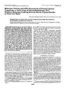
CAT was isolated from baker’s yeast with a subunit molecular 100 and disrupted by agitation with glass beads. The suspension was weight of approximately 65,000 as determined by SDS-poly- centrifuged at 3,000 X g for 10 min, and the supernatantwas used for enzyme assay. Mitochondria were isolated from complete medium/ acrylamide gel electrophoresis. Antibodies were raised against galactose-grown cells following the procedure of Daum et al. (19). this proteininrabbit,whichwerespecificandsensitive CAT assay was carried outin 50 mM Tris/HCl buffer (pH 7.4) enough to be used in screening a Xgtll yeast cDNA library. containing 100 mM NaCl using the 5,5’-dithiobis(nitrobenzoic acid) The identification and sequencing of cDNA and nuclear method (4).Procedures for SDS-polyacrylamide gel electrophoresis, DNA encoding for YCAT a n d t h e phenotype of C A T cells transfer of proteins from polyacrylamide gels to nitrocellulose membrane, and processing of immunoblots were described previously (20). are described in this report. C13 MNR studies were carried out on cells grown in complete medium/galactose to mid-log phase. Cells were centrifuged, and 1 g, MATERIALS AND METHODS wet weight, wastransferred to 10 ml of 50 mM phosphate buffer (pH The yeast strains used were: PSY 142; Mat CY,ura 3-52, leu 2-2, 7.4), containing 5 mM [3-’3C]pyruvate (MSD Isotopes). Cellswere ade2-I, leu 2-3, leu 2-112, his incubated under continuous shaking at 30 “C for 30 min, and the leu2-112, lys 2-801, M M Y 03; Mat CY, 3-11, his 3-15, trp 1-1, ura 3-1. Cells were grown at 30 “C in complete reaction was stopped by the addition of perchloric acid at a final medium (2% peptone, 1%yeast extract supplemented with carbon concentration of 5%. The mixture was subjected to a freeze-thaw sources, glucose, glycerol,pyruvate, lactate, acetate at 2 % concentra- cycle and centrifuged at 3,000 X g for 10 min. The supernatant was tion, or in minimal medium containing 0.75% yeast nitrogen base neutralized with KOH, and the potassium perchlorate precipitate was with ammonium sulfate (DIFCO) with the appropriate supplemen- removed by centrifugation. The clear supernatant was lyophilized. tation and carbon source. The 13CNMR spectrum of perchloric acid-soluble phase was obtained For identification of the cDNA clone encoding for YCAT, the on a GN-500 spectrometer at 11.75 tesla. All spectra reported in this cDNA library from S. cerevisiae in the expression vector Xgtll was work were acquired by using a 45” carbon pulse and 6-s delay between obtained from Clontech. The library was plated on 150-mm Petri pulses to ensurenonsaturating conditions. Samples were kept at dishes at 20,000 plaques/plate and replicas were loaded onto nitro25 “C during data acquisition. cellulose membrane from fiveplates. Filters were screened using antiThe carnitine and acetylcarnitine contents of yeast cellswere YCAT antibody raised in rabbit (12). The primary immunoreaction measured in the perchloric acid extract of complete medium/galacwas detected with alkaline phosphatase-conjugated anti-rabbit IgG tose-grown cell as described previously (21). phosphate/nitro blue and developed with 5-bromo-4-chloro-3-indolyl tetrazolium phosphatase reaction system (Kirkegard and Perry LabRESULTS oratories). Phage DNAfrom positive clones was isolated by the method of Helms et al. (13), and the inserts obtained after EcoRI Approximately IO5 plaqueswerescreened in a yeast S. digestion were subcloned in the plasmid pUC18. cerevisiae Xgtll cDNA library with antibody raised against Nuclear DNA encoding for YCATwas isolated from a plasmid YCAT in rabbit. Eleven positive signals were obtained from YEP 13 library containing 6-8 kb nuclear DNA Sau3Al fragments cloned into the unique BarnHI site of the plasmid (14). Bacteria were which the inserts were liberated and their size determined. plated at a cell density of 10,000/150-mm plate, and replicas were The longest insert, 2.2 kb, was subcloned in pUC18 plasmid made from eight plates onto Hybond nylon membrane (Amersham vector andanalyzedfurther.Restrictionenzymemapping Corp.). Filters were hybridized and washed according to the manu- (Fig. L4)allowed us to subclone overlapping fragments of this facturer’s protocol. As a probe for screening we used the 5’ EcoRI- cDNA t o M13 phage vector for sequencing. BarnHI fragment of that cDNA clone that had already been sequenced Sequencingthe2,125-bpinsertrevealedonelongopen A Trandom P (Fig. 1A). This fragment was labeled using [ c Y - ~ * P ] ~ by 1and 1994 bases and a 118-bp untransreading frame between primers (Boehringer), hybridized to theprepared filters, and exposed t o Kodak XAR films. Plasmids from the positive clones were isolated lated region followed by the poly(A) tail (Fig. 2). The supposed and subjected to restriction analysis and were subcloned in pGEM4 polyadenylation signal (AATACA) was found between 20 a n d (Promega) plasmid vector. 15 bases upstream of the poly(A) tail. To find the translation DNA sequences of cDNA and nuclear DNA were obtained after initiation codon we had to isolate the nuclear DNA encoding subcloning overlapping DNA fragments in M13mp18 and 19 vectors by the dideoxynucleotide method (15) using the T7 polymerase kit fortheYCAT,sincethecDNAwasnotlongenoughto ascertain the presenceof initiation codon at the 5‘ end. (Amersham). We used a YEP 13 nuclear DNA plasmid library, and t h e High molecular weightnuclear DNA from yeast cells for Southern blot were prepared as described in (16). Complete restriction endo- 5’ EcoRI-BanHI fragment of the cDNA (see Fig. 1 A ) was nuclease digestions were performed with a &fold excess of enzyme used as a probe. Eight x lo4 colonies were examined from under the conditions suggested by the suppliers. Digested DNA sam- which seven positives were isolated and subjected to restricples were run in 1% agarose gels, denatured,andtransferred to Hybond-N nylon membrane. Probes were labeled with [cY-”P]~ATP tion enzyme mapping.We found the characteristic restriction and hybridized as described before. The final washing conditions were sites on all of these clones, which proved the identity of t h e 42 “C in 2 X SSC, 0.1% SDS for 15 min. isolated nuclear DNAs (Fig. 1B). One of these nuclear DNAs Isolation of plasmids from Escherichia coli cells, restriction enzyme was sequenced from the unique PvuII site in the upstream mapping, subcloning ofDNA fragments, and transformation of E. direction. No intron in this DNA segment was found. This coli DH5n were carried out as described by Maniatis et al. (17). Transformation of yeast cells was done as described by Ito et al. (18). sequence revealed complete identity with the 5’ 300-bp seYCAT enzyme activity was measured in the cell free lysate or in quence of cDNA in the overlapping region. Moreover,a new isolated mitochondria. Cell-free lysate was prepared from cells grow- translation initiation codon was found 13 bp upstream from ing in complete medium with the carbon source as indicated in the t h e 5’ e n d of the cDNA preceded bya long repeat of TA. text to midlog phase, chilled, and harvested by centrifugation. Cells The full-length open reading frame encodes a polypeptide were resuspended in 50 mM Tris/HCl buffer (pH 7.4) containing 100 of 670 amino acids with a molecular size of 77.3 kDa. We mM NaC1, 1 mM EDTA, 1 mM benzamidine HC1, and 1%Triton Xsearched the PIR protein data base (release 28.0) for homologous proteins using the FASTA program (22). The highly homologous proteins were the rat carnitine octanoyltransferase (3) and r a t and human carnitine palmitoyltransferase (23, 24). Surprisingly, choline acetyltransferase (pig and Drosophila) (25, 26) showed high amino acid sequence homology to B ”--It YCAT, as well all over the protein sequence, not only in the FIG. 1. Restriction endonuclease map of yeast CAT cDNA already known LPXLPXPXL (24) motif. Besides this motif ( A ) and nuclear DNA fragment ( B ) . Thick lines represent the we found four others that are conserved in choline and carcoding region. nitine acyltransferases, as well, suggesting an important role
cDNA for Carnitine Acetyltransferase
1826
cCA.'F"'1'A:fA~"L'1'ATA~ATCCCTTAAAAACTAAAAAGAAAGCACTCATGAGGATCTGTCAT
60
' *
XIHKSSFM L XIBRCXXLa
HetArgIleCyeHIs TCGACAAC~CTCTCA~ACTTAAAGCA~'CTTCCGATAACGTCAAGGAGAGCAATGCATTCG 120
YCAT PCLAT RCOT HCPT RVDXAXFX L R V D X A X F X a RCPT
290-328 245-283 280-318 291-328
YCAT PCALT RCOT HCPT RCPT
455-478 412-435 445-468 452-475 452-475
KIQ-SSLXVXXX
6 ~er~rB~lrrLeuScrAclrLcuLy~AcpLr.uProlleTl~rSerArgArpAl~lietH~eSer
GCCdTTG'I'CAATfAC1'CCAC~CAAAAGGCCCAATTTCCCGTAGAGACAAATAATGGGGAA 180 26 ~~~~1c~nlAcnl'Y~SerTl~ffilnLysAlaGlnPheProValGluThrAsnAsnClyGlu
291-328
CACCI\'ITGGGCGGAAF.nCCGAACAAATTCTACCAGAACAAAAGGCCCAATTTTCAAGGC 240 46 IIIsTYrT~pAlnCluLy~P~~AcnLyoPb~TyrClrlAsnLysbrgProAenPheCInGly A'CTACCTTPCCTAAACAACAAGACTTACCATCATTACCCGTGCCCGAATTGAAGTCTACA 66 IleTl~rPl~rAlaLYt;OInGlnAcpLeuProSerLfuProValProGluLeuLyeSerThr
CTTCACAAGTATTT(~CAAACCATCCGCCCATTTTGCAATGATGTAGAAACTTTTGAAAGA 8 6 ~euAsPLYETYrLeuGlnTl~rlleAmProPheCyoAsnAlspV~1GluThrPheGluArg
300 360
CACCAGCTGTTATGTAAGGACTTCTCGGAGCACATGGGGCCTATCTTACAAGACCGATTG 420 106 ~1~~GlnLL.ULCuCysLysAByPheSelOlUHl.lletClyP~o~leLeu~lnAspArgL~u AAACAGTATGCCAACCATAAAAGAAdCTGGATGGCCAAGTTTTGGGATGAACAATCCTAT 126 LYDGluTYrAleAol~EcpLysArgAenTrpnetdlaLyePl~eTrpAspcluClnSerTyr
400
YCAT 488-502 PCLAT 445-459 RCOT 478-492
TTACAATACAACCAT~CTATTGTTCCATACGTC~CTTATTTTTATTCTCATATGCCATTA 540
HCPT RCPT
146 L C U G l n T Y r A s n A ~ p P r o I l e V ~ l P ~ ~ T y r V a l S ~ ~ T y ~ P h ~ T y ~ S a r ~ l ~ m ~ t p ~ ~ L ~ ~
CCGAATCATTTATCGAAGATCGATAATGATCCTTTGATTAAGGCTACTGCGATTATCTCA 600 166 ~ r o A ~ n H I ~ L e U S E ~ L y e I 1 e A o p b s n A E P P r o L e ~ l l ~ L y ~ A l ~ T h ~ A l ~ l l ~ l l ~ s ~ ~
485-499 485-499
FIG.3. Alignment of highly homologous regions present in choline and carnitine acyltransferases. RCOT, rat carnitine ocGGTATGCCATTfTGTnTGAAl'AGTTTTTCATTGATGTTTAACACTTCGAGATTGCCTGGT tanoyltransferase; HCPT, human carnitine palmitoyltransferase 11; 720 GlYli~tP~OPI~cCYBM~tA~~S~~Ph~S~~L~~~~tPl~~A~~Th~Se~A~gL~uP~~Gly RCPT, rat carnitine palmitoyltransferase;PCLAT, pig choline aceAAGCCACA(l'Gb7AAC(:AAGATACAAATATT1'TTTATTCAGTTTATGAGAACAACTTTGTA 780 tyltransferase. Bones indicate sequence identity; homologous amino I~YCPruB1UABPABn~1~~ADPTI~~AG~~IlePheTYrSerValTyrCluAenAenPheVal acids are shownby a one-letter code, and nonhomologous amino acids ACTATCCCl'l'ATAAACGGA.ZGrTTTACAAACTGATGACCCATGACGGQAATCACAAACCG 840 TbrIleAleTYrLYaUlYL~sPl~eTyrLYcLeu~etThrHIeAepClyAsnHIsLy~Pro are substitutedby X . Numbers show the positions of selected peptide fragments inthe respective protein. CTTTCCGAAAACGAAP.TCTGGAGGCAACTGTACTCTGTGGTATTCCAAGGATCGCAGTCC 900
ACCGTGGTTAAATTC.ZTCGAAGCTATTAAAG~CGAATCTTTACCCGTAGAAATTATCAAA 660 186 ThrValValLy~Pl~elleGluAlaIleLysAcpOluSsrLeuProV~1G1~I1~IleLys 206
226
246 266
LeuSerCluAEnCluIleTrPA~~Cl~LeuT~rSerYalV~lPh~Gl~Glys~ffil~s~~
GAl'CCCAAACTAGCTGGCATTGGTTCTCTCACCTCTTTACCTCGTGATCAATGGCGTGAA
286 ABPProl~yELCUGly(ilyIleGlySerLeuTl~rSerLeuProArgAe~lnTrpl\rg0lu GTACATAl'GGAGCTTATCAA~GATCCTATTTCTCAGGATTCACTAGAAACAATCCATAAG
306 ValII1c~etCluLeu~letLysA~pProIleScffilnAspSerL~uGluThrIleHIsLys 326 TCTTCCTTTATGCTATCTTTGGATCTTGACCAATCCCCTGTCACTTTGGAAGAAAAGTCA
9GO
A
8
"
1020
2
3 4
1
2
3
4
1080
SerSerPhenetLeuCyeLe~A~pL~~A~pOlnSerProValThrLeuGluGluLYaSer ACAAATTGCTGGCACGGTGATGGTATTAACAGATTCTACGATAAGTCTTTACAGTTCCTA 1140 346 AraA~nCyeTrpllIsG1yA~pC1YIleA~nAnlPheTY~A~pLysSsrLeuClnPheLeu GTCACCGGTAATGGTTCATCAGGTTTCTTAGCTGAACACTCGAAGATGGATGGTACGCCA
1200
kb
366 ValTllrClyAsnGlySerSerGlyPheLeuAlaGluHI8SerLY~netABffilYThrPro
ACATTCTTTTfAAATAACTACGTTTGTCAGCAGTTGAATAAACTAGATGTGGATGACTTC 1260 386 T h r L E u P h e L r u A s n A s n ' C y ~ V ~ l C y s C l n C l n L e u n s y ~ L ~ " A ~ p V ~ 1 A ~ p A ~ p P h a ATCACAAAAGTAATTACGCCATCATCTACGGTGGCAATGAAACCTATGGAACTGCCCTTC
1320
406 I ~ o t A r C L y a V a l I l s T h r P r o S e r S e r T h r Y a l d l a n s r L y ~ P ~ ~ H ~ t G l ~ L ~ ~ P ~ ~ P h ~ AfTA'fCACACCGAACATTCATAAAGCAATCGAATCTGCCCAACTACAATTTAAGGAAACA
1380
426 1leIleTl~rProLyclleHIeLysAlellcGluSerAlaGlnLeuGlnPheLyeGluThr ATTGGTGAGCATGACCTACGTGTTTGGCACTACAACAAATATGGAAAAACGTTTATAAAA 1440 446 I l f G l Y G l ~ H I ~ A c p L t ~ A r ~ V ~ l T ~ p H I ~ T Y ~ A ~ ~ L Y ~ T Y r G l Y L Y ~ T h r P h e ~ l e L Y ~
CGCCATGGCATGTCACCTGATCCATTTATTCAACAAGTTATCCAACTGGCGGTTTTCAAA 1500
72.2-
PC-
-
6.1 - 0 0 5,O-
4.63.0-
9
Ii q-/
20-
466 AL'~III~GlylietSerProAspAlsPheIleGlnGlnVal1leClnLeuAlaValPheLYs
TATCTGAAACGACAACTACCAACTTACGAGGCTGCTTCCACGAGAAAATACTTCAAAGGC 1560 486 Ty~L~"Ly~ArgClnLe"ProThrTyffiluAl~AlaSerThrArgLYnTYrPh~LY~G1Y
FIG.4. Southern blotting analysis of PSY 142 and MMY 03 parental and C A T nuclear DNA. Total nuclear DNA from AATCCCGATGTTCCTACTGCAGAAAAGATTCAGGC~TTGAAACATTCTGCAAAAGAGCAT 1680 yeast cells was digested with BamHI, separated on 1%agarose, and 526 A ~ ~ G l y A ~ p V a l P ~ o I l ~ A l a C l u L y ~ l l e G l n n l a L e u L Y ~ H I ~ S e r A l ~ L Y ~ G l u ~ I ~ blotted to Hybond nylon membrane. Lane 1 , PSY I42 parental; lane TCGACGTACCTGAAAAATGCTGCAAATGGTAATGGTGTCGATCGTCATTTCTTCGGTCTA 1740 2, PSY 142 C A T ; lane 3, M M Y 03 parental; lane 4, M M Y 03 C A T . 616 SerTl~rTyrLeuLysAonAlaAlnAsnG1YAenG1YVa1AopAr~HIsPhePhsClYLeu Probes were the random prime-labeled Ura3 gene (panel A ) and the AAGAATATGCTAAAATCTAATGATGACCAAATTCCGCCCCTTTTCAAAGATCCCTTATTT 1800 686 L y a A s n ~ r t L e u L y 8 S e r A ~ " A ~ p A ~ p C l " l l ~ P ~ o P r o L ~ " P h ~ L y ~ A ~ p P ~ ~ L e u P h e 5'end EcoRI-BamHI fragment of YCAT cDNA (panel B ) . Hybridizationandwashingconditionswere AATTATTCTTCAACTTGGTTGATCI'CCACATCTCAACTATCTTCGGAATATTTTGAC~T as described. The film was 1860 586 A a n T y r S e r S e r T h r T r p L e u I l s S s r T h r S e r C l n L a u S ~ ~ S ~ ~ G l ~ T y ~ P h ~ A ~ ~ G l ~ exposed for14 h. Positions of molecular weight markersare indicated TATGGTTGGTCCCAAGTAAATGACAACGGGTTTGGACTGGCATACATGTTGAATAACGAG 1920 on the left side. 606 lYrGlyTrpSerGlnVelAenAegAsnClYPl~eGlvLeuAleTyrnetLeuAsnAsnClu CGTACTGAAACTGGTAGATCTGTGTCCACCGCCTCCTTAGAATTTGTTTCTAAATGGCAA 1620 506 A r g T h r G l ~ T h r G l y A r g S ~ ~ V ~ l S ~ ~ T h ~ A l ~ S ~ ~ L ~ ~ G l ~ P h ~ V ~ l S ~ ~ L Y ~ T ~ p G l ~
TUGCTGCATATCAATATTGTCAACAAACCAGCCAAGAGTGGAGCCAGTGTTAACAGATTA 1980 626 TepLc~llloIlrAsnIleValAsnLyoProAlsLY~S~~GlYAlaSerValAenAr~Leu
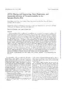
Ura 3 into the yeastgenome was examined by Southern blot probes (Fig. 4). Hybridusing the Ura3 and YCAT cDNA as ization with the Uragene revealed one additional banda t 5.9 CGAAAAGCAAAGTTATGAATTTTTCACCTTTCCTCTTCAATTAATTTGTGAATATTTTTG 2100 666 ArgLyoAlaLysLeu*** kb besides the endogenous Ura 3 gene that was a t 6.1 in PSY TTAAGTATCTACACTTACATACGTATAAATAGGGGAGAAAAAAGGGCTTACG~TA 2160 142 and at12 kb in M M Y 03 strains. Using the YCAT cDNA 2173 CTCATCATTACTAn as probe hybridization was found in the transformed cells FIG.2. Complete sequence of yeast CAT DNA. Nucleotides are numbered on the right and amino acids on the left. Nucleotides only in one band with the same molecular size as the extra underlined in the 5'- and 3'noncoding regionare the supposed TATA Ura gene. These results show that the Ura3 gene integrated Cat of yeast chromosome. Since box and polyadenylation signal, respectively. An asterisk marks the into only one site to the locus the finalwashing conditions were done at very low stringency 5' end of the cDNA. conditions (2 x SSC, 0.1% SDS at 42 "C) the simple YCAT fortheseamino acid segments (Fig. 3). These conserved cDNAhybridization pattern suggested that this Cat gene regions are found in thesecond half of the proteinsequences. isolated by us is present only in one copy per haploid yeast The YCAT cDNA was used to generate disruption of mu- genome. CAT enzyme activity was measured in the whole cell lysate, tant yeast cells of M M Y 03 and PSY 142 strains. The NdeIBamHI fragment of cDNA was replaced by the yeast Ura 3 isolated mitochondria, and in themitochondrial supernatant gene (27), and this constructwas liberated from the plasmid (cytosol) of the transformed C A T PSY 142 cells grown in by digestion with EcoRI and HpaI restriction enzymes. The complete medium/galactose. In thewhole cell lysate of C A T released fragment wasused to transform yeast cells from cells the enzyme activity decreased to less than 5% of the which Ura+ transformants were selected. The integration of parental value. An evenmore pronounced decreasein the CACTA1"PATTTATCTCAAGCTGCTGA~AAAl'TTTTGACGCCTTGGAAAATGAGAATAAA 2040 646 H l ~ T Y r T Y r L ~ ~ S ~ ~ C l ~ A l s d l a d o p c l u I l e P h e A s p A l ~ L ~ ~ G l ~ A ~ ~ G l ~ A ~ ~ L y ~
Acetyltransferase Carnitine cDNA for CAT activity was observed in theisolated C A T mitochondria; however, in the cytosol we found no change in the enzyme activity(TableI).The low residual CATactivityinthe mitochondria of CAT- cells may be contamination from the cytosol. Immunoblot analysisof the mitochondrial and cytosol fractions of parental and C A T PSY 142 cells revealed that ouranti-CATantibody recognizes only themitochondrial CAT; no immunoreactive material was found in the cytosol of parental or C A T cells. Moreover, no CAT protein was found in mitochondria of C A T cells (Fig. 5). Free carnitine and short chain acid-soluble carnitine esters were measured in the perchloricacid extract of parental and C A T cells (Table 11). In C A T cells without any significant change in t h e free carnitine level, the short chain carnitine ester concentration decreased below the detection limit of our radioactive carnitineassay. This result shows that after theelimination of CAT from the mitochondria no othermajor carnitine acetyltransferase activity remained in the yeast cells. The phenotypeof C A T cells wasstudied by comparing the growth of mutant cells with the parental strain on different carbon sources. C A T cells were able to grow on complete or minimal medium plates containing fermentable or nonfermentable carbon sources such as glycerol, lactate, pyruvate, and acetate (data not shown). No difference was found in the time required for the colonies to reach the visible size, in the TABLE I CAT activities in total homogenate and subcellular fractions of P S Y 142parental andC A T cells Cells were grown in complete medium containing 2% galactose to midlog phase. Total cell homogenate was prepared by disrupting cells with glassbeads. Mitochondriaand cytosolwere separatedafter lyticase treatment asdescribed by Daum et al. (19). CAT activity in thesesamples wasmeasuredusing the 5,5'-dithiobis(nitrobenzoic acid) assay (4). Milliunits/mg protein meansnmol of substrate converted/min/mg of protein a t 25 "C. Values represent means zk S.D. for three separatecultures. CAT activity Total homogenate
1827 TABLE I1 Carnitine andacetylcarnitine concentration in PSY 142 parental and C A T cells Perchloric acid extract was prepared from cellsgrowing in complete medium/galactose. Carnitineandacetylcarnitinedeterminations were carried out asdescribed in (21). Values represent means of two determinations. Concentration Acetvlcarnitine Carnitine ~~
~
nmol/g wet weight
Parental
73.5
43.5
CAT-
63.8
0.0
1 2
67 8
i ii
I I
9
10
III I I
A
B
3 50
40
30
20
PPM
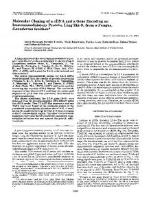
FIG.6. P y r u v a t e m e t a b o l i s m of PSY 142 p a r e n t a l a n d C A T cells. Cellswere grown in completemedium/galactose to [3-"CC]pyruvate-~ontaining midlog phaseandthentransferredto medium. After a 30-min incubation a t 30 "C perchloric acid extract was prepared from the cells and subjected to NMR spectroscopy. Panel A, parental; panel R, C A T cells. The assigned peaks were as follows: I, Glu C2; 2, Asp C2; 3, malate C3; 4, Asp C3; 5, Glu C4; 6, Glu C3; 7, pyruvate C3; 8; acetate C2; 9, lactate C3; 10, Ala C3.
Mitochondria cytosol
logarithmic growth rate, orin the yield of growth between the parental and C A T cells. Parental 250 f 10 550 zk 10 30 2 5 The pyruvatemetabolism of mutant cells was investigated using NMRspectroscopy after labeling the metabolite pool of CAT12 f 3 3 f 2 30 2 7 complete medium/galactose-grown cells with[3-"CC]pyruvate. The NMR spectrum of the perchloric acid extract obtained MW 7 2 3 4 from the labeled parental and CAT- cells is shown in Fig. 6. x103 , This experiment revealed a striking difference in the metabolism of pyruvate between parental and C A T cells. In the 94parental cells a significant amount of CI3 label still remained inpyruvate. Ahigh amount of added label was found in acetate,alanine,andas expected inthe citric acid cycle intermediates, glutamate and malate, and in oxaloacetate the derivative, aspartate. A signal in the lactate peak could not be detected. In C A T cells a very high signal intensity was 43found in the lactatepeak in addition to acetate.Alanine was labeled much less compared with the acetate peak, and much less pyruvate was converted to the citric acid cycle intermediate in these cells. The decreased I3C incorporation into citric FIG.5. Immunoblot analysisof PSY 142 parental and C A T acid cycle intermediatesis obvious if the acetate C2 and cells. Complete medium/galactose-grown cells were subjected to cell glutamate C4 or even when alanine C3 and glutamateC4 are fractionation as described by Daum et al. (19) and analyzed on SDS- compared. Comparison of glutamate C3 and C4 peak intenpolyacrylamide gel electrophoresis. Lanes 1 and 2 are parental mito- sities can be used as a rough estimation for the turnover rate chondria and cytosol. Lanes 3 and 4 are C A T mitochondria and cytosol, respectively. Separated proteinswere transferred to nitrocel- of the citric acid cycle assuming identical pool size, because cycle through acetyl-coA,label appears lulose membrane and were immunodecorated by antiserum raised as pyruvate enters the against yeast CAT in rabbit. The migration position of pure YCAT first in glutamate at C4 and then scrambling in the second is shown by an arrow. part of the cycle transfers the label from C4 to the position milliunits/mg protein
1828
cDNA forCarnitine Acetyltransferase
of C3 and C2. The C3/C4 ratio was 0.50 and 0.45 in parental transferases. A highly homologous motif (LPXLPXPXL) (24) and C A T cells, respectively, suggestingthat the turnover ratein choline and carnitine acetyltransferases has alreadybeen of the citricacid cycle is not affected by the absenceof CAT. described, which is present in YCAT as well. This motif was suggested to be the choline or carnitine binding site, since DISCUSSION this fragment is presentonly in this group of proteins and is To begin to investigate the function of CAT we set out to missing fromother acyltransferases. Comparing carnitine and determine the primary structure of this enzyme. We chose choline acyltransferaseswith YCAT we found fourmore this protein because despite the fact that it is present with highly conserved motifs in the second half of the proteins we think that high activity and amount in eukaryotic cells no clear-cut in which was present only in these proteins. Thus, it is very uncertain to assign a specific function to any of vivo function could be assigned t o it. We decided to isolate the cDNA encodingCAT from a yeast these fragments a t present. Based on the high homology between YCAT-choline aceX g t l l library becausewe had alreadyisolated CAT from yeast tyltransferases, and YCAT-carnitine acyltransferases we proS.cerevisiae and raised antibody against the enzyme in rabbit, which was specific and sensitive enough toscreen an expres- pose that CAT of primitive eukaryotic cells might be the sion library. A 2.2-kb insert of one of the positive clones was common ancestor of both enzymescatalyzing acyltransfer sequenced and used as a probe t o screen a YEP 13 library from CoA to a quaternary amine-containing substrate. The in vivo function of YCAT was examined with the help containing 6-8-kb yeast nuclear DNA fragments. Thecoding sequence was localized on the nuclear DNA by restriction of mutant cells in which the mitochondrial CATwas deleted mapping andsequenced in thatregion that corresponds to theby disruptingthe CAT gene withUra 3. Cells withthe 5’ end of cDNA. The overlapping region of this nuclear DNA disrupted gene were able to grow on different fermentable and nonfermentable carbonsources, evenon acetate,showing that fragment was completely identicaltothatfoundinCAT CAT has no indispensable function in acetate metabolism. cDNA.Reading the nuclear DNA upstream we found the supposed initiation codon 16 basesfrom the 5’ end of cDNA. The significance of the previously suggested role of CAT in Upstream from this ATG between 2 and 17 bases the long transport of acetyl group in or outof the mitochondria can be repeat of TA can be assumedto be the TATA box, the questioned based on these results. Participation of CAT in transcription initiation site. The ATG found on the nuclear the mitochondrial acetate transport cannot be ruled out in wild type cell; however, yeast cells have enough capacity to DNA is suggested as the translation initiation site, since yeast bypass a supposed block of acetate metabolism in theabsence favors the initiationcodon closest to the TATAbox. The one long open reading frame obtained this way encodes of mitochondrial CATusing other metabolic pathways. Surprising differences were inthepyruvate metabolism a polypeptide of 670 amino acids witha molecular weight of studied by NMR after labeling cells with [3-13C]pyruvate. In 77,300. This molecular weight is approximately 12,000 larger parental cells pyruvate is converted mostly t o acetate, alanine, than that of the isolated YCAT enzyme (65,000). The measC A T cells the entry and citricacid cycle intermediates. In the ured size of isolated YCAT, however, cannot be used for the of pyruvate to alanine and citric acid cycle intermediates prediction of the amino terminusof mature enzyme because decreasedsignificantly; instead,pyruvate was converted of the uncertainty in the determination of molecular weight mostly to lactate and acetate. The turnover rate of the citric by SDS-polyacrylamide gel electrophoresis, but it suggests acid cycle was not affected in the C A T cells. that theenzyme undergoes post-translational processing. Similar functions of CAT in insect flight muscle (29) and The identityof this isolated cDNA was proved by the fact later in rat (30) and human (31) mitochondria have been that we could use it to disrupt the yeast chromosomal CAT demonstrated. CAT, in the presence of carnitine, was able to gene, thus giving rise to C A T cells. The elimination of CAT promote the pyruvate dehydrogenase activity in these orgaactivity from the mitochondria of the transformed cells was nisms supposedly by decreasing the pyruvate dehydrogenase shown by enzyme activity determination andby immunoblot. inhibitoracetyl-coA level. Rapidadaptationto a sudden T h e resultthattheacetylcarnitinecontent of these cells increase of energy demand on the initiation of muscle activity decreased significantly showsthat no othermajor CAT activ- was suggested as the physiological role of this indirect reguity remainedin the cell. The residual low CAT activity in the latory effect. cytosol of mutant cells did not change, suggesting that this In yeast, growing on galactose pyruvate can be converted cDNA codes for the mitochondrial isoenzyme of CAT. to citric acid cycle intermediate via pyruvate dehydrogenase CAT isencoded by the nuclear genome, thus this protein is or by the cytosolic pyruvate decarboxylase and aldehyde oxisynthesized in the cytoplasm as precursor and then imported dase, in which acetate is formed first and than converted to to the matrix space. Importis followed by aproteolytic acetyl-coA or directly to a citric acid cycle intermediate by cleavage by matrix processing proteasesthat remove the the glyoxylate cycle. Based on these resultswe cannot decide mitochondrial targetingsequence from the amino terminus of which pathway is affected most by the absence of CAT, but the protein; consequently the mature protein that can be we clearly show that CAT has an important function in the isolated is usually smaller than its precursor form (28). We metabolism which will be studied furtherusing this C A T cell assume that the found nucleotide sequence encodes the pre- line. cursor of YCAT (pre-YCAT) containinga targeting sequence. REFERENCES T h e amino terminusof the pre-YCAT contains mostly apolar and positively charged amino acids resembling that of mito1. Hoppel, C. L. (1976) in The Enzymes of Biological Membranes (Martonosi, A., ed.) Vol. 2, pp. 119-143, Plenum Publishing Corp., New York chondrial targeting sequences, giving further support to our 2. Fritz, I. B. (1963) Adu. Lipid Res. 1,285-334 idea. 3. Chattejee, B., Song, C. S., Kim, J. M., and Roy, A. K. (19&3)Biochemistry 27,9000-9006 A search of the PIR protein data base yielded homologies 4. Fritz, I. B., Schultz, S., and Srere, P. A. (1963) J. Biol. Chem. 2 3 8 , 2509between the alreadyknown carnitine acyltransferases rat and 2517 Marquis, N. R., and Fritz, I. B. (1965) J. Biol. Chem. 240,2193-2196 human carnitine palmitoyltransferase 11, rat carnitine octan- 5. 6. Bremer, J. (1962) J. Biol. Chem. 237,2228-2231 7. Chalmers, R. A,, Roe, C. R., Tracey, B. M., Stacey, T. E., Hoppel, C. L., oyltransferase giving an additional proof for the identity of and Millineton. D. S. (1983) Biochem. SOC.Trans. 11.724-725 this isolated cDNA. Two other proteins appeared with the 8. DiDonato, S.: Rimoldi, M., Garavaglia, B., and Uziel G: (1979) Neurology 2 9 , 1578-1583 same degree of homology, pig and Drosophila choline acetyl-
cDNA forCarnitine Acetyltransferase 9. 10. 11. 12. 13. 14. 15. 16.
Kohlaw, B. B., and Tan-Wilson, A. (1977) J. Bacteriol. 129,1159-1161 Emaus, R. K., and Bieber, L. L. (1983) J. Biol. Chem. 258,13160-13165 Rothstein, R. J. (1983) Methods Enzymol. 1 0 1 , 202-211 Kispal, G., Cseko, J., Alkonyi, I., and Sandor, A. (1991) Biochim. Biophys. Acta 1086,217-222 Helms, C., Dutchik, J. E., and Olson, M. V. (1987) Methods Enzymol. 1 5 3 , 69-82 Broach, J. R., Strathern, J. N., and Hicks, J. B. (1979) Gene (Amst.) 8, 121-127 Sanger, F., Nicklen, S., and Culson, A. R. (1977) Proc. Natl. Acad. Sci. U. 5. A. 74,5463-5467 Davis. R. W.. Thomas. M.. Cameron. J. R.. St. John. T. P.. Scherer.. S... and Padgett, R . A. (1980) !dethods Enzymol: 66,4041411 Maniatis, T., Fritsch, E. F., and Sambrook, J. (1982) Molecular Cloning:A Laboratory Manual, Cold Spring Harbor Laboratory, Cold Spring Harbor, '
17.
NY
18. Iti,.H., Fukada, Y., Murata, K., and Kimura, A. (1983) J. Bacteriol. 1 6 3 , 163-168 19. Daum, G., Bohni, P. C., and Schatz, G. (1972) J. Biol. Chem. 2 6 7 , 1302813033 20. DeBlas, A. L., and Cherwinsky, M.M. (1983) Anal. Biochem. 1 3 3 , 214219
1829
21. Sandor, A., Cseko, J., and Alkonyi, I. (1989) J. Chromatogr. 4 9 7 , 250-257 22. Pearson, W. R., and Lipman, D. L. (1988) Proc. Natl. Acad. Sci. U. S. A. 86,2444-2448 23. Woeltje, K. F., Esser, V., Weis, B. C., Sen, A,, Cox, W. F. McPhaul, M. J., Slaughter, C. A., Foster, D. W., and McGarry, J. D. (1990) J. Biol. Chem. 265,10720-10725 24. Finocchiaro, G., Taroni, F., Rocchi, M., Martin, A. L., Colombo, I., Torri Tarelli, G., and DiDonato, S. (1991) Proc. Natl. Acad. Sci. U. S. A. 88, 661-665 25. Berrard, S., Brice, A,, Lottspeich, F., Braun, A., Barde, Y., and Mallet, J. (1987) Proc. Natl. Acad. Sci. U. S. A. 84,9280-9284 26. Brice, A,, Berrard, S., Raynaud, B., Ansieau, S., Coppola, T., Weber, M. J., and Mallet, J. (1989) J. Neurosci. Res. 2 3 , 266-273 27. Botstein, D.,Falco, S. C., Stewart, S. E., Breenan, M., Scberer, S., Stinchcomb, D. T., Struhl, K., and Davis, R. (1979) Gene (Amst.)8,17-25 28. Pfanner, N., and Neupert, W. (1990) Annu. Reu. Biochem. 69,331-353 29. Childress, C. C., Sacktor, B., and Traynor,D.R. (1966) J. Biol. Chem. 2 4 2 , 754-760 30. Lysiak, W., Lilly, K., Toth, P. P., and Bieber, L.L. (1988) Nutrition 4 , 215-219 31. Uziel, G., Garavaglia, B., and DiDonato, S. (1988) Muscle Nerve 1 1 , 720724