Jun 23, 1986 - length copy of a functional human gene for ferritin heavy chain has been isolated. The gene consists of four exons spanning approximately 3 ...
Proc. Natl. Acad. Sci. USA Vol. 83, pp. 7226-7230, October 1986 Biochemistry
Cloning, characterization, expression, and chromosomal localization of a human ferritin heavy-chain gene (iron-storage protein)
MATTHIAS W. HENTZE*, STEPHEN KEIM*, PANOS PAPADOPOULOSt, STEPHEN O'BRIENt, WILLIAM MODIt, JIM DRYSDALEt, WARREN J. LEONARD*, JOE B. HARFORD*, AND RICHARD D. KLAUSNER*§ *Cell Biology and Metabolism Branch, National Institute of Child Health and Human Development, National Institutes of Health, Bethesda, MD 20892; tDepartment of Biochemistry and Pharmacology, Tufts University School of Medicine, Boston, MA 02111; and tLaboratory of Viral Carcinogenesis, National Cancer Institute, National Institutes of Health, Frederick, MD 21701
Communicated by Gilbert Ashwell, June 23, 1986
(15, 16). Several genomic clones for ferritin heavy and light chains have been described. Many of these genes appear to be pseudogenes that do not contain introns and that may have arisen by reverse transcription of ferritin mRNA in germ-line cells (17, 18). However, Costanzo et al. (17) have recently described a heavy-chain genomic clone that has three introns. This gene contains potential regulatory promoter sequences characteristic of a functional gene but was not shown to be expressed. We have cloned what appears to be the same gene. We show here that this gene is derived from chromosome 11 and demonstrate its functionality by expression of human mRNA in both transiently transfected and stably transformed murine fibroblasts.
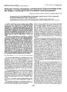
A genomic phage clone containing a fullABSTRACT length copy of a functional human gene for ferritin heavy chain has been isolated. The gene consists of four exons spanning approximately 3 kilobases and has been localized to chromosome 11. The functionality of the gene was demonstrated by the fact that both transient transfectants and stable transformants of murine fibroblasts actively transcribe human ferritin heavychain mRNA.
Ferritin is the major iron-storage protein found in all eukaryotic cells. It is a spherical complex of 24 subunits. Human ferritins consist of various combinations of heavy (Mr 21,000) and light (Mr 19,000) chains. These two subunits share extensive sequence homology but are derived from different genes (1, 2). Ferritin may serve several functions in iron metabolism. A major function may well be to sequester and thus detoxify iron that is taken up by cells but not utilized for metabolic requirements. High iron uptake results in a high fractional delivery of iron to ferritin and low iron uptake results in a high fractional delivery of iron to non-ferritin pools (3). The level of ferritin itself seems to determine this fractional distribution within the cell. In addition, ferritin iron can also be released for cellular utilization, especially under conditions of iron need. The amount of ferritin in cells is highly regulated; iron leads to a large increase in the synthesis of both heavy and light chains (4). The relative synthesis of the two chains varies greatly in different tissues, as a function of development and pathological state (5-7). Regulation by iron seems to occur at both transcriptional and translational levels (8, 9), whereas developmental changes seem to occur at the transcriptional level (2). Because of ferritin's central role in iron metabolism, understanding the molecular basis for ferritin regulation is essential to an understanding of cellular iron homeostasis. A major step toward this understanding has been taken with the cloning of cDNAs encoding human ferritin heavy and light chains (2, 10, 11). Analysis of the genes encoding these proteins will extend these studies and allow us to begin to examine regulatory control elements. Genomic cloning of active ferritin genes is somewhat confounded by Southern blot hybridization studies, which show that the genes for each subunit are part of multigene families (1). In situ hybridization demonstrates ferritin heavy-chain genes on at least eight different chromosomes and light-chain genes on at least three chromosomes (12-14). Which of these represent functional genes and which, pseudogenes is unclear. Detection of human ferritin by immunologic techniques in stable rodent-human hybrid cell lines has pointed to chromosomes 11 and 19 as the source of heavy and light chains, respectively
MATERIALS AND METHODS Cloning of a Human Genomic Ferritin Heavy-Chain Gene. Clones for the human ferritin heavy-chain gene were isolated from a previously described total human genomic liver library (19). Three hundred thousand plaques were screened by plaque hybridization (20) using the 496-base-pair (bp) Pst I 3' fragment and the 308-bp Pst I 5' fragment from the ferritin heavy-chain cDNA clone pHF16 (10). The fragments were labeled by the random priming method (33). Thirty-three positive clones were plaque-purified, and DNA was isolated according to standard procedures (20). One of the clones, M11, gave rise to two EcoRI fragments, of length 5.2 and 10.6 kilobases (kb), which hybridized to the 5' or 5' and 3' probes of the cDNA, respectively. A 7.2-kb HindIII fragment spanning the entire hybridizing region was isolated and subcloned into the plasmid vector pUC8 (Bethesda Research Laboratories) for further characterization (see Fig. 1). Restriction Fragment Characterization and Sequence Analysis. The recombinant plasmid pUCM11 was amplified in Escherichia coli strain HB101, and plasmid DNA was isolated by an alkaline-lysis protocol (20). Aliquots (1 ,ug) were digested to completion with various restriction enzymes (Bethesda Research Laboratories, or International Biotechnologies, New Haven, CT) and analyzed by electrophoresis in 0.7% agarose gels and Southern blotting (21). Several fragments were gel-isolated by use of NA-45 DEAE membranes (Schleicher & Schuell) according to the manufacturer's protocol and subcloned into the replicative form of the M13 vectors mp18 or mpl9 (Fig. 1). DNA sequencing was carried out by the dideoxy chain-termination method of Sanger et al. (22), using the 15-bp M13 primer (Pharmacia) and [a-32P]dATP (Amersham). Abbreviations: bp, base pair(s); kb, kilobase(s). §To whom reprint requests should be addressed at: Cell Biology and Metabolism Branch, NICHD, NIH, 9000 Rockville Pike, Bethesda, MD 20892.
The publication costs of this article were defrayed in part by page charge payment. This article must therefore be hereby marked "advertisement" in accordance with 18 U.S.C. §1734 solely to indicate this fact.
7226
Biochemistry: Hentze et al.
Proc. Natl. Acad. Sci. USA 83 (1986)
Expression of pUCM11 in Murine B6 Fibroblasts. The recombinant plasmid pUCM11 was transfected into mouse B6 fibroblasts. Briefly, in each experiment 20 jig of RNase A-treated plasmid DNA was introduced into 1-2 x 10⁶ host cells by use of the calcium phosphate precipitation method as described by Graham and van der Erb (23). For assay of transient expression, the cells were harvested with a cell scraper 48 hr after transfection and washed with phosphatebuffered saline, and total cellular RNA was prepared by the guanidinium/CsCl method (20). For assay of stable transformation, the plasmid pUCM11 was coprecipitated with the herpes simplex virus thymidine kinase gene at a molar ratio of 30:1, and transformants were selected and grown in medium containing hypoxanthine, aminopterin, and thymidine (HAT medium) for 3-4 weeks. A single-stranded 546-bp "antisense" Sac I fragment (465 bp of the Sac I fragment plus 81 bp of the M13 mpl8 polylinker and primer DNA; Fig. 1) was uniformly labeled with [a-32P]dATP, using the appropriate M13 template and the Klenow fragment of DNA polymerase, and isolated by electrophoresis in an 8 M urea/6% polyacrylamide sequencing gel. This fragment (=3 x 10⁴ cpm) was hybridized to 50 Ag of total cellular RNA (20) and treated with 200 units of nuclease S1 (Boehringer Mannheim) for 30 min at 45°C. The samples were phenol-extracted, ethanol-precipitated, resuspended, denatured, and analyzed in an 8 M urea/6% polyacrylamide gel. Genomic Southern Analysis and Chromosomal Localization. High molecular weight genomic DNA was purified (20) from either peripheral blood lymphocytes or Epstein-Barr virustransformed cultured lymphocytes. Aliquots (8-15 ug) were digested to completion with EcoRI, HindIll, or BamHI and single- or double-blotted onto Biotrans nylon membranes (ICN) for not less than 15 hr. After the membranes were baked for 2 hr at 80°C, prehybridization and hybridization were carried out according to the manufacturer's protocol. DNA probes were 32P-labeled by random priming, to a specific activity of -3 x 10⁸ cpm/4ug, and 106 cpm/ml was added to the prehybridization solution for overnight incubation. The blots were washed three times in changes of 2x SSC/0.5% NaDodSO4 at room temperature and subsequently 0.1x SSC/0.5% NaDodSO4 at 65°C for 20 min each. (lx SSC is 150 mM NaCl/14 mM sodium citrate.) The blots were subjected to autoradiography with a Lightning Plus screen (DuPont) by incubation at -70°C for 1-5 days. For chromosomal localization, a panel of 44 different human-rodent cell hybrids was analyzed by Southern blotting. The hybrid cell lines used have been described in detail (24). As a hybridization probe an upstream genomic fragment was isolated from pUCM11 (see Fig. 1). This probe did not cross-hybridize with the DNA of mouse RAG and Chinese hamster E36 cells. The chromosomal assignment was made by statistical discordance analysis according to published methods (25). H
P
P S
!
I
I
a a
a a m
I
r
7227
RESULTS Cloning of a Ferritin Heavy-Chain Gene. A human liver genomic library was screened with a 32P-labeled 3' fragment of the heavy-chain cDNA. This 3' probe covered bases 309 to 804 of the cDNA according to the numbering of Boyd et al. (10). Thirty-three clones were chosen by plaque hybridization, purified, and partially characterized. One ofthese, M11, was chosen for further characterization based on the following criteria: the phage DNA also hybridized to the 5' cDNA fragment (bases 1-308), but the 5' and 3' coding regions of the phage insert could be separated by a restriction enzyme (in this case EcoRI) that does not cut the cDNA, thus suggesting that the clone might contain an intron. This resulted in two EcoRI fragments of 5.2 and 10.6 kb. The 5.2-kb fragment strongly hybridized only to the 5' cDNA probe, whereas the 10.6-kb fragment strongly hybridized to both the 5' and the 3' cDNA probe. The entire coding region is contained within a 7.2-kb HindIII fragment, and this was subcloned in pUC8 for further mapping (Fig. 1). Restriction fragments of this clone (pUCM11) were subcloned in M13 for sequencing by the dideoxy chain-termination method (see Fig. 1). Sequencing demonstrated that this genomic clone contains the cDNA sequence previously reported (11) without a single nucleotide change (Fig. 2). The gene is organized as four exons and three introns. The major transcription start site is located 24 bases downstream of the first T of the "TATA box" (ref. 27; see below). This start site will be referred to as nucleotide + 1 of the predicted mRNA. Intron 1 is about 1.65 kb long and is placed between mRNA bases 331 and 332. Intron 2 is 256 bases long, between bases 478 and 479; intron 3 is 96 bases long, between bases 604 and 605. The entire gene covers about 3 kb. Our sequence differs from that of Costanzo et al. (17) in a few places but is otherwise identical in intron placement and in the putative promoter region. The 3' noncoding region is identical to the published cDNA, including the consensus polyadenylylation signal AATAAA. Our sequencing agrees with that of Boyd et al. (10) and the corrected cDNA sequence reported by Costanzo et al. (17, 28). Expression of the Ferritin Heavy-Chain Gene. Examination of the sequence of the ferritin gene suggested that it represented a functional gene. In order to test the functionality of the cloned gene, murine B6 fibroblasts were transformed with the 7.2-kb HindIII fragment. Stable transformants were made by cotransfecting with the herpes thymidine kinase gene and selecting transformants in hypoxanthine/aminopterin/ thymidine (HAT) medium for 4 weeks. RNA prepared from nontransformed B6 cells, ferritin transformants, and human K562 cells was hybridized to the Sac I fragment derived from the 5' end of the gene (see Fig. 1). These DNARNA hybrids were subjected to nuclease S1 digestion to establish (i) the presence of human ferritin gene transcripts and (ii) the transcription initiation site. The results (Fig. 3) show that human ferritin mRNA was produced in the genomic
SB
P
P
I-
UI
I
Ri PvB Sp K I
H I< ^
AAA -
1 kb I
-
-
i
FIG. 1. Restriction map of pUCMll. One-microgram samples of plasmid DNA were digested to completion with each of the following enzymes: HindIII (H), Pst I (P), Sac I (S), Bal I (B), EcoRI (Ri), Eco RV (Rv), Sph I (Sp), and Kpn I (K). The samples were analyzed by electrophoresis in 0.7% agarose gels and Southern blotting. Duplicate blots were probed separately with two random-primed nonoverlapping fragments of the ferritin heavy-chain cDNA (10). For sequence analysis, multiple DNA fragments were isolated by gel electrophoresis and subcloned in the M13 vectors mpl8 and mpl9. Arrows indicate the 5' to 3' direction of transcription of the single-stranded M13 template. Open triangles indicate the 465-bp Sac I fragment used to construct a DNA probe for the nuclease S1 protection assay. The 0.7-kb Pst I fragment (solid squares) represents the genomic probe isolated for the chromosomal-localization experiments. Exons are represented by boxes.
7228
Proc. Natl. Acad. Sci. USA 83 (1986)
Biochemistry: Hentze et al.
GAGCTCCGCCAGAGCGCGCGAGGGCCTCCAGCGGCCGCCCCTCCCCCACAGCAGGGGCGG GGTCCCGGCCCACCGGAAGGAGCGGGCTCGGGGCGGGCGGCGCTGATTGGCCGGGGCGGG +1 Exon 1 CCTGACGCCGACGCGGCIIAI GAGACCACAAGCGACCCGCEIjGGCCAGACGTTCTTCGC CGAGAGTCGTCGGGGTTTCCTGCTTCAACAGTGCTTGGACGGAACCCGGCGGCTCGTTCC
CCACCCCGGCCGGCCGCCCATAGCCAGCCCTCCGTCACCTCTTCACCGCAGCCCTCGGGA GCTGCCCCAAGGCCCCCGCCGCCGCTCCAGCGCCGCGCAGCCACCGCCGCCGCCGCCGCC TCTCCTTAGTCGCCGCCATGACGACCGCGTCCACCTCGCAGGTGCGCCAGAACTACCACC
MetThrThrAlaSerThrSerGinVolAr8GlnAsnTyrHisG
AGGACTCAGAGGCCGCCATCAACCGCCAGATCAACCTGGAGCTCTACGCCTCCTACGTTT InAspSerGIuAIaAaI IeAsnArgGInIIeAsnLeuGluLeuT~rAlaSerTyrVaIT
ACCTGTCCA:IGTGAGCGCGGGCGGGCCTAAGCGGTGGC ..................... yr LeuSe rMe t
*
approx. 1.5kb ................................
AGCTTCTCTGATCCCTAGTATAAACACT
Exon 2
TCAGTGTTCCCCTTTCAGIaTTACTACTTTGACCGCGATGATGTGGCTTTGAAGAACTTT
SerTyrTyrPheAspArgAspAspVaIAiaLeuLgsAsnPhe
GCCAAATACTTTCTTCACCAATCTCATGAGGAGAGGGAACATGCTGAGAAACTGATGAAG AlaLysTyrPheLeuHisGInSerHisGiuGluArgGluHisAlaGluLysLeuMetLys 60 70
CTGCAGAACCAACGAGGTGGCCGAATCTTCCTTCAGGATATCAWGGTGAACAAAAGATCC LeuGInAsnGInArgGlyGlyAr8iIePheLeuGInAspIleLys 8
TAGGGGTGTCATACTTCATCATCTGGCAGTGTTCGGGTATCAGAAATCACTTAAACTAGC AATTGCCCTTATAAAGTGATGATACACTGGGCTTTTGCCTTTTGTGCTTTTTTAGGCTTA CCATCTAAACTAAATTAGGCAAATAGTAATGTCCCTTTT GCCAAAACGTGGTGGTTAGAG ATGATGGGCTTGCTGACTTCTAGGTTAGTTGGTAGAGATGCATTAACCTATTCTCATTCA Exon 3 GPAACCAGACTGTGATGACTGGGAGAGCGGGCTGAATGCAATGGAGTGTGCATTACATTT LysProAspCysAspAspTrpGluSerGlyLeuAsnAlaMetGluCysAlaLeuHisLe 90 100 GGAAAAAAATGTGAATCAGTCACTACTGGAACTGCACAAACTGGCCACTGACAAAAATGA uGluLysAsnValAsnGInSerLeuLeuGluLeuHisLysLeuAlaThrAspLysAsnAs 110 120
CCCCCATIGTGAGTATTGGAACCCCAGGAAATAAATGGAGGAAATCATTTGCCTTAGGGAT pProHi s
Exon 4 TGGGAAAGCTGCCCACTAACTGTCTTGCCCCATTGTTTTGCAG[=GTGTGACTTCATTGA LeuCysAspPhel leGl 1 30
GACACATTACCTGAATGAGCAGGTGAAAGCCATCAAAGAATTGGGTGACCACGTGACCAA uThrHisTyrLeuAsnGluGInValLysAlolIeLysGIuLeuGlyAspHisValThrAs
140 150 CTTGCGCAAGATGGGAGCGCCCGAATCTGGCTTGGCGGAATATCTCTTTGACAAGCACAC nLeuArgLysMetGlyAlaProGIuSerGlyLeuAlaGluTyrLeuPheAspLysHisTh 160 170
CCTGGGAGACAGTGATAATGAAAGCTAAGCCTCGGGCTAATTTCCCCATAGCCGTGGGGT rLeuGlyAspSerAspAsnGIuSerEnd
180 GACTTCCCTGGTCACCAAGGCAGTGCATGCATGTTGGGGTTTCCTTTACCTTTTCTATAA GTTGTACCAAAACATCCACTTAAGTTCTTTGATTTGTACCATTCCTTCAIATAGAAAT TTGGTACC
transformants. One major start site for transcription was observed for the ferritin transformants. This site is located at the sequence GCAGGG, with the initiating A placed 24 bases downstream of the TATA box. A minor start site was observed at the sequences CCAGAC placed 32 bases downstream of the TATA box. K562 cells produced transcripts from both of these start sites, in equivalent amounts. In addition, a minor protected fragment of about 480 bases was seen variably in the transfectants. No protected 5' ferritin gene fragment was observed in the host murine B6 cells. Nuclease S1 protection experiments also demonstrated ferritin gene expression in transiently transfected B6 cells (data not shown). Localization of the Ferritin Heavy-Chain Gene. To establish the chromosomal localization of this gene, we subcloned a 0.7-kb Pst I segment whose 3' end is about 0.5 kb upstream
FIG. 2. Sequence of the cloned human ferritin heavy-chain gene. The nucleotide sequence was determined by the dideoxy chaintermination method (22) according to the sequencing strategy outlined in Fig. 1. The major transcription initiation site is marked +1, a minor start site (at +9) is underlined. The TATA box and the polyadenylylation signals are boxed. The presence or absence of the bases underscored with an asterisk could not be determined with certainty. The sequence as displayed was prepared using the DNA-DRAW program (26) on a Dec 10 computer (Digital Equipment, Maynard, MA). The encoded amino acid sequence is shown below the nucleotide sequence.
of the TATA box (Fig. 1). This fragment therefore lacked coding sequence and was determined not to contain highly repetitive sequences as evaluated by hybridization to total genomic DNA. When total genomic DNA samples from normal individuals were digested with EcoRI or HindIII and analyzed by Southern blot hybridization to a ferritin heavychain cDNA probe, a complex pattern of at least 15 bands was seen (Fig. 4). In contrast, when the subcloned Pst I fragment was used, only a single band was identified under high-stringency conditions with HindIII (7.2 kb) and with BamHI (4.3 kb) (data not shown), as predicted by restriction mapping of the clone. The genomic EcoRI fragment of 6.3 kb was larger than predicted by the map of the clone. This is probably due to truncation of this fragment by Hae III or Alu I in the construction of the library. Utilizing this Pst I fragment, we attempted to identify the
Proc. Natl. Acad. Sci. USA 83 (1986)
Biochemistry: Hentze et al. a
b
B
A
B6
K562
c
d
Eco RI Eco RI
do"
7229
Hind
Hind III
_R
III
17-
5466
10.6 -^
5.7
-
7.2
40
tW t..
31 Oo-
OM =:304 280-
27 13-
296
_
4.0 3.1
-
234D-
FIG. 3. pUCM11 is actively transcribed in transformed murine fibroblasts. Mouse B6 fibroblasts were stably transformed with pUCM11 as described under Materials and Methods. Total cellular RNA was prepared from the transformed and nontransformed host cells and from the human erythroleukemia cell line K562. The Sac I fragment (see Fig. 1) was uniformly labeled with [a-32P]dATP, hybridized to 50 Ag of total cellular RNA, and treated with 200 units of nuclease S1 at 370C for 45 min. (Left) Two protected fragments of 304 bases and 296 bases are indicative of two independent start sites in human K562 cells. The 546-base fragment represents undigested full-length probe. (Right) Comparison between transformed (lanes a and b) and nontransformed (lanes c and d) B6 fibroblasts with (lanes b and d) and without (lanes a and c) addition of nuclease S1. Total cellular RNA from nontransformed cells fails to protect any fragment of the labeled probe (lane d). In contrast, RNA from pUCMlltransformed cells protects 304- and 296-base fragments, representing the major and minor start sites, respectively (lane b), also seen in K562 cells. The S1 protection assays of K562 and B6 RNA were analyzed in separate polyacrylamide gels. Lengths in nucleotides are indicated. Sizes were determined by extrapolation from molecular size markers.
chromosome containing this gene. This was accomplished by analyzing rodent-human hybrid cell lines whose complement of human chromosomes is well-characterized (24). No crosshybridization of this probe was seen to either hamster or mouse DNA. Discordance analysis of positive hybridization to HindIII digests of hybrid cell genomic DNA localized this gene to chromosome 11 (Fig. 5).
DISCUSSION The cloning and expression of this structural gene for human ferritin will allow us to begin to examine the molecular mechanisms involved in ferritin gene expression and regulation. The critical role that this molecule plays in intracellular iron metabolism underscores the importance of being able to evaluate the regulatory aspects of ferritin gene expression. Human ferritin heavy-chain gene sequences represent a large multigene family dispersed over at least eight different human chromosomes. It has been hypothesized that some of these are processed pseudogenes that have arisen by reverse transcription of mRNA in germ-line cells (17). It is therefore important to emphasize that the gene we report here has a coding region sequence identical to human ferritin heavychain cDNA sequences reported from several tissue sources (2, 10, 28) (with minor sequence discrepancies likely due to
FIG. 4. The 0.7-kb Pst I probe recognizes a single member of the human heavy-chain multigene family. Total genomic DNA samples (10 ,ug) from unrelated healthy individuals were digested to completion with EcoRI or HindIII, electrophoresed in 0.7% agarose gels, and blotted onto nylon membranes. The blots were probed with the 496-bp 3' Pst I fragment from the ferritin heavy-chain cDNA (A) or the 0.7-kb Pst I probe (see Fig. 1) from pUCM11 (B) and then washed at high stringency. A shows that the cDNA recognizes various members of a multigene family. In contrast, the 0.7-kb Pst I probe from pUCM11 recognizes a single member of that family and represents a unique fragment in the human genome (B). Sizes (in kb) determined by comparison to standards of known lengths are given.
technical reasons). Recently, Costanzo et al. (17) also described a human ferritin heavy-chain gene that has an identical intron/exon structure, the same apparent intron sizes, and essentially identical sequences, including the promoter and the 5' and 3' noncoding regions. Using primerextension analysis, Costanzo et al. suggested a transcription start site at the cytosine 29 bases downstream of the TATA box. Our nuclease S1 protection experiments indicate two transcription initiation sites in either K562 cells or stably transformed B6 cells. Based on these data, we suggest the adenines at either 24 or 32 bases downstream from the 5' end of the TATA box as transcription start sites. This is in agreement with previous observations that show that the majority of mRNAs begin with an adenine (29). The existence of a multigene family, probably including several pseudogenes, necessitates the demonstration of the functionality of the cloned genomic fragment. Our demonstration that pUCMll gives rise to human ferritin heavychain RNA, as revealed by the nuclease S1 protection experiments using RNA from stably transformed mouse cells, proves that it contains a functional gene. The localization of this gene to human chromosome 11 confirms the assignment made by Worwood et al. (15), who used immunologic techniques for the detection of human ferritin in rodent-human hybrid cells. A discordance value of 4% is not unusual and is likely to be explained by minor chromosomal rearrangements in a few of the hybrid cell lines used. This gene has several interesting structural features. Several G+C-rich regions are present upstream of the TATA box. Three contain the hexanucleotide Spl core binding sequence and possess, respectively, 10, 9, and 8 bases of the decanucleotide Spl consensus sequence as defined by Dynan
7230
Biochemistry: Hentze et al. c
Proc. Natl. Acad. Sci. USA 83 (1986)
50
.r
IL
40
m-
0D
30
0 U co (a)
20
10 a) 0.
1
2
3
4
5 6
7
8
9 10 11 T